Skip to main content Accessibility help
×
Hostname: page-component-76fb5796d-25wd4 Total loading time: 0 Render date: 2024-04-26T23:28:14.566Z Has data issue: false hasContentIssue false

Chapter 3 - Cellular and Molecular Biology of Hypoxic-Ischemic Encephalopathy

from Section 1 - Epidemiology, Pathophysiology, and Pathogenesis of Fetal and Neonatal Brain Injury

Published online by Cambridge University Press:  13 December 2017

David K. Stevenson
Affiliation:
Stanford University, California
William E. Benitz
Affiliation:
Stanford University, California
Philip Sunshine
Affiliation:
Stanford University, California
Susan R. Hintz
Affiliation:
Stanford University, California
Maurice L. Druzin
Affiliation:
Stanford University, California
Get access

Summary

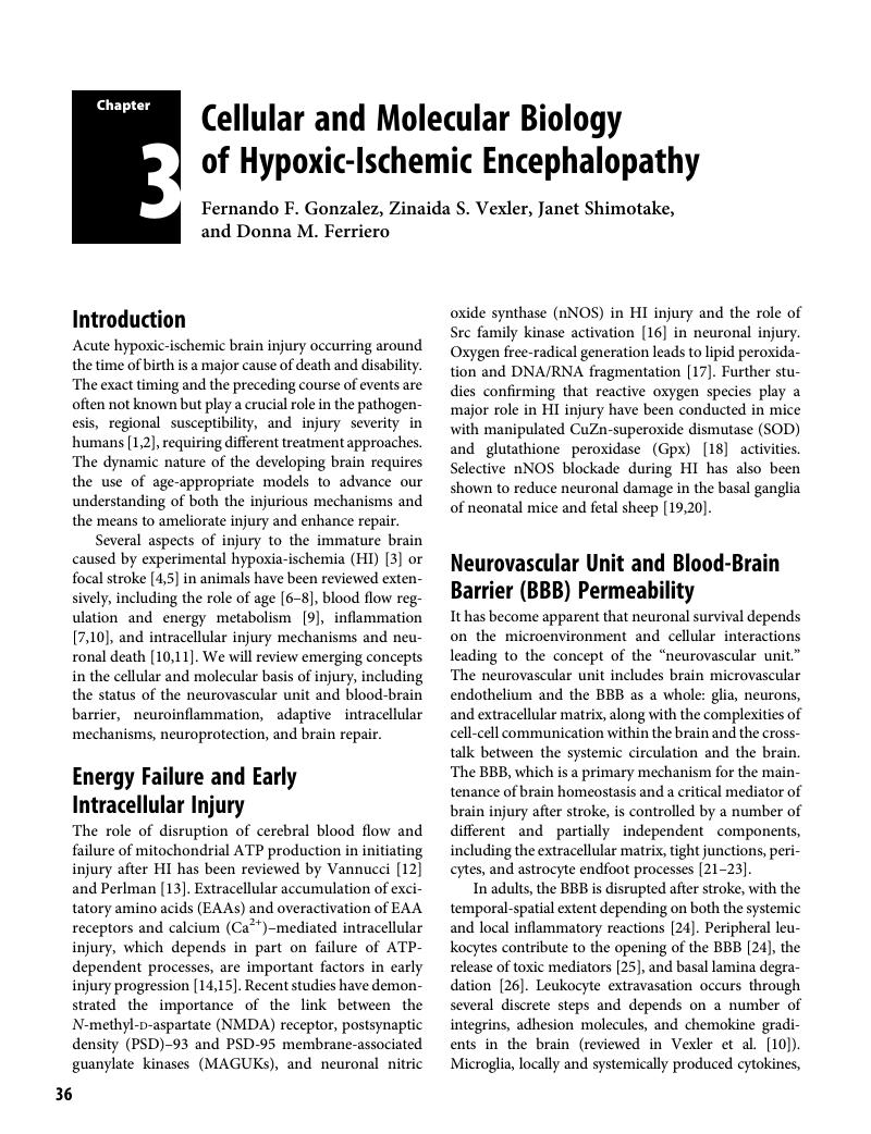
Image of the first page of this content. For PDF version, please use the ‘Save PDF’ preceeding this image.'
Type
Chapter
Information
Publisher: Cambridge University Press
Print publication year: 2017

Access options

Get access to the full version of this content by using one of the access options below. (Log in options will check for institutional or personal access. Content may require purchase if you do not have access.)

References

Volpe, JJ. Brain injury in the premature infant: overview of clinical aspects, neuropathology, and pathogenesis. Semin Pediatr Neurol 1998; 5(3): 135–51.Google Scholar
Ferriero, DM. Neonatal brain injury. N Engl J Med 2004; 351(19): 1985–95.Google Scholar
Vannucci, RC, Vannucci, SJ. A model of perinatal hypoxic-ischemic brain damage. Ann NY Acad Sci 1997; 835: 234–49.Google Scholar
Derugin, N, Ferriero, DM, Vexler, ZS. Neonatal reversible focal cerebral ischemia: a new model. Neurosci Res, 1998; 32(4)349–53.CrossRefGoogle ScholarPubMed
Ashwal, S, et al. Core and penumbral nitric oxide synthase activity during cerebral ischemia and reperfusion. Stroke 1998; 29(5)1037–46; discussion 1047.CrossRefGoogle ScholarPubMed
McQuillen, PS, Ferriero, DM. Selective vulnerability in the developing central nervous system. Pediatr Neurol 2004; 30(4)227–35.Google Scholar
Hagberg, H, Mallard, C. Effect of inflammation on central nervous system development and vulnerability. Curr Opin Neurol 2005; 18(2): 117–23.CrossRefGoogle Scholar
Vexler, ZS. Glia and inflammation in neurodegenerative disease. In Yenari, MA, Giffard, RG, eds., Inflammation and Ischemia in the Developing Brain. Hauppauge, NY: Nova Science Publishers, 2006.Google Scholar
Vannucci, SJ, Hagberg, H. Hypoxia-ischemia in the immature brain. J Exp Biol 2004; 207(18): 3149–54.Google Scholar
Vexler, ZS, Tang, D, Yenari, M. Inflammation in adult and neonatal stroke. Clin Neurosci Res 2006: 6(5): 293313.Google Scholar
Northington, FJ, et al. Failure to complete apoptosis following neonatal hypoxia-ischemia manifests as “continuum” phenotype of cell death and occurs with multiple manifestations of mitochondrial dysfunction in rodent forebrain. Neuroscience 2007; 149(4)822–33.Google Scholar
Vannucci, RC, Vannucci, SJ. Glucose metabolism in the developing brain. Semin Perinatol 2000; 24(2): 107–15.CrossRefGoogle ScholarPubMed
Perlman, JM. Intervention strategies for neonatal hypoxic-ischemic cerebral injury. Clin Ther 2006; 28(9): 1353–65.Google Scholar
Vexler, ZS, Ferriero, DM. Mechanisms of ischemic cell death in the developing brain, in Chan, P, ed., Handbook of Neurochemistry and Molecular Neurobiology. New York: Springer, 2006.Google Scholar
Rousset, CI, et al. A dual role for AMP-activated protein kinase (AMPK) during neonatal hypoxic-ischaemic brain injury in mice. J Neurochem 2015; 133(2)242–52.Google Scholar
Jiang, X, et al. Activated Src kinases interact with NMDA receptor after neonatal brain ischemia. Ann Neurol 2008; 63(5):632–41.CrossRefGoogle ScholarPubMed
Fraser, M, et al. Extracellular amino acids and lipid peroxidation products in periventricular white matter during and after cerebral ischemia in preterm fetal sheep. J Neurochem 2008; 105(6): 2214–23.Google Scholar
Sheldon, RA, Christen, S, Ferriero, DM. Genetic and pharmacologic manipulation of oxidative stress after neonatal hypoxia-ischemia. Int J Dev Neurosci 2007; 26(1): 8792.CrossRefGoogle ScholarPubMed
Ferriero, DM, et al. Neonatal mice lacking neuronal nitric oxide synthase are less vulnerable to hypoxic-ischemic injury. Neurobiol Dis 1996; 3(1): 6471.CrossRefGoogle ScholarPubMed
Drury, PP, et al. Partial neuroprotection by nNOS inhibition during profound asphyxia in preterm fetal sheep. Exp Neurol 2013; 250: 282–92.CrossRefGoogle ScholarPubMed
Bolton, SJ, Anthony, DC, Perry, VH. Loss of the tight junction proteins occludin and zonula occludens-1 from cerebral vascular endothelium during neutrophil-induced blood-brain barrier breakdown in vivo. Neuroscience 1998; 86(4)1245–57.Google Scholar
Bazzoni, G, et al. Interaction of junctional adhesion molecule with the tight junction components ZO-1, cingulin, and occludin. J Biol Chem 2000; 275(27): 20520–6.CrossRefGoogle ScholarPubMed
Kratzer, I, Chip, S, Vexler, ZS. Barrier mechanisms in neonatal stroke. Front Neurosci 2014; 8: 359.Google Scholar
Garcia, JH, et al. Influx of leukocytes and platelets in an evolving brain infarct (Wistar rat). Am J Pathol 1994; 144(1)188–99.Google Scholar
Hallenbeck, JM. Significance of the inflammatory response in brain ischemia. Acta Neurochir Suppl 1996; 66: 2731.Google Scholar
Rosenberg, GA. Matrix metalloproteinases in neuroinflammation. Glia 2002; 39(3)279–91.Google Scholar
Allan, SM, Tyrrell, PJ, Rothwell, NJ. Interleukin-1 and neuronal injury. Nat Rev Immunol 2005; 5(8): 629–40.Google Scholar
Pan, W, et al. Stroke upregulates TNF-α transport across the blood-brain barrier. Exp Neurol 2006; 198(1): 222–33.Google Scholar
Kniesel, U, Risau, W, Wolburg, H. Development of blood-brain barrier tight junctions in the rat cortex. Brain Res Dev Brain Res 1996; 96(1–2): 229–40.Google Scholar
Engelhardt, B. Development of the blood-brain barrier. Cell Tissue Res 2003; 314(1): 119–29.Google Scholar
Blamire, AM, et al. Interleukin-1β -induced changes in blood-brain barrier permeability, apparent diffusion coefficient, and cerebral blood volume in the rat brain: a magnetic resonance study. J Neurosci 2000; 20(21): 8153–9.Google Scholar
Palmer, C, Roberts, RL, Young, PI. Timing of neutrophil depletion influences long-term neuroprotection in neonatal rat hypoxic-ischemic brain injury. Pediatr Res 2004; 55(4): 549–56.CrossRefGoogle ScholarPubMed
Bona, E, et al. Chemokine and inflammatory cell response to hypoxia-ischemia in immature rats. Pediatr Res 1999; 45(4 Pt 1): 500–9.Google Scholar
Denker, S, et al. Macrophages are comprised of resident brain microglia not infiltrating peripheral monocytes acutely after neonatal stroke. J Neurochem 2007; 100(4): 893904.Google Scholar
Svedin, P, et al. Matrix metalloproteinase-9 gene knock-out protects the immature brain after cerebral hypoxia-ischemia. J Neurosci 2007; 27(7): 1511–8.Google Scholar
Ranasinghe, HS, et al. Inhibition of MMP-9 activity following hypoxic ischemia in the developing brain using a highly specific inhibitor. Dev Neurosci 2012; 34(5): 417–27.CrossRefGoogle ScholarPubMed
Raivich, G, et al. Neuroglial activation repertoire in the injured brain: graded response, molecular mechanisms and cues to physiological function. Brain Res Brain Res Rev 1999; 30(1): 77105.Google Scholar
Monje, ML, Toda, H, Palmer, TD. Inflammatory blockade restores adult hippocampal neurogenesis. Science 2003; 302(5651): 1760–5.Google Scholar
Walton, NM, et al. Microglia instruct subventricular zone neurogenesis. Glia 2006; 54(8): 815–25.Google Scholar
Britschgi, M, Wyss-Coray, T. Immune cells may fend off Alzheimer disease. Nat Med 2007; 13(4): 408–9.Google Scholar
Marin-Teva, JL, et al. Microglia promote the death of developing Purkinje cells. Neuron 2004; 41(4): 535–47.Google Scholar
Derugin, N, et al. Evolution of brain injury after transient middle cerebral artery occlusion in neonatal rat. Stroke 2000; 31: 1752–61.Google Scholar
McRae, A, et al. Microglia activation after neonatal hypoxic-ischemia. Brain Res Dev Brain Res 1995; 84(2): 245–52.Google Scholar
Dommergues, MA, et al. Early microglial activation following neonatal excitotoxic brain damage in mice: a potential target for neuroprotection. Neuroscience 2003; 121(3): 619–28.Google Scholar
Tikka, T, et al. Minocycline, a tetracycline derivative, is neuroprotective against excitotoxicity by inhibiting activation and proliferation of microglia. J Neurosci 2001; 21(8): 2580–8.Google Scholar
van den Tweel, ER, et al. Long-term neuroprotection with 2-iminobiotin, an inhibitor of neuronal and inducible nitric oxide synthase, after cerebral hypoxia-ischemia in neonatal rats. J Cereb Blood Flow Metab 2005; 25(1): 6774.Google Scholar
Dingman, A, et al. Aminoguanidine inhibits caspase-3 and calpain activation without affecting microglial activation following neonatal transient ischemia. J Neurochem 2006; 96: 1467–79.Google Scholar
Faustino, JV, et al. Microglial cells contribute to endogenous brain defenses after acute neonatal focal stroke. J Neurosci 2011; 31(36): 12992–3001.Google Scholar
Dong, Y, Benveniste, EN. Immune function of astrocytes. Glia 2001; 36(2): 180–90.CrossRefGoogle ScholarPubMed
Zhang, Y, et al. Notch-1 signaling regulates astrocytic proliferation and activation after hypoxia exposure. Neurosci Lett 2015; 603: 1218.Google Scholar
Benveniste, EN. Cytokine actions in the central nervous system. Cytokine Growth Factor Rev 1998; 9(3–4): 259–75.Google Scholar
Teo, JD, Morris, MJ, Jones, NM. Hypoxic postconditioning reduces microglial activation, astrocyte and caspase activity, and inflammatory markers after hypoxia-ischemia in the neonatal rat brain. Pediatr Res 2015; 77(6): 757–64.CrossRefGoogle ScholarPubMed
Iadecola, C, Zhang, F, Xu, X. Inhibition of inducible nitric oxide synthase ameliorates cerebral ischemic damage. Am J Physiol 1995; 268(1 Pt 2): R286–92.Google Scholar
Nawashiro, H, et al. High susceptibility to cerebral ischemia in GFAP-null mice. J Cereb Blood Flow Metab 2000; 20(7): 1040–4.Google Scholar
Benjelloun, N, et al. Inflammatory responses in the cerebral cortex after ischemia in the P7 neonatal rat. Stroke 1999; 30(9): 1916–23; discussion 1923–4.CrossRefGoogle ScholarPubMed
Benjelloun, N, et al. Apoptotic mitochondrial pathway in neurones and astrocytes after neonatal hypoxia-ischaemia in the rat brain. Neuropathol Appl Neurobiol 2003; 29(4): 350–60.Google Scholar
Ducrocq, S, et al. Poly(ADP-ribose) synthase inhibition reduces ischemic injury and inflammation in neonatal rat brain. J Neurochem 2000; 74(6): 2504–11.Google Scholar
Joly, LM, et al. Distribution of poly(ADP-ribosyl)ation and cell death after cerebral ischemia in the neonatal rat. Pediatr Res 2003; 53(5): 776–82.Google Scholar
Jin, Y, Silverman, AJ, Vannucci, SJ. Mast cell stabilization limits hypoxic-ischemic brain damage in the immature rat. Dev Neurosci 2007; 29(4–5): 373–84.Google Scholar
Patkai, J, et al. Deleterious effects of IL-9-activated mast cells and neuroprotection by antihistamine drugs in the developing mouse brain. Pediatr Res 2001; 50(2): 222–30.Google Scholar
Biran, V, et al. Stroke induces histamine accumulation and mast cell degranulation in the neonatal rat brain. Brain Pathol 2008; 18(1): 19.Google Scholar
Mesples, B, et al. Neuronal TGF-β1 mediates IL-9/mast cell interaction and exacerbates excitotoxicity in newborn mice. Neurobiol Dis 2005; 18(1): 193205.Google Scholar
Hedtjarn, M, Mallard, C, Hagberg, H. Inflammatory gene profiling in the developing mouse brain after hypoxia-ischemia. J Cereb Blood Flow Metab 2004; 24(12): 1333–51.Google Scholar
Szaflarski, J, Burtrum, D, Silverstein, FS. Cerebral hypoxia-ischemia stimulates cytokine gene expression in perinatal rats. Stroke 1995; 26(6): 1093–100.Google Scholar
Dommergues, MA, et al. Proinflammatory cytokines and interleukin-9 exacerbate excitotoxic lesions of the newborn murine neopallium. Ann Neurol 2000; 47(1): 5463.Google Scholar
Fox, C, et al. Minocycline confers early but transient protection in the immature brain following focal cerebral ischemia-reperfusion. J Cereb Blood Flow Metab 2005; 25(9): 1138–49.Google Scholar
Mesples, B, Plaisant, F, Gressens, P. Effects of interleukin-10 on neonatal excitotoxic brain lesions in mice. Brain Res Dev Brain Res, 2003; 141(1–2): 2532.Google Scholar
Gerard, C, Rollins, BJ. Chemokines and disease. Nat Immunol 2001; 2(2): 108–15.CrossRefGoogle ScholarPubMed
Hedtjarn, M, et al. Global gene expression in the immature brain after hypoxia-ischemia. J Cereb Blood Flow Metab 2004; 24(12): 1317–32.Google Scholar
Galasso, JM, et al. Acute excitotoxic injury induces expression of monocyte chemoattractant protein-1 and its receptor, CCR2, in neonatal rat brain. Exp Neurol 2000; 165(2): 295305.Google Scholar
Cowell, RM, et al. Hypoxic-ischemic injury induces macrophage inflammatory protein-1alpha expression in immature rat brain. Stroke 2002; 33(3): 795801.CrossRefGoogle ScholarPubMed
Cowell, RM, Plane, JM, Silverstein, FS. Complement activation contributes to hypoxic-ischemic brain injury in neonatal rats. J Neurosci 2003; 23(28): 9459–68.Google Scholar
Hu, BR, et al. Involvement of caspase-3 in cell death after hypoxia-ischemia declines during brain maturation. J Cereb Blood Flow Metab 2000; 20(9): 1294–300.Google Scholar
Han, BH, et al. Selective, reversible caspase-3 inhibitor is neuroprotective and reveals distinct pathways of cell death following neonatal hypoxic- ischemic brain injury. J Biol Chem 2002; 277(33): 30128–36.Google Scholar
Yakovlev, AG, et al. Differential expression of apoptotic protease-activating factor-1 and caspase-3 genes and susceptibility to apoptosis during brain development and after traumatic brain injury. J Neurosci 2001; 21(19): 7439–46.Google Scholar
Zhu, C, et al. The influence of age on apoptotic and other mechanisms of cell death after cerebral hypoxia-ischemia. Cell Death Differ 2005; 12(2): 162–76.Google Scholar
Manabat, C, et al. Reperfusion differentially induces caspase-3 activation in ischemic core and penumbra after stroke in immature brain. Stroke 2003; 34(1): 207–13.Google Scholar
Bittigau, P, et al. Apoptotic neurodegeneration following trauma is markedly enhanced in the immature brain. Ann Neurol 1999; 45(6): 724–35.Google Scholar
Cheng, Y, et al. Caspase inhibitor affords neuroprotection with delayed administration in a rat model of neonatal hypoxic-ischemic brain injury [see comments]. J Clin Invest 1998; 101(9): 1992–9.Google Scholar
West, T, Atzeva, M, Holtzman, DM. Caspase-3 deficiency during development increases vulnerability to hypoxic-ischemic injury through caspase-3-independent pathways. Neurobiol Dis 2006; 22(3): 523–37.Google Scholar
Zhu, C, et al. X chromosome-linked inhibitor of apoptosis protein reduces oxidative stress after cerebral irradiation or hypoxia-ischemia through up-regulation of mitochondrial antioxidants. Eur J Neurosci 2007; 26(12): 3402–10.Google Scholar
Matsumori, Y, et al. Hsp70 overexpression sequesters AIF and reduces neonatal hypoxic/ischemic brain injury. J Cereb Blood Flow Metab 2005; 25(7): 899910.Google Scholar
Blomgren, K, Leist, M, Groc, L. Pathological apoptosis in the developing brain. Apoptosis 2007; 12(5): 9931010.Google Scholar
Carloni, S, et al. Extended role of necrotic cell death after hypoxia-ischemia-induced neurodegeneration in the neonatal rat. Neurobiol Dis 2007; 27(3): 354–61.Google Scholar
Chavez-Valdez, R, Martin, LJ, Northington, FJ. Programmed necrosis: a prominent mechanism of cell death following neonatal brain injury. Neurol Res Int 2012; 2012: 257563.Google Scholar
Johnston, MV, Hagberg, H. Sex and the pathogenesis of cerebral palsy. Dev Med Child Neurol 2007; 49(1): 74–8.Google Scholar
Zup, SL, Edwards, NS, McCarthy, MM. Sex- and age-dependent effects of androgens on glutamate-induced cell death and intracellular calcium regulation in the developing hippocampus. Neuroscience 2014; 281C: 7787.Google Scholar
Hagberg, H, et al. PARP-1 gene disruption in mice preferentially protects males from perinatal brain injury. J Neurochem 2004; 90(5)1068–75.Google Scholar
Renolleau, S, Fau, S, Charriaut-Marlangue, C. Gender-related differences in apoptotic pathways after neonatal cerebral ischemia. Neuroscientist 2008; 14(1): 4652.Google Scholar
Del Pino Sans, J. et al. Microarray analysis of neonatal rat anteroventral periventricular transcriptomes identifies the proapoptotic Cugbp2 gene as sex-specific and regulated by estradiol. Neuroscience 2015; 303: 312–22.Google Scholar
Renolleau, S, et al. Sex, neuroprotection, and neonatal ischemia. Dev Med Child Neurol 2007; 49(6): 477; author reply 477–8.Google Scholar
Nijboer, CH, et al. Gender-specific neuroprotection by 2-iminobiotin after hypoxia-ischemia in the neonatal rat via a nitric oxide independent pathway. J Cereb Blood Flow Metab 2007; 27(2): 282–92.Google Scholar
Nijboer, CH, et al. Gender-dependent pathways of hypoxia-ischemia-induced cell death and neuroprotection in the immature P3 rat. Dev Neurosci 2007; 29(4–5): 385–92.Google Scholar
Wang, GL, Semenza, GL. Characterization of hypoxia-inducible factor 1 and regulation of DNA binding activity by hypoxia. J Biol Chem 1993; 268(29): 21513–8.Google Scholar
Bergeron, M, et al. Induction of hypoxia-inducible factor-1 (HIF-1) and its target genes following focal ischaemia in rat brain. Eur J Neurosci 1999; 11(12): 4159–70.Google Scholar
Chavez, JC, LaManna, JC. Activation of hypoxia-inducible factor-1 in the rat cerebral cortex after transient global ischemia: potential role of insulin-like growth factor-1. J Neurosci 2002; 22(20): 8922–31.Google Scholar
Bruick, RK, McKnight, SL. A conserved family of prolyl-4-hydroxylases that modify HIF. Science 2001; 294(5545): 1337–40.Google Scholar
Semenza, GL. Hypoxia-inducible factor 1: master regulator of O2 homeostasis. Curr Opin Genet Dev 1998; 8(5): 588–94.Google Scholar
Baranova, O, et al. Neuron-specific inactivation of the hypoxia inducible factor 1 alpha increases brain injury in a mouse model of transient focal cerebral ischemia. J Neurosci 2007; 27(23): 6320–32.Google Scholar
Zaman, K, et al. Protection from oxidative stress-induced apoptosis in cortical neuronal cultures by iron chelators is associated with enhanced DNA binding of hypoxia-inducible factor-1 and ATF-1/CREB and increased expression of glycolytic enzymes, p21(waf1/cip1), and erythropoietin. J Neurosci 1999; 19(22): 9821–30.Google Scholar
Bruick, RK. Expression of the gene encoding the proapoptotic Nip3 protein is induced by hypoxia. Proc Natl Acad Sci USA 2000; 97(16): 9082–7.Google Scholar
Van Hoecke, M, et al. Evidence of HIF-1 functional binding activity to caspase-3 promoter after photothrombotic cerebral ischemia. Mol Cell Neurosci 2007; 34(1): 40–7.Google Scholar
Helton, R, et al. Brain-specific knock-out of hypoxia-inducible factor-1alpha reduces rather than increases hypoxic-ischemic damage. J Neurosci 2005; 25(16): 4099–107.Google Scholar
Bergeron, M, et al. Role of hypoxia-inducible factor-1 in hypoxia-induced ischemic tolerance in neonatal rat brain. Ann Neurol 2000; 48(3): 285–96.Google Scholar
Mu, D, et al. Regulation of hypoxia-inducible factor 1alpha and induction of vascular endothelial growth factor in a rat neonatal stroke model. Neurobiol Dis 2003; 14(3): 524–34.Google Scholar
Mu, D, et al. Hypoxia-inducible factor 1alpha and erythropoietin upregulation with deferoxamine salvage after neonatal stroke. Exp Neurol 2005; 195(2):407–15.Google Scholar
Li, L, et al. The involvement of phosphoinositid 3-kinase/Akt pathway in the activation of hypoxia-inducible factor-1alpha in the developing rat brain after hypoxia-ischemia. Brain Res 2008; 1197:152–8.Google Scholar
Li, L, et al. The requirement of extracellular signal-related protein kinase pathway in the activation of hypoxia inducible factor 1alpha in the developing rat brain after hypoxia-ischemia. Acta Neuropathol 2008; 115(3): 297303.Google Scholar
Jones, NM, Bergeron, M. Hypoxic preconditioning induces changes in HIF-1 target genes in neonatal rat brain. J Cereb Blood Flow Metab 2001; 21(9): 1105–14.Google Scholar
Bernaudin, M, et al. Brain genomic response following hypoxia and re-oxygenation in the neonatal rat. Identification of genes that might contribute to hypoxia-induced ischemic tolerance. J Biol Chem 2002; 277(42): 39728–38.Google Scholar
Jelkmann, W. Erythropoietin: structure, control of production, and function. Physiol Rev 1992; 72(2): 449–89.Google Scholar
Chen, ZY, et al. Endogenous erythropoietin signaling is required for normal neural progenitor cell proliferation. J Biol Chem 2007; 282(35): 25875–83.Google Scholar
Shingo, T, et al. Erythropoietin regulates the in vitro and in vivo production of neuronal progenitors by mammalian forebrain neural stem cells. J Neurosci 2001; 21(24): 9733–43.Google Scholar
Wu, H, et al. Generation of committed erythroid BFU-E and CFU-E progenitors does not require erythropoietin or the erythropoietin receptor. Cell 1995; 83(1): 5967.Google Scholar
Kilic, E, et al. Brain-derived erythropoietin protects from focal cerebral ischemia by dual activation of ERK-1/-2 and Akt pathways. FASEB J 2005; 19(14): 2026–8.Google Scholar
Digicaylioglu, M, Lipton, SA. Erythropoietin-mediated neuroprotection involves cross-talk between Jak2 and NF-kappaB signalling cascades. Nature 2001; 412(6847): 641–7.Google Scholar
Kawakami, M, et al. Erythropoietin receptor-mediated inhibition of exocytotic glutamate release confers neuroprotection during chemical ischemia. J Biol Chem 2001; 276(42): 39469–75.Google Scholar
Marti, HH, et al. Neuroprotection and Angiogenesis: Dual Role of Erythropoietin in Brain Ischemia. News Physiol Sci 2000; 15: 225–9.Google Scholar
Solaroglu, I, et al. Erythropoietin prevents ischemia-reperfusion from inducing oxidative damage in fetal rat brain. Childs Nerv Syst 2003; 19(1): 1922.Google Scholar
Sola, A, et al. Potential for protection and repair following injury to the developing brain: a role for erythropoietin? Pediatr Res 2005; 57(5 Pt 2): 110 R–17 R.Google Scholar
Wang, L, et al. Treatment of stroke with erythropoietin enhances neurogenesis and angiogenesis and improves neurological function in rats. Stroke 2004; 35(7): 1732–7.Google Scholar
Ribatti, D, et al. Human erythropoietin induces a pro-angiogenic phenotype in cultured endothelial cells and stimulates neovascularization in vivo. Blood 1999; 93(8): 2627–36.Google Scholar
Sun, Y, Calvert, JW, Zhang, JH. Neonatal hypoxia/ischemia is associated with decreased inflammatory mediators after erythropoietin administration. Stroke 2005; 36(8): 1672–8.Google Scholar
Dzietko, M, et al. Erythropoietin protects the developing brain against N-methyl-D-aspartate receptor antagonist neurotoxicity. Neurobiol Dis 2004; 15(2): 177–87.Google Scholar
Xiong, T, et al. Erythropoietin for neonatal brain injury: opportunity and challenge. Int J Dev Neurosci 2011; 29(6): 583–91.Google Scholar
Kellert, BA, McPherson, RJ, Juul, SE. A comparison of high-dose recombinant erythropoietin treatment regimens in brain-injured neonatal rats. Pediatr Res 2007; 61(4): 451–5.Google Scholar
Spandou, E, et al. Hypoxia-ischemia affects erythropoietin and erythropoietin receptor expression pattern in the neonatal rat brain. Brain Res 2004; 1021(2): 167–72.Google Scholar
McClure, MM, Threlkeld, SW, Fitch, RH. Auditory processing and learning/memory following erythropoietin administration in neonatally hypoxic-ischemic injured rats. Brain Res 2007; 1132(1): 203–9.Google Scholar
Iwai, M, et al. Erythropoietin promotes neuronal replacement through revascularization and neurogenesis after neonatal hypoxia/ischemia in rats. Stroke 2007; 38(10): 2795–803.Google Scholar
Kumral, A, et al. Erythropoietin improves long-term spatial memory deficits and brain injury following neonatal hypoxia-ischemia in rats. Behav Brain Res 2004; 153(1): 7786.Google Scholar
Chang, YS, et al. Erythropoietin improves functional and histological outcome in neonatal stroke. Pediatr Res 2005; 58(1): 106–11.Google Scholar
Gonzalez, F, et al. Erythropoietin enhances long-term neuroprotection and neurogenesis in neonatal stroke. Dev Neurosci 2007; 29(4–5): 321–30.Google Scholar
Gonzalez, FF, et al. Erythropoietin increases neurogenesis and oligodendrogliosis of subventricular zone precursor cells after neonatal stroke. Stroke 2013; 44(3): 753–8.Google Scholar
Zhu, C, et al. Erythropoietin improved neurologic outcomes in newborns with hypoxic-ischemic encephalopathy. Pediatrics 2009; 124(2): e218–26.Google Scholar
Elmahdy, H, et al. Human recombinant erythropoietin in asphyxia neonatorum: pilot trial. Pediatrics 2010; 125(5): e1135–42.Google Scholar
Ferrara, N, Gerber, HP. The role of vascular endothelial growth factor in angiogenesis. Acta Haematol 2001; 106(4): 148–56.Google Scholar
Carmeliet, P, et al. Abnormal blood vessel development and lethality in embryos lacking a single VEGF allele. Nature 1996; 380(6573): 435–9.Google Scholar
Dougher, M, Terman, BI. Autophosphorylation of KDR in the kinase domain is required for maximal VEGF-stimulated kinase activity and receptor internalization. Oncogene 1999; 18(8): 1619–27.Google Scholar
Takahashi, T, et al. A single autophosphorylation site on KDR/Flk-1 is essential for VEGF-A-dependent activation of PLC-gamma and DNA synthesis in vascular endothelial cells. EMBO J 2001; 20(11): 2768–78.Google Scholar
Fujio, Y, Walsh, K. AKT mediates cytoprotection of endothelial cells by vascular endothelial growth factor in an anchorage-dependent manner. J Biol Chem 1999; 274(23): 16349–54.Google Scholar
Shibuya, M. Structure and dual function of vascular endothelial growth factor receptor-1 (Flt-1). Int J Biochem Cell Biol 2001; 33(4): 409–20.Google Scholar
Vogel, C, et al. Flt-1, but not Flk-1 mediates hyperpermeability through activation of the PI3-K/Akt pathway. J Cell Physiol 2007; 212(1): 236–43.Google Scholar
Rahimi, N, Dayanir, V, Lashkari, K. Receptor chimeras indicate that the vascular endothelial growth factor receptor-1 (VEGFR-1) modulates mitogenic activity of VEGFR-2 in endothelial cells. J Biol Chem 2000; 275(22): 16986–92.Google Scholar
Autiero, M, et al. Role of PlGF in the intra- and intermolecular cross talk between the VEGF receptors Flt1 and Flk1. Nat Med 2003; 9(7): 936–43.Google Scholar
Jin, KL, Mao, XO, Greenberg, DA. Vascular endothelial growth factor: direct neuroprotective effect in in vitro ischemia. Proc Natl Acad Sci USA 2000; 97(18): 10242–7.Google Scholar
Shweiki, D, et al. Vascular endothelial growth factor induced by hypoxia may mediate hypoxia-initiated angiogenesis. Nature 1992; 359(6398): 843–5.Google Scholar
Sun, Y, et al. VEGF-induced neuroprotection, neurogenesis, and angiogenesis after focal cerebral ischemia. J Clin Invest 2003; 111(12): 1843–51.Google Scholar
Wang, Y, et al. VEGF-overexpressing transgenic mice show enhanced post-ischemic neurogenesis and neuromigration. J Neurosci Res, 2007. 85(4): p. 740–7.Google Scholar
Zhang, Z.G., et al. VEGF enhances angiogenesis and promotes blood-brain barrier leakage in the ischemic brain. J Clin Invest 2000; 106(7): 829–38.Google Scholar
Manoonkitiwongsa, PS, et al. Neuroprotection of ischemic brain by vascular endothelial growth factor is critically dependent on proper dosage and may be compromised by angiogenesis. J Cereb Blood Flow Metab 2004; 24(6): 693702.Google Scholar
Ogunshola, OO, et al. Neuronal VEGF expression correlates with angiogenesis in postnatal developing rat brain. Brain Res Dev Brain Res 2000; 119(1): 139–53.Google Scholar
Laudenbach, V, et al. Neonatal hypoxic preconditioning involves vascular endothelial growth factor. Neurobiol Dis 2007; 26(1): 243–52.Google Scholar
Shimotake, J, et al. Vascular endothelial growth factor receptor-2 inhibition promotes cell death and limits endothelial cell proliferation in a neonatal rodent model of stroke. Stroke 2010; 41(2): 343–9.Google Scholar
Waterhouse, EG, Xu, B. New insights into the role of brain-derived neurotrophic factor in synaptic plasticity. Mol Cell Neurosci 2009; 42(2): 81–9.Google Scholar
Zheng, F, Wang, H. NMDA-mediated and self-induced bdnf exon IV transcriptions are differentially regulated in cultured cortical neurons. Neurochem Int 2009; 54(5–6): 385–92.Google Scholar
Chen, K, et al. Creating a neurogenic environment: the role of BDNF and FGF2. Mol Cell Neurosci 2007; 36(1): 108–20.Google Scholar
Rivera, C, et al. BDNF-induced TrkB activation down-regulates the K+-Cl- cotransporter KCC2 and impairs neuronal Cl extrusion. J Cell Biol 2002; 159(5): 747–52.Google Scholar
Gluckman, PD, et al. Selective head cooling with mild systemic hypothermia after neonatal encephalopathy: multicentre randomised trial. Lancet 2005; 365(9460): 663–70.Google Scholar
Shankaran, S, et al. Whole-body hypothermia for neonates with hypoxic-ischemic encephalopathy. N Engl J Med 2005; 353(15): 1574–84.Google Scholar
Wyatt, JS, et al. Determinants of outcomes after head cooling for neonatal encephalopathy. Pediatrics 2007; 119(5): 912–21.Google Scholar
Yager, JY, et al. Preventing hyperthermia decreases brain damage following neonatal hypoxic-ischemic seizures. Brain Res 2004; 1011(1): 4857.Google Scholar
Mishima, K, et al. Effects of hypothermia and hyperthermia on attentional and spatial learning deficits following neonatal hypoxia-ischemic insult in rats. Behav Brain Res 2004; 151(1–2): 209–17.Google Scholar
Zhu, C, et al. Post-ischemic hypothermia-induced tissue protection and diminished apoptosis after neonatal cerebral hypoxia-ischemia. Brain Res 2004; 996(1): 6775.Google Scholar
Wagner, BP, Nedelcu, J, Martin, E. Delayed postischemic hypothermia improves long-term behavioral outcome after cerebral hypoxia-ischemia in neonatal rats. Pediatr Res 2002; 51(3): 354–60.Google Scholar
Liu, Y, et al. Topiramate extends the therapeutic window for hypothermia-mediated neuroprotection after stroke in neonatal rats. Stroke 2004; 35(6): 1460–5.Google Scholar
Thoresen, M, et al. A piglet survival model of posthypoxic encephalopathy. Pediatr Res 1996; 40(5): 738–48.Google Scholar
Bona, E, et al. Protective effects of moderate hypothermia after neonatal hypoxia-ischemia: short- and long-term outcome. Pediatr Res 1998; 43(6): 738–45.Google Scholar
Taylor, DL, et al. Improved neuroprotection with hypothermia delayed by 6 hours following cerebral hypoxia-ischemia in the 14-day-old rat. Pediatr Res 2002; 51(1): 13–9.Google Scholar
Gunn, AJ, et al. Dramatic neuronal rescue with prolonged selective head cooling after ischemia in fetal lambs. J Clin Invest 1997; 99(2): 248–56.Google Scholar
Gunn, AJ, et al. Cerebral hypothermia is not neuroprotective when started after postischemic seizures in fetal sheep. Pediatr Res 1999; 46(3): 274–80.Google Scholar
Jatana, M, et al. Combination of systemic hypothermia and N-acetylcysteine attenuates hypoxic-ischemic brain injury in neonatal rats. Pediatr Res 2006; 59(5): 684–9.Google Scholar
Hobbs, CT, Tucker, M, Aquilina, A, et al. Xenon and hypothermia combine additively. offering long-term functional and histopathologic neuroprotection after neonatal hypoxia/ischemia. Stroke 2008; 39(4):1307–13.Google Scholar
Wu, YW, et al. Erythropoietin for neuroprotection in neonatal encephalopathy: safety and pharmacokinetics. Pediatrics 2012; 130(4): 683–91.Google Scholar
Loren, DJ, et al. Maternal dietary supplementation with pomegranate juice is neuroprotective in an animal model of neonatal hypoxic-ischemic brain injury. Pediatr Res 2005; 57(6): 858–64.Google Scholar
West, T, Atzeva, M, Holtzman, DM. Pomegranate polyphenols and resveratrol protect the neonatal brain against hypoxic-ischemic injury. Dev Neurosci 2007; 29(4–5): 363–72.Google Scholar
Feng, Y, et al. Grape seed extract given three hours after injury suppresses lipid peroxidation and reduces hypoxic-ischemic brain injury in neonatal rats. Pediatr Res 2007; 61(3): 295300.Google Scholar
Parent, JM, et al. Rat forebrain neurogenesis and striatal neuron replacement after focal stroke. Ann Neurol 2002; 52(6): 802–13.Google Scholar
Lindvall, O, Kokaia, Z. Recovery and rehabilitation in stroke: stem cells. Stroke 2004; 35(11 Suppl. 1): 2691–4.Google Scholar
Plane, JM, et al. Neonatal hypoxic-ischemic injury increases forebrain subventricular zone neurogenesis in the mouse. Neurobiol Dis 2004; 16(3): 585–95.Google Scholar
Arvidsson, A, et al. Neuronal replacement from endogenous precursors in the adult brain after stroke. Nat Med 2002; 8(9): 963–70.Google Scholar
Zhang, R, et al. Activated neural stem cells contribute to stroke-induced neurogenesis and neuroblast migration toward the infarct boundary in adult rats. J Cereb Blood Flow Metab 2004; 24(4): 441–8.Google Scholar
Zhang, R, et al. Stroke transiently increases subventricular zone cell division from asymmetric to symmetric and increases neuronal differentiation in the adult rat. J Neurosci 2004; 24(25): 5810–5.Google Scholar
Ohab, JJ, et al. A neurovascular niche for neurogenesis after stroke. J Neurosci 2006; 26(50): 13007–16.Google Scholar
Romanko, MJ, et al. Death effector activation in the subventricular zone subsequent to perinatal hypoxia/ischemia. J Neurochem 2007; 103(3): 1121–31.Google Scholar
Yang, Z, et al. Sustained neocortical neurogenesis after neonatal hypoxic/ischemic injury. Ann Neurol 2007; 61(3): 199208.Google Scholar
Greenberg, DA, Jin, K. From angiogenesis to neuropathology. Nature 2005; 438(7070): 954–9.Google Scholar
Distler, JH, et al. Angiogenic and angiostatic factors in the molecular control of angiogenesis. Q J Nucl Med 2003; 47(3): 149–61.Google Scholar
van Velthoven, CT, Kavelaars, A, Heijnen, CJ. Mesenchymal stem cells as a treatment for neonatal ischemic brain damage. Pediatr Res 2012; 71(4 Pt 2): 474–81.Google Scholar
van Velthoven, CT, et al. Stem cells for neonatal stroke: the future is here. Front Cell Neurosci 2014; 8: 207.Google Scholar

Save book to Kindle

To save this book to your Kindle, first ensure coreplatform@cambridge.org is added to your Approved Personal Document E-mail List under your Personal Document Settings on the Manage Your Content and Devices page of your Amazon account. Then enter the ‘name’ part of your Kindle email address below. Find out more about saving to your Kindle.

Note you can select to save to either the @free.kindle.com or @kindle.com variations. ‘@free.kindle.com’ emails are free but can only be saved to your device when it is connected to wi-fi. ‘@kindle.com’ emails can be delivered even when you are not connected to wi-fi, but note that service fees apply.

Find out more about the Kindle Personal Document Service.

Available formats
×

Save book to Dropbox

To save content items to your account, please confirm that you agree to abide by our usage policies. If this is the first time you use this feature, you will be asked to authorise Cambridge Core to connect with your account. Find out more about saving content to Dropbox.

Available formats
×

Save book to Google Drive

To save content items to your account, please confirm that you agree to abide by our usage policies. If this is the first time you use this feature, you will be asked to authorise Cambridge Core to connect with your account. Find out more about saving content to Google Drive.

Available formats
×