Skip to main content Accessibility help
×
Hostname: page-component-848d4c4894-r5zm4 Total loading time: 0 Render date: 2024-06-16T01:45:44.708Z Has data issue: false hasContentIssue false

2 - Functional studies of isolated GPCR-G protein complexes in the membrane bilayer of lipoprotein particles

from PART I - ADVANCES IN GPCR PROTEIN RESEARCH

Published online by Cambridge University Press:  05 June 2012

Adam J. Kuszak
Affiliation:
University of Michigan
Xiao Jie Yao
Affiliation:
Stanford University
Sören G.F. Rasmussen
Affiliation:
Stanford University
Brian K. Kobilka
Affiliation:
Stanford University
Roger K. Sunahara
Affiliation:
University of Michigan
Sandra Siehler
Affiliation:
Novartis Institute for Biomedical Research
Graeme Milligan
Affiliation:
University of Glasgow
Get access

Summary

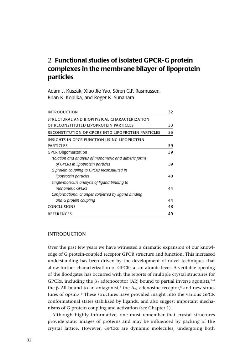
Image of the first page of this content. For PDF version, please use the ‘Save PDF’ preceeding this image.'
Type
Chapter
Information
G Protein-Coupled Receptors
Structure, Signaling, and Physiology
, pp. 32 - 52
Publisher: Cambridge University Press
Print publication year: 2010

Access options

Get access to the full version of this content by using one of the access options below. (Log in options will check for institutional or personal access. Content may require purchase if you do not have access.)

References

Cherezov, V., D.Rosenbaum, M., et al. (2007). “High-resolution crystal structure of an engineered human beta2-adrenergic G protein-coupled receptor.”Science 318(5854): 1258–65.CrossRefGoogle Scholar
Rasmussen, S. G., Choi, H. J., et al. (2007). “Crystal structure of the human beta2 adrenergic G-protein-coupled receptor.”Nature 450(7168): 383–7.CrossRefGoogle Scholar
Rosenbaum, D. M., Cherezov, V., et al. (2007). “GPCR engineering yields high-resolution structural insights into beta2-adrenergic receptor function.”Science 318(5854): 1266–73.CrossRefGoogle Scholar
Hanson, M. A., Cherezov, V., et al. (2008). “A specific cholesterol binding site is established by the 2.8 A structure of the human beta2-adrenergic receptor.”Structure 16(6): 897–905.CrossRefGoogle Scholar
Warne, T., Serrano-Vega, M. J., et al. (2008). “Structure of a beta1-adrenergic G-protein-coupled receptor.”Nature 454(7203): 486–91.CrossRefGoogle Scholar
Jaakola, V. P., Griffith, M. T., et al. (2008). “The 2.6 angstrom crystal structure of a human A2A adenosine receptor bound to an antagonist.”Science 322(5905): 1211–7.CrossRefGoogle Scholar
Park, J. H., Scheerer, P., et al. (2008). “Crystal structure of the ligand-free G-protein-coupled receptor opsin.”Nature 454(7201): 183–7.CrossRefGoogle Scholar
Scheerer, P., Park, J. H., et al. (2008). “Crystal structure of opsin in its G-protein-interacting conformation.”Nature 455(7212): 497–502.CrossRefGoogle Scholar
Shimamura, T., Hiraki, K., et al. (2008). “Crystal structure of squid rhodopsin with intracellularly extended cytoplasmic region.”J Biol Chem 283(26): 17753–6.CrossRefGoogle Scholar
Brouillette, C. G., Anantharamaiah, G. M., et al. (2001). “Structural models of human apolipoprotein A-I: a critical analysis and review.”Biochim Biophys Acta 1531(1–2): 4–46.CrossRefGoogle Scholar
Jonas, A., J. Wald, H., et al. (1990). “Apolipoprotein A-I structure and lipid properties in homogeneous, reconstituted spherical and discoidal high density lipoproteins.”J Biol Chem 265(36): 22123–9.Google Scholar
Klon, A. E., Jones, M. K., et al. (2000). “Molecular belt models for the apolipoprotein A-I Paris and Milano mutations.”Biophys J 79(3): 1679–85.CrossRefGoogle Scholar
Jonas, A. (1986). “Reconstitution of high-density lipoproteins.”Methods Enzymol 128: 553–82.CrossRefGoogle Scholar
Rogers, D. P., Brouillette, C. G., et al. (1997). “Truncation of the amino terminus of human apolipoprotein A-I substantially alters only the lipid-free conformation.”Biochemistry 36(2): 288–300.CrossRefGoogle Scholar
Klon, A. E., Segrest, J. P., et al. (2002). “Comparative models for human apolipoprotein A-I bound to lipid in discoidal high-density lipoprotein particles.”Biochemistry 41(36): 10895–905.CrossRefGoogle Scholar
Li, Y., A. Kijac, Z., et al. (2006). “Structural analysis of nanoscale self-assembled discoidal lipid bilayers by solid-state NMR spectroscopy.”Biophys J 91(10): 3819–28.CrossRefGoogle Scholar
Shih, A. Y., Denisov, I. G., et al. (2005). “Molecular dynamics simulations of discoidal bilayers assembled from truncated human lipoproteins.”Biophys J 88(1): 548–56.CrossRefGoogle Scholar
Shaw, A. W., McLean, M. A., et al. (2004). “Phospholipid phase transitions in homogeneous nanometer scale bilayer discs.”FEBS Lett 556(1–3): 260–4.CrossRefGoogle Scholar
Bayburt, T. H., Leitz, A. J., et al. (2007). “Transducin activation by nanoscale lipid bilayers containing one and two rhodopsins.”J Biol Chem 282(20): 14875–81.CrossRefGoogle Scholar
Whorton, M. R., Bokoch, M. P., et al. (2007). “A monomeric G protein-coupled receptor isolated in a high-density lipoprotein particle efficiently activates its G protein.”Proc Natl Acad Sci U S A 104(18): 7682–7.CrossRefGoogle Scholar
Civjan, N. R., Bayburt, T. H., et al. (2003). “Direct solubilization of heterologously expressed membrane proteins by incorporation into nanoscale lipid bilayers.”Biotechniques 35(3): 556–60, 562–3.Google Scholar
Katzen, F., J. Fletcher, E., et al. (2008). “Insertion of membrane proteins into discoidal membranes using a cell-free protein expression approach.”J Proteome Res 7(8): 3535–42.CrossRefGoogle Scholar
Segrest, J. P. (1977). “Amphipathic helixes and plasma lipoproteins: thermodynamic and geometric considerations.”Chem Phys Lipids 18(1): 7–22.CrossRefGoogle Scholar
Munford, M. L., Lima, V.R., Vieira, T.O., Heinzelmann, G., Creczynski-Pasa, T.B., Pasa, A.A. (2005). “AFMIn-Situ Characterization of Supported Phospholipid Layers Formed by Vesicle Fusion.” Microscopy and Microanalysis11(Suppl. 3): 90–3.Google Scholar
Keough, K. (2003). “How thin can glass be? New ideas, new approaches.”Biophys J 85(5): 2785–6.CrossRefGoogle Scholar
Kuszak, A. J., Pitchiaya, S., et al. (2009). “Purification and functional reconstitution of monomeric mu-opioid receptors: allosteric modulation of agonist binding by Gi2.”J Biol Chem 284(39): 26732–41.CrossRefGoogle Scholar
Yao, X. J., Velez Ruiz, G., et al. (2009). “The effect of ligand efficacy on the formation and stability of a GPCR-G protein complex.”Proc Natl Acad Sci U S A 106(23): 9501–6.CrossRefGoogle Scholar
Whorton, M. R., Jastrzebska, B., et al. (2008). “Efficient coupling of transducin to monomeric rhodopsin in a phospholipid bilayer.”J Biol Chem 283(7): 4387–94.CrossRefGoogle Scholar
Denisov, I. G., Grinkova, Y. V., et al. (2004). “Directed self-assembly of monodisperse phospholipid bilayer Nanodiscs with controlled size.”J Am Chem Soc 126(11): 3477–87.CrossRefGoogle Scholar
Bayburt, T. H., Grinkova, Y. V., et al. (2006). “Assembly of single bacteriorhodopsin trimers in bilayer nanodiscs.”Arch Biochem Biophys 450(2): 215–22.CrossRefGoogle Scholar
Banerjee, S., Huber, T., et al. (2008). “Rapid incorporation of functional rhodopsin into nanoscale apolipoprotein bound bilayer (NABB) particles.”J Mol Biol 377(4): 1067–81.CrossRefGoogle Scholar
Pin, J. P., Kniazeff, J., et al. (2005). “Allosteric functioning of dimeric class C G-protein-coupled receptors.”Febs J 272(12): 2947–55.CrossRefGoogle Scholar
Hebert, T. E. and Bouvier, M. (1998). “Structural and functional aspects of G protein-coupled receptor oligomerization.”Biochem Cell Biol 76(1): 1–11.CrossRefGoogle Scholar
Angers, S., A. Salahpour, , et al. (2000). “Detection of beta 2-adrenergic receptor dimerization in living cells using bioluminescence resonance energy transfer (BRET).”Proc Natl Acad Sci U S A 97(7): 3684–9.Google Scholar
Overton, M. C. and Blumer, K. J. (2000). “G-protein-coupled receptors function as oligomers in vivo.”Curr Biol 10(6): 341–4.CrossRefGoogle Scholar
Rios, C. D., Jordan, B. A., et al. (2001). “G-protein-coupled receptor dimerization: modulation of receptor function.”Pharmacol Ther 92(2–3): 71–87.CrossRefGoogle Scholar
Dalrymple, M. B., Pfleger, K. D., et al. (2008). “G protein-coupled receptor dimers: functional consequences, disease states and drug targets.”Pharmacol Ther 118(3): 359–71.CrossRefGoogle Scholar
Leitz, A. J., Bayburt, T. H., et al. (2006). “Functional reconstitution of Beta2-adrenergic receptors utilizing self-assembling Nanodisc technology.”Biotechniques 40(5): 601–2, 604, 606, passim.CrossRefGoogle Scholar
Wald, G. (1968). “Molecular basis of visual excitation.”Science 162(850): 230–9.CrossRefGoogle Scholar
Waldhoer, M., Bartlett, S. E., et al. (2004). “Opioid receptors.”Annu Rev Biochem 73: 953–90.CrossRefGoogle ScholarPubMed
Pineyro, G. and Archer-Lahlou, E. (2007). “Ligand-specific receptor states: implications for opiate receptor signalling and regulation.”Cell Signal 19(1): 8–19.CrossRefGoogle Scholar
Cvejic, S. and Devi, L. A. (1997). “Dimerization of the delta opioid receptor: implication for a role in receptor internalization.”J Biol Chem 272(43): 26959–64.CrossRefGoogle Scholar
Jordan, B. A. and Devi, L. A. (1999). “G-protein-coupled receptor heterodimerization modulates receptor function.”Nature 399(6737): 697–700.CrossRefGoogle Scholar
Wang, D., Sun, X., et al. (2005). “Opioid receptor homo- and heterodimerization in living cells by quantitative bioluminescence resonance energy transfer.”Mol Pharmacol 67(6): 2173–84.CrossRefGoogle Scholar
George, S. R., Fan, T., et al. (2000). “Oligomerization of mu- and delta-opioid receptors. Generation of novel functional properties.”J Biol Chem 275(34): 26128–35.CrossRefGoogle Scholar
Martin, N. A. and Prather, P. L. (2001). “Interaction of co-expressed mu- and delta-opioid receptors in transfected rat pituitary GH(3) cells.”Mol Pharmacol 59(4): 774–83.CrossRefGoogle Scholar
Levac, B. A., O ' Dowd, B. F., et al. (2002). “Oligomerization of opioid receptors: generation of novel signaling units.”Curr Opin Pharmacol 2(1): 76–81.CrossRefGoogle Scholar
Kuszak, A. J. (2009). The function of the monomeric form of the mu-opioid receptor: G protein-mediated allosteric regulation of agonist binding and stimulation of nucleotide exchange. Department of Pharmacology. Ann Arbor, University of Michigan. Ph.D. Thesis: 122 p.Google Scholar
Chabre, M. and Maire, M. (2005). “Monomeric G-protein-coupled receptor as a functional unit.”Biochemistry 44(27): 9395–403.CrossRefGoogle Scholar
Liang, Y., Fotiadis, D., et al. (2003). “Organization of the G protein-coupled receptors rhodopsin and opsin in native membranes.”J Biol Chem 278(24): 21655–62.CrossRefGoogle Scholar
Fahmy, K. and Sakmar, T. P. (1993). “Light-dependent transducin activation by an ultraviolet-absorbing rhodopsin mutant.”Biochemistry 32(35): 9165–71.CrossRefGoogle Scholar
Jastrzebska, B., Fotiadis, D., et al. (2006). “Functional and structural characterization of rhodopsin oligomers.”J Biol Chem 281(17): 11917–22.CrossRefGoogle Scholar
Lean, A., Stadel, J. M., et al. (1980). “A ternary complex model explains the agonist-specific binding properties of the adenylate cyclase-coupled beta-adrenergic receptor.”J Biol Chem 255(15): 7108–17.Google Scholar
Kenakin, T. (2004). “G-protein coupled receptors as allosteric machines.”Receptors Channels 10(2): 51–60.CrossRefGoogle Scholar
Park, P. S., Filipek, S., et al. (2004). “Oligomerization of G protein-coupled receptors: past, present, and future.”Biochemistry 43(50): 15643–56.CrossRefGoogle Scholar
Baneres, J. L. and Parello, J. (2003). “Structure-based analysis of GPCR function: evidence for a novel pentameric assembly between the dimeric leukotriene B4 receptor BLT1 and the G-protein.”J Mol Biol 329(4): 815–29.CrossRefGoogle Scholar
Ferre, S., Baler, R., et al. (2009). “Building a new conceptual framework for receptor heteromers.”Nat Chem Biol 5(3): 131–4.CrossRefGoogle Scholar
Park, P. S., Sum, C. S., et al. (2002). “Cooperativity and oligomeric status of cardiac muscarinic cholinergic receptors.”Biochemistry 41(17): 5588–604.CrossRefGoogle Scholar
Park, P. S. and Wells, J. W. (2003). “Monomers and oligomers of the M2 muscarinic cholinergic receptor purified from Sf9 cells.”Biochemistry 42(44): 12960–71.CrossRefGoogle Scholar
Park, P. S. and Wells, J. W. (2004). “Oligomeric potential of the M2 muscarinic cholinergic receptor.”J Neurochem 90(3): 537–48.CrossRefGoogle Scholar
Damian, M., Mary, S., et al. (2008). “G protein activation by the leukotriene B4 receptor dimer. Evidence for an absence of trans-activation.”J Biol Chem 283(30): 21084–92.CrossRefGoogle Scholar
Gurevich, V. V. and Gurevich, E. V. (2008). “How and why do GPCRs dimerize?”Trends Pharmacol Sci 29(5): 234–40.CrossRefGoogle Scholar
Farrens, D. L., Altenbach, C., et al. (1996). “Requirement of rigid-body motion of transmembrane helices for light activation of rhodopsin.”Science 274(5288): 768–70.CrossRefGoogle Scholar
Sheikh, S. P., Vilardarga, J. P., et al. (1999). “Similar structures and shared switch mechanisms of the beta2-adrenoceptor and the parathyroid hormone receptor. Zn(II) bridges between helices III and VI block activation.”J Biol Chem 274(24): 17033–41.CrossRefGoogle Scholar
Ballesteros, J. A., A. Jensen, D., et al. (2001). “Activation of the beta 2-adrenergic receptor involves disruption of an ionic lock between the cytoplasmic ends of transmembrane segments 3 and 6.”J Biol Chem 276(31): 29171–7.CrossRefGoogle Scholar
Greasley, P. J., Fanelli, F., et al. (2002). “Mutagenesis and modelling of the alpha(1b)-adrenergic receptor highlight the role of the helix 3/helix 6 interface in receptor activation.”Mol Pharmacol 61(5): 1025–32.CrossRefGoogle Scholar
Shapiro, D. A., Kristiansen, K., et al. (2002). “Evidence for a model of agonist-induced activation of 5-hydroxytryptamine 2A serotonin receptors that involves the disruption of a strong ionic interaction between helices 3 and 6.”J Biol Chem 277(13): 11441–9.CrossRefGoogle Scholar
Lodowski, D. T., Angel, T. E., et al. (2009). “Comparative analysis of GPCR crystal structures.”Photochem Photobiol 85(2): 425–30.CrossRefGoogle Scholar
Gether, U., S. Lin, , et al. (1997). “Agonists induce conformational changes in transmembrane domains III and VI of the beta2 adrenoceptor.”Embo J 16(22): 6737–47.CrossRefGoogle Scholar
Yao, X., Parnot, C., et al. (2006). “Coupling ligand structure to specific conformational switches in the beta2-adrenoceptor.”Nat Chem Biol 2(8): 417–22.CrossRefGoogle Scholar
Swaminath, G., Xiang, Y., et al. (2004). “Sequential binding of agonists to the beta2 adrenoceptor. Kinetic evidence for intermediate conformational states.”J Biol Chem 279(1): 686–91.CrossRefGoogle Scholar
Swaminath, G., X. Deupi, et al. (2005). “Probing the beta2 adrenoceptor binding site with catechol reveals differences in binding and activation by agonists and partial agonists.”J Biol Chem 280(23): 22165–71.CrossRefGoogle Scholar
Ghanouni, P., Gryczynski, Z., et al. (2001). “Functionally different agonists induce distinct conformations in the G protein coupling domain of the beta 2 adrenergic receptor.”J Biol Chem 276(27): 24433–6.CrossRefGoogle Scholar
Ghanouni, P., Steenhuis, J. J., et al. (2001). “Agonist-induced conformational changes in the G-protein-coupling domain of the beta 2 adrenergic receptor.”Proc Natl Acad Sci U S A 98(11): 5997–6002.CrossRefGoogle Scholar
Nobles, M., Benians, A., et al. (2005). “Heterotrimeric G proteins precouple with G protein-coupled receptors in living cells.”Proc Natl Acad Sci U S A 102(51): 18706–11.CrossRefGoogle Scholar
Gales, C., J. J. Durm, , et al. (2006). “Probing the activation-promoted structural rearrangements in preassembled receptor-G protein complexes.”Nat Struct Mol Biol 13(9): 778–86.CrossRefGoogle Scholar
Audet, N., Gales, C., et al. (2008). “Bioluminescence resonance energy transfer assays reveal ligand-specific conformational changes within preformed signaling complexes containing delta-opioid receptors and heterotrimeric G proteins.”J Biol Chem 283(22): 15078–88.CrossRefGoogle Scholar

Save book to Kindle

To save this book to your Kindle, first ensure coreplatform@cambridge.org is added to your Approved Personal Document E-mail List under your Personal Document Settings on the Manage Your Content and Devices page of your Amazon account. Then enter the ‘name’ part of your Kindle email address below. Find out more about saving to your Kindle.

Note you can select to save to either the @free.kindle.com or @kindle.com variations. ‘@free.kindle.com’ emails are free but can only be saved to your device when it is connected to wi-fi. ‘@kindle.com’ emails can be delivered even when you are not connected to wi-fi, but note that service fees apply.

Find out more about the Kindle Personal Document Service.

Available formats
×

Save book to Dropbox

To save content items to your account, please confirm that you agree to abide by our usage policies. If this is the first time you use this feature, you will be asked to authorise Cambridge Core to connect with your account. Find out more about saving content to Dropbox.

Available formats
×

Save book to Google Drive

To save content items to your account, please confirm that you agree to abide by our usage policies. If this is the first time you use this feature, you will be asked to authorise Cambridge Core to connect with your account. Find out more about saving content to Google Drive.

Available formats
×