Skip to main content Accessibility help
×
  • Cited by 4
Publisher:
Cambridge University Press
Online publication date:
March 2013
Print publication year:
2013
Online ISBN:
9781139088268

Book description

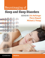
This up-to-date, superbly illustrated book is a practical guide to the effective use of neuroimaging in the patient with sleep disorders. There are detailed reviews of new neuroimaging techniques – including CT, MRI, advanced MR techniques, SPECT and PET – as well as image analysis methods, their roles and pitfalls. Neuroimaging of normal sleep and wake states is covered plus the role of neuroimaging in conjunction with tests of memory and how sleep influences memory consolidation. Each chapter carefully presents and analyzes the key findings in patients with sleep disorders indicating the clinical and imaging features of the various sleep disorders from clinical presentation to neuroimaging, aiding in establishing an accurate diagnosis. Written by neuroimaging experts from around the world, Neuroimaging of Sleep and Sleep Disorders is an invaluable resource for both researchers and clinicians including sleep specialists, neurologists, radiologists, psychiatrists, psychologists.

Reviews

“…Invaluable resource for researchers and clinicians in…sleep medicine, neurology, radiology, psychiatry, and psychology. A particular strength of the book is the incorporation of color figures and graphs of neuroimaging results.”

- Doody's Review Service

Refine List

Actions for selected content:

Select all | Deselect all
  • View selected items
  • Export citations
  • Download PDF (zip)
  • Save to Kindle
  • Save to Dropbox
  • Save to Google Drive

Save Search

You can save your searches here and later view and run them again in "My saved searches".

Please provide a title, maximum of 40 characters.
×

Contents


Page 1 of 3


  • Chapter 8 - Fundamentals of magnetoencephalography
    pp 62-71
  • View abstract

    Summary

    Greater insights into the mechanisms and consequences of sleep and sleep disorders have been achieved through advances in brain imaging methods that describe various aspects of neural function. These are collectively referred to as functional neuroimaging. These include techniques such as PET, fMRI, single-photon emission computed tomography (SPECT), transcranial sonography, magnetoencephalography (MEG), low-resolution brain electromagnetic tomography (LORETA), and combined methods such as combined EEG and fMRI. Extensive applications of brain imaging have been made to help clarify the changes in regional brain function that result from perturbations in either homeostatic or circadian processes, and also have clarified the relationship between these brain changes and the behavioral consequences of these disruptions. The earliest applications of neuroimaging to the study of sleep disorders were those of functional neuroimaging methods to study the global brain states of waking, NREM, and REM sleep. Brain imaging studies have been utilized in narcolepsy and the hypersomnias.
  • Chapter 10 - Methodology of combined EEG and fMRI
    pp 82-90
  • View abstract

    Summary

    This chapter examines possible neuronal networks and mechanisms responsible for the switch from waking to non-rapid eye movement (NREM) and REM sleep. The activated cortical state during waking is induced by the activity of multiple waking neurochemical systems. In contrast to the complex and extensive neurochemical network involved in waking, the neurons inducing slow-wave sleep (SWS) are localized in the lateral preoptic area and the adjacent basal forebrain. A cluster of these neurons is localized in a small nucleus called the ventrolateral preoptic nucleus (VLPO), which is situated above the optic chiasm. Neurons specifically active during paradoxical sleep (PS) were recorded in the posterior hypothalamus (PH) of cats or head-restrained rats. One-third of these GABAergic neurons were immunoreactive for the neuropeptide melanin concentrating hormone (MCH). PS onset would be due to the activation of glutamatergic PS-on neurons from the sublaterodorsal tegmental nucleus (SLD).
  • Chapter 11 - Neuroimaging of wakefulness
    pp 91-95
  • View abstract

    Summary

    This chapter provides an overview of the fundamental elements of magnetic resonance imaging (MRI). Four terms describe the magnetic properties of materials, such as contrast agents, used in MRI. These terms are diamagnetism, paramagnetism, superparamagnetism, and ferromagnetism. The persistence of magnetization when the external magnetic field is removed distinguishes ferromagnetic materials from paramagnetic materials. To be useful for MRI, the proton must have spin angular momentum, in addition to the nuclear magnetism. Echo time (TE) and repetition time (TR) are basic parameters of image acquisition. Improvement in the magnitude of the MR signal can improve signal-to-noise ratio (SNR). Magnetic resonance angiography (MRA) uses the same MRI system and methods to make images of blood vessels. The most common MRA technique is based on the time-of-flight (TOF) effect, where blood protons flowing into the slice during the acquisition yield very high signal, but signal from stationary protons is suppressed.
  • Chapter 12 - Neuroimaging of phasic and non-phasic NREM activities
    pp 96-104
  • View abstract

    Summary

    This chapter reviews three of the most important and topical advanced magnetic resonance imaging (MRI) techniques. Functional MRI (fMRI) permits dynamic evaluation of neural activity in specific brain regions. To understand normal and pathological brain function, one must understand the structural arrangement of white matter tracts, and how this arrangement varies between individuals. This goal can be approached in living subjects with diffusion tensor imaging (DTI), a variant of diffusion-weighted imaging (DWI), which permits the non-invasive evaluation of regional white matter structure. Standard anatomical MRI depicts the structural features of the brain, while fMRI demonstrates regional brain activity. Magnetic resonance spectroscopy (MRS), in contrast, allows non-invasive examination of the chemical composition of the brain. The advanced neuroimaging techniques described in this chapter allow non-invasive evaluation of the intact brain at biochemical, network, and functional levels.
  • Chapter 13 - Functional connectivity in wakefulness and sleep
    pp 105-113
  • View abstract

    Summary

    Positron emission tomography (PET) and more recently PET/computed tomography (CT) has been utilized as a measure of functional imaging for many decades. The manufacture of PET radiotracers for imaging molecular processes in the human brain begins with the production of the PET radioisotope using a cyclotron. A nuclear reaction takes place in the target between the particle and the atom of the target material that gives rise to the PET radioisotope. The radioisotope is then sent to the radiopharmacy where it is used in the preparation of a radiopharmaceutical. Different patient protocols for PET imaging with 18F fluorodeoxyglucose (18F-FDG) are present for oncological and cardiac indications. PET regional cerebral blood flow (rCBF) imaging with 15O-water can be quantified using a mathematical model using a diffusible tracer technique method. Software has been developed to assess imaging more quantitatively with comparison to normal subjects.
  • Chapter 14 - Functional neuroimaging of human REM sleep
    pp 114-120
  • View abstract

    Summary

    Radiotracer imaging methods such as single-photon emission computed tomography (SPECT) and positron emission tomography (PET) are well suited to provide information about the functional, metabolic, and molecular status of tissues and organs. Brain SPECT has a well-established role for a number of clinical indications. Cerebral perfusion studies are used in the evaluation of dementias, epilepsy, cerebrovascular disease, trauma, brain death, and to assist with neuropsychiatric evaluation. Brain function is evaluated at baseline, before and after pharmacotherapy or psychotherapy, and following a number of activation tasks to examine a large number of psychiatric conditions. The integration of SPECT and CT in a single imaging device facilitates anatomical localization of the radiopharmaceutical to differentiate physiological uptake from that associated with disease. SPECT and SPECT/CT is continuing to evolve with the introduction of new technologies that have the potential to improve performance beyond that possible with Anger's pioneering approach.
  • Chapter 15 - Complementarity of dream research and neuroimaging of sleep
    pp 121-128
  • View abstract

    Summary

    Transcranial B-mode sonography (TCS) is a widely available, non-invasive and cost-effective diagnostic instrument. By convention and due to the resolution of the ultrasound waves in proximity of the probe, structures that are close to the midline are assessed from the ipsilateral side whereas structures that are located distant to the midline are examined from the contralateral side. Most data on TCS in disorders associated with insomnia or parasomnias have been evaluated from the movement disorder perspective and did not address diagnosis or differential diagnosis of sleep disorders directly. Some interesting findings indicate that TCS is valuable for the evaluation of sleep disorder patients with suspected restless legs syndrome (RLS), depressive disorder, or rapid eye movement (REM) sleep behavior disorder (RBD). In a patient complaining about disturbed sleep a TCS demonstrating substantia nigra (SN) hypoechogenicity, raphe hypoechogenicity, and red nucleus (RN) hyperechogenicity may help to support a suspected diagnosis of RLS.
  • Chapter 16 - Functional neuroimaging of sleep deprivation
    pp 129-136
  • View abstract

    Summary

    This chapter outlines basic aspects of magnetoencephalography (MEG) technology, and provides an example of how the method is used clinically. In the clinical arena, MEG is most commonly used for presurgical planning purposes with the goal of localizing epileptogenic regions in patients with medically refractory seizures. In many of these cases, MEG provides unique clinical information that alters clinical care in a positive manner. Resective surgery was performed in the epileptogenic zone as originally identified by MEG and the patient has been seizure free for more than 2.5 years. The chapter describes how MEG is being used in sleep research. MEG is starting to play an increased role in the understanding of how brain circuits are modulated during sleep, with the most significant insights coming in relationship to the origins of sleep spindles and the complex modulation of neural interactions during rapid eye movement (REM) sleep.
  • Chapter 17 - Neuroimaging of attention and alteration of processing capacity in sleep-deprived persons
    pp 137-144
  • View abstract

    Summary

    Low-resolution brain electromagnetic tomography (LORETA) was one of the first attempts to solve both the inverse problem and the reference electrode problem. It is able to localize deep sources as well by minimizing the squared three-dimensional (3D) spatial Laplacian operator to determine the unique solution. In 2001, the authors published the first study that applied LORETA to sleep electroencephalogram (EEG) data. They selected artifact-free epochs with sleep spindles and determined LORETA power in the frequency domain via the EEG cross-spectral matrix. LORETA revealed cortical spindle sources predominantly medially in the frontal and parietal lobe. The cortical generators localized for delta waves in slow-wave sleep (SWS) showed considerable overlap with the spindle generators. LORETA was applied to reveal changes in brain activity due to chronic hypoxia in patients with obstructive sleep apnea syndrome (OSAS).
  • Chapter 18 - Economic decision-making and the sleep-deprived brain
    pp 145-153
  • View abstract

    Summary

    Electroencephalogram (EEG) in combination with a fast whole-brain imaging method such as functional magnetic resonance imaging (fMRI) is an ideal tool to study sleep. This chapter covers practical aspects when planning and conducting a simultaneous EEG-fMRI experiment. It describes the choice of hardware to provide patient safety and comfort, while delivering high quality EEG and fMRI data. The chapter examines the choice of post processing methods applied to the electrophysiological data for scanner and subject-induced artifact reduction. Two empirical approaches have been mainly used to integrate fMRI and EEG data; first, using fMRI for the better determination of the source of the measured electrical EEG signal and second, trying to find the common neural origin of both the EEG and fMRI signals in a broader sense. EMG recordings are feasible but await further improved artifact reduction methods dealing with motion in the magnetic field.
  • Chapter 19 - Functional imaging of inter-individual differences in response to sleep deprivation
    pp 154-162
  • View abstract

    Summary

    This chapter reports results on spontaneous brain activity during wakeful rest. It focuses on findings obtained with electroencephalography (EEG) and functional magnetic resonance imaging (fMRI), either acquired separately or simultaneously. The chapter discusses two approaches to analyze resting state activity with fMRI, namely reverse subtraction (activity correlated with task deactivation) and independent component analysis (ICA)-based region analysis. A third way to explore resting state blood oxygen level-dependent (BOLD) activity is to add information obtained with independent recording modalities, in particular EEG. When EEG and fMRI are recorded simultaneously, fMRI activity patterns associated with EEG-defined brain states can be analyzed. Relaxed wakefulness in the EEG is characterized by alpha and beta band oscillations. A thorough analysis of EEG and fMRI patterns during wakeful rest yields a complex relationship where certain EEG patterns can be associated with different BOLD maps and vice versa.

Page 1 of 3


Metrics

Full text views

Total number of HTML views: 0
Total number of PDF views: 0 *
Loading metrics...

Book summary page views

Total views: 0 *
Loading metrics...

* Views captured on Cambridge Core between #date#. This data will be updated every 24 hours.

Usage data cannot currently be displayed.