


No CrossRef data available.
Article contents
Increased and decreased cortical thickness and unaltered amygdala nuclei in patients at clinical-high risk of psychosis
Published online by Cambridge University Press: 19 July 2023
Abstract
Core share and HTML view are not available for this content. However, as you have access to this content, a full PDF is available via the ‘Save PDF’ action button.
Introduction
There is much evidence of grey matter alterations in subjects at clinical-high risk of psychosis (CHR). Although, to the best of our knowledge, no studies have analyzed both cerebral cortex and amygdala nuclei morphometry alterations in CHR individuals.
Objectives
The aim of the study was to explore cortical thickness and amygdala nuclei morphometric characteristics in CHR patients.
Methods
Nineteen right-handed male patients (17-24 years, mean age 21.1 ± 2.1) fulfilling CHR criteria and 20 matched healthy controls (18-24 years, mean age 21.1 ± 1.8) underwent T1-weighted structural MRI at 3T Philips scanner. Images were processed via FreeSurfer 7.0. Cortical thickness (according to Desikan atlas) and volumes of 9 separate amygdala nuclei bilaterally were compared between groups. The morphometry data, SOPS, HDRS (Hamilton Depression Rating Scale) scores and chlorpromazine equivalents were included in correlation analysis.
Results
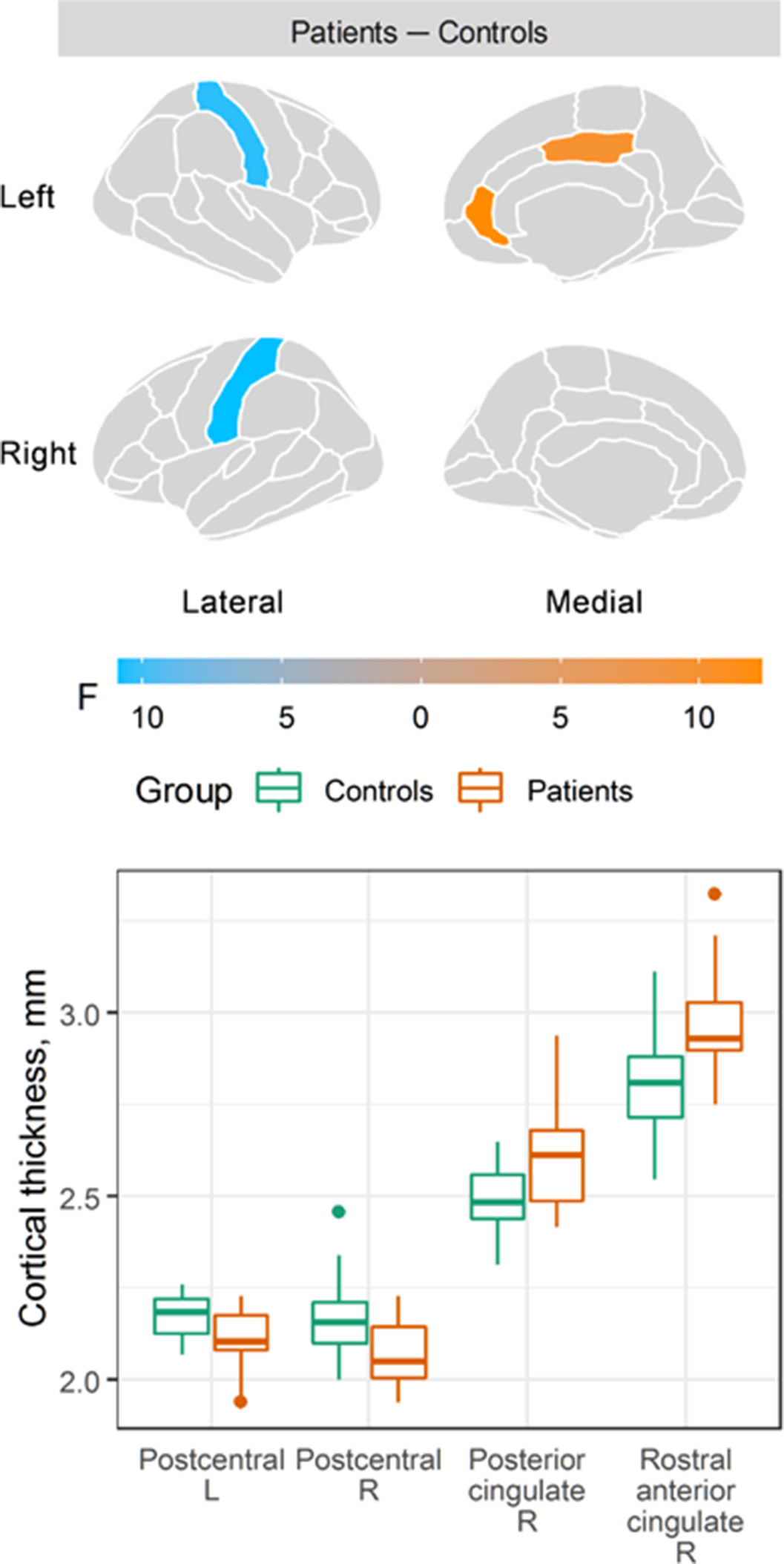
Compared to healthy controls, patients showed decreased cortical thickness in the left [F(1, 36) = 10.8, p = 0.002; Cohen’s d = −1.1, 95% CI: −1.8 to −0.4] and right [F(1, 36) = 10.5, p = 0.003; Cohen’s d = −1.0, 95% CI: −1.7 to −0.3] postcentral gyri, and increased cortical thickness in the right posterior cingulate [F(1, 36) = 9.9, p = 0.003; Cohen’s d = 1.0, 95% CI: 0.3 to 1.6] and the right rostral anterior cingulate gyri [F(1, 36) = 12.2, p = 0.001; Cohen’s d = 1.1, 95% CI: 0.4 to 1.8]. No changes in any amygdala nuclei were detected. No correlations between altered cortical thickness, HDRS, SOPS or chlorpromazine equivalents were revealed.
Image:
Conclusions
The current findings suggest that volumetric characteristics of amygdalar complex are unaffected in the CHR state. The results have some inconsistency with our previous findings (Tomyshev et al. Psychiatry Res Neuroimaging. 2019; 289 26-36), which revealed only a decrease in cortical thickness in CHR individuals. However, the cross-sectional design of the current study and the lack of correlations between cortical thickness and clinical symptoms do not allow to conclude definitely whether the revealed higher cortical thickness can represents some resilience mechanisms, which will be elucidated via further research.
This study was supported by RSFBR 22-15-00437
Disclosure of Interest
None Declared
- Type
- Abstract
- Information
- European Psychiatry , Volume 66 , Special Issue S1: Abstracts of the 31st European Congress of Psychiatry , March 2023 , pp. S548 - S549
- Creative Commons

 This is an Open Access article, distributed under the terms of the Creative Commons Attribution licence (https://creativecommons.org/licenses/by/4.0/), which permits unrestricted re-use, distribution, and reproduction in any medium, provided the original work is properly cited.
- Copyright
- © The Author(s), 2023. Published by Cambridge University Press on behalf of the European Psychiatric Association
 You have
Access
You have
Access
 Open access
Comments
No Comments have been published for this article.