
Hostname: page-component-89b8bd64d-rbxfs Total loading time: 0 Render date: 2026-05-11T08:38:07.911Z Has data issue: false hasContentIssue false

Formation of aligned core/sheath microfiber scaffolds with a poly-L-lactic acid (PLLA) sheath and a conductive poly(3,4-ethylenedioxythiophene) (PEDOT) core

Published online by Cambridge University Press:  04 June 2019

Rachel A. Martin
Affiliation:
Chemical Engineering, Michigan Technological University, Houghton, Michigan 49931, USA
Marie Wendling
Affiliation:
Bachelor’s Degree Student, Biomedical Engineering, Michigan Technological University, Houghton, Michigan 49931, USA
Bailey Mohrenweiser
Affiliation:
Bachelor’s Degree Student, Chemical Engineering, Michigan Technological University, Houghton, Michigan 49931, USA
Zichen Qian
Affiliation:
Ph.D. Student, Biomedical Engineering, Michigan Technological University, Houghton, Michigan 49931, USA
Feng Zhao
Affiliation:
Associate Professor, Ph.D., Biomedical Engineering, Michigan Technological University, Houghton, Michigan 49931, USA
Michael E. Mullins*
Affiliation:
Professor, Ph.D., Chemical Engineering, Michigan Technological University, Houghton, Michigan 49931, USA
*
a)Address all correspondence to this author. e-mail: memullin@mtu.edu
Get access

Abstract

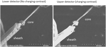
Electrospun coaxial fibers are used to create core/sheath fiber structures to act as growth-promoting scaffolds for in vitro dorsal root ganglia (DRG) cell cultures. The core was a conducting polymer, poly(3,4-ethylenedioxythiophene):poly(styrene sulfonate) (PEDOT:PSS), and the sheath was poly-L-lactic acid (PLLA), which created coaxial fibers with a conductive core and an insulating sheath. SEM analysis confirmed the conductivity of the core and insulation of the sheath. Several coaxial spinneret designs were tested with the best results obtained by using various annular spinning needle combinations. Using a 22G/16G and 22G/17G combination, fibers with diameters of 6.1 ± 2.4 µm and 3.3 ± 0.9 µm were spun, respectively. The fibers showed a Young’s modulus and hardness of 0.16 ± 0.13 and 0.02 ± 0.01 GPa for the larger diameters, and 0.7 ± 0.4 and 0.03 ± 0.03 GPa for the smaller diameter fibers. In vitro test cultures showed the fibers successfully directed chick DRG axonal outgrowth with low biotoxicity.

Information

Type
Article
Copyright
Copyright © Materials Research Society 2019 

Access options

Get access to the full version of this content by using one of the access options below. (Log in options will check for institutional or personal access. Content may require purchase if you do not have access.)

Article purchase

Temporarily unavailable