
5 results
Dysfunction of microstructure and metabolism in corpus callosum in juvenile schizophrenia
-
- Journal:
- European Psychiatry / Volume 66 / Issue S1 / March 2023
- Published online by Cambridge University Press:
- 19 July 2023, pp. S616-S617
-
- Article
-
- You have access
- Open access
- Export citation
-
Introduction
The corpus callosum (CC) is one of the important structures responsible for communication between the brain hemispheres. Its role is particularly important in cognitive tasks performance, information processing, concentration of attention, in mnestic processes. The corresponding dysfunctions are the major symptoms of schizophrenia, and hence, structural characteristics of CC in schizophrenics are in the focus of attention.
ObjectivesThe aim of the study was to analyze the microstructural and metabolic features of the corpus callosum in recently onset schizophrenia.
MethodsThe study was carried out in 13 men with juvenile endogenous paroxysmal psychosis (disease standing ≤ 5 years after first manifestation) aged 17-27 years (median 22.0±3.1 years). The studies were carried out during unfolding remission or in remission. Control group consisted of 15 mentally healthy young men (18-28 years). MRI and 1H-MRS studies were carried out on Achieva 3T MRI scanner device (Phillips). Diffuse tensor images were obtained in the axial plane using echo-planar pulse sequence. Diffuse gradients were applied in 32 noncolinear vectors. The spectra were recorded by single voxel 1H-MRS. The spectroscopic voxel (2×1×1 cm) was placed in the CC genu region. The PRESS sequence was used (TR/TE=1500/40 msec).
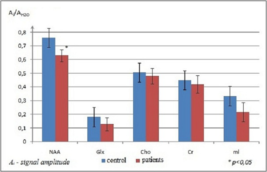
ResultsStatistical analysis showed no abnormal diffusion values in the CC splenium in the patients. Significant changes in the parameters were found in the CC genu. The values of ADC and RD increased, while FA coefficient decreased in the CC genu of patients with the initial stage of schizophrenia; PD values were normal. The increase of RD in the presence of unchanged PD indicated a decrease of water diffusion velocity and anisotropy in the direction perpendicular to the axon orientation. A typical 1H-MRS of the CC genu was presented in Figure 1. The results of statistical analysis of metabolite signal intensities in the CC genu of patients and normal subjects were presented in Figure 2. NAA level was reduced significantly in the patients. No appreciable changes in Cho values in the CC genu were detected in the patients vs. normal subjects.
Image:
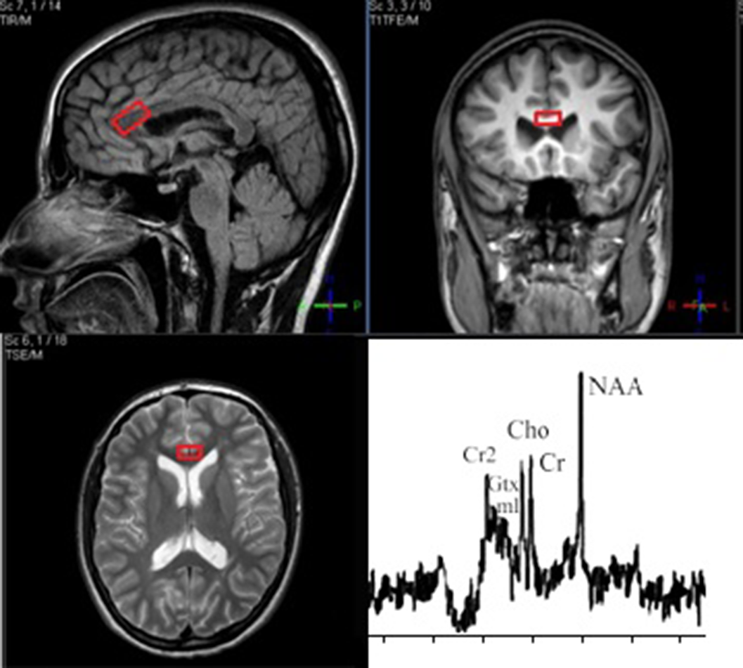
Image 2:
 Conclusions
The increase of RD could be caused by several factors: impairment of myelin membranes, axon damage because of impairment of axon cytoskeleton, and changed organization of fibrils. Our results showed that RD increase in patients with early schizophrenia did not conform to active demyelination, which was proven by the normal level of Cho, while axon damage, shown by low level of NAA, did not lead to PD reduction.
The decrease of NAA level detected in our study indicated axonal damage in the CC genu of patients in the early stage of schizophrenia. The increase of RD in the presence of normal Cho level seemed to indicate disorders in the axon cytoskeleton damage, but not active demyelination.
Disclosure of InterestNone Declared


Decrease in anterior cingulate cortex GABA in schizophrenia at early stage
-
- Journal:
- European Psychiatry / Volume 66 / Issue S1 / March 2023
- Published online by Cambridge University Press:
- 19 July 2023, pp. S609-S610
-
- Article
-
- You have access
- Open access
- Export citation
-
Introduction
There is evidence that the concentrations of the main inhibitory neurotransmitter (GABA) may be altered in schizophrenia. The purpose of this study is to find the changes in the GABA concentration in the area of anterior and posterior cingulate cortex of patients with early-stage schizophrenia using the spectral-edited magnetic resonance spectroscopy.
ObjectivesTo measure the cerebral concentrations of the gamma-aminobutyric acid in schizophrenia patients at early stage.
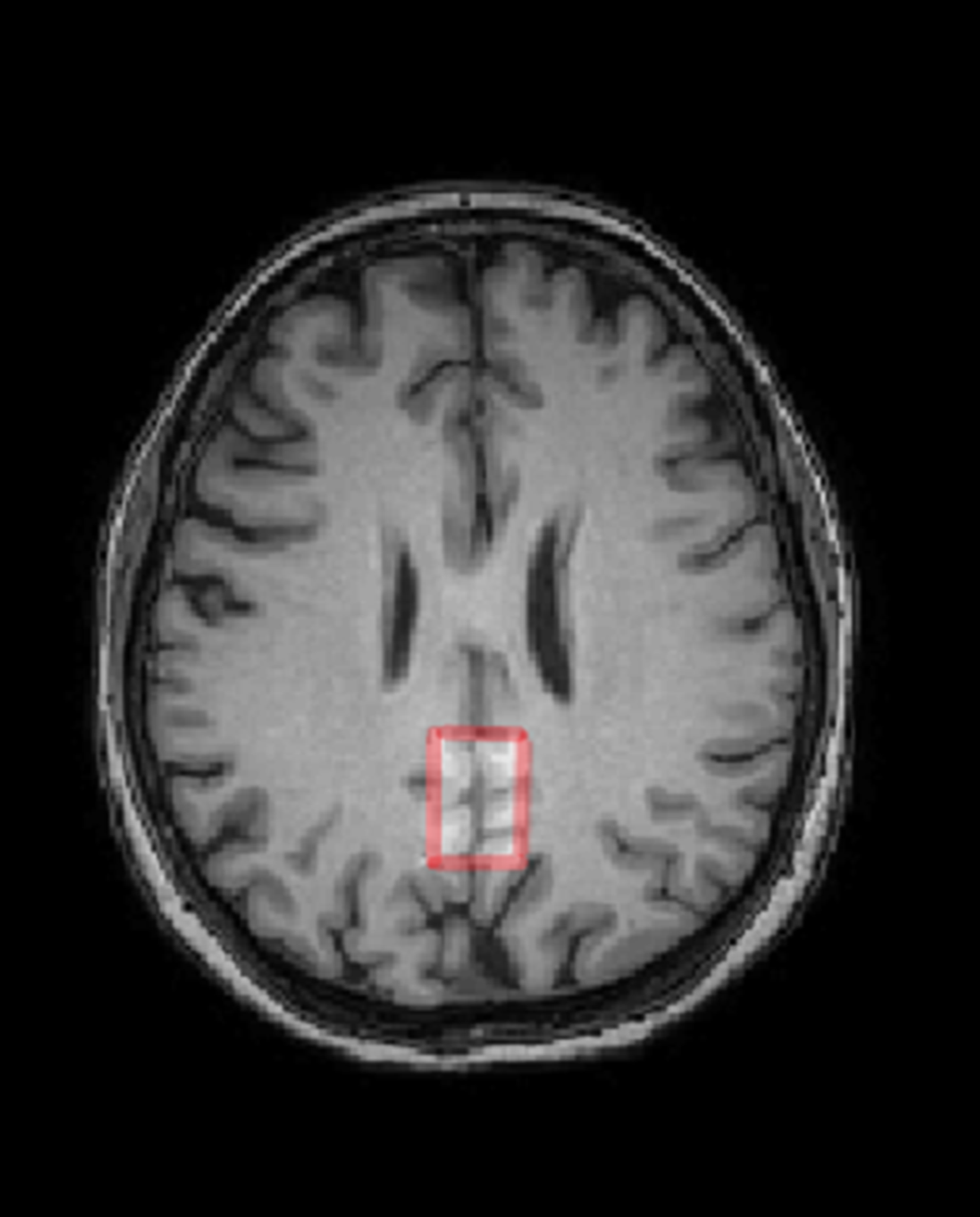
MethodsThirty-one subject, 18 controls (11m+7f, 29.6±5.7 y.o.) and 13 schizophrenia patients (F20.0, 8m+5f, 27.5±3.1 y.o.). Philips Achieva dStream 3T MRI scanner, standard head coil. The 3D T1w head images and MEGA-PRESS GABA spectra in ACC and PCC areas were acquired with the following parameters: 50x25x25 mm, TR = 2 s, TE = 64 ms, 180-editing pulses applied at 1.9 ppm and 7.6 ppm, NSA = 288 (acq.time ~10 min). GABA spectra were processed in Gannet program. The intensities of the GABA, Glutamate+glutamine (Glx), creatine (Cr) and unsuppressed water signals were acquired. T-test was used in search for between-group differences.
ResultsIn ACC region, significant reduction of the GABA/Water was observed (by ~15%, p=0.02) as well as a trend to a decrease in GABA/Cr (by ~10%, p=0.07) in schizophrenia. In PCC, no significant GABA/Water or GABA/Cr differences were observed. Glx/Water and Glx/Cr in both areas were also unchanged.
Image:
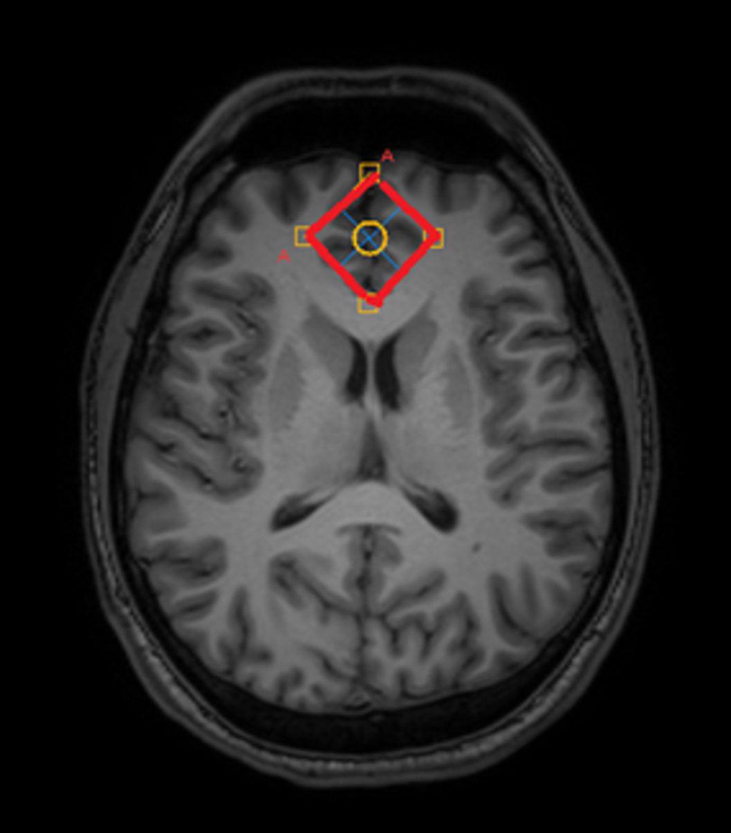
Image 2:
 Conclusions
This study provides insight into neurotransmitter alterations at early-stage schizophrenia. The results demonstrate the region-specific changes in the balance of the main neurotransmitters. Since this balance is crucial for the normal cerebral functioning, the results may facilitate better understanding of the dynamics of the pathological process and provide additional information for understanding the biological mechanisms of the schizophrenia development.
Disclosure of InterestNone Declared


Alterations in brain myelination at early-stage schizophrenia detected by macromolecular proton fraction MRI
-
- Journal:
- European Psychiatry / Volume 66 / Issue S1 / March 2023
- Published online by Cambridge University Press:
- 19 July 2023, pp. S134-S135
-
- Article
-
- You have access
- Open access
- Export citation
-
Introduction
There is evidence that cerebral myelination is impaired in schizophrenia. The purpose of this study is to find the myelin content changes in the brain structures of patients with early-stage schizophrenia using the macromolecular proton fraction (MPF) method, and also to evaluate the differences in the myelination of these structures.
ObjectivesTo measure MPF in the brain structures of schizophrenia patients
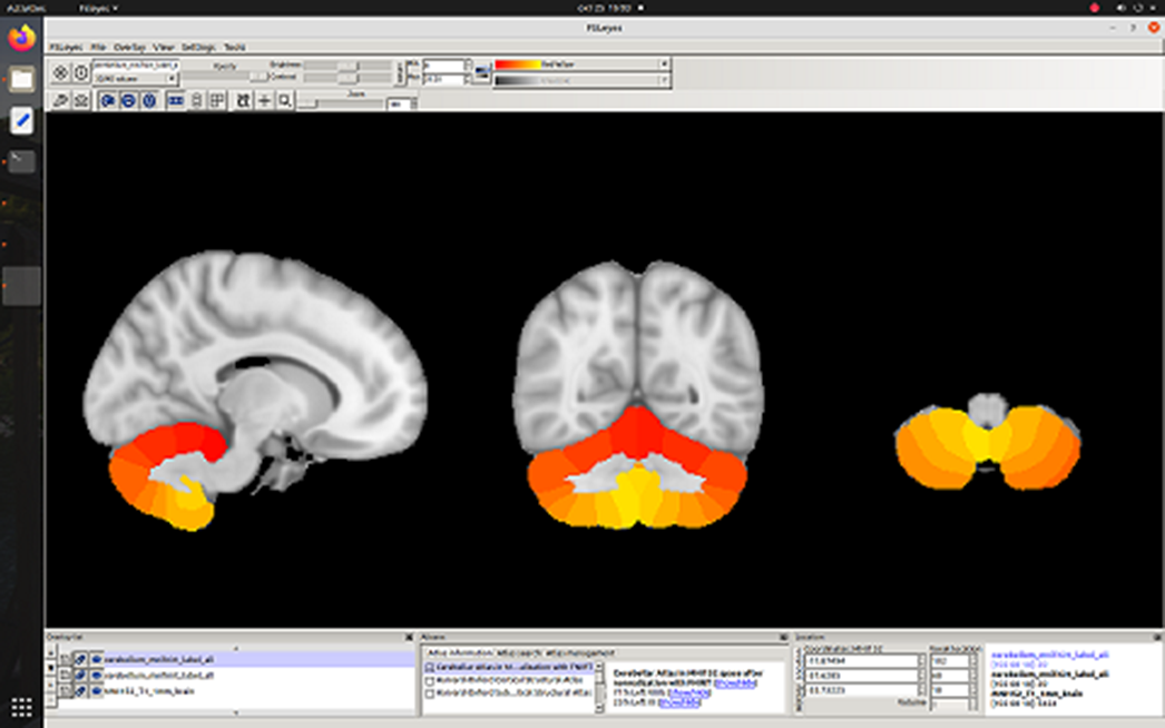
MethodsForty-five subjects, 22 controls (10m+12f, 31.6±9.7 y.o.) and 23 schizophrenia patients (F20.0, 11m+12f, 31.5±5.1 y.o.). Philips Achieva dStream 3T MRI scanner, standard head coil. The magnetization transfer (TR=20 ms, TE=4.60 ms, FA=10°), T1-weighted (TR=20 ms, TE=4.60 ms, FA=20°) and PD-weighted (TR=20 ms, TE=4.60 ms, FA=4°) were acquired. The MPF maps were reconstructed using home-made software. In FSL, non-brain structures were removed and MPF maps were registered to a standard MNI152 1 mm atlas. Harvard Oxford Cortical and Subcortical atlases were used to select areas of interest. T-test was used in search for between-group differences.
ResultsA 3% decrease in myelination in schizophrenia was observed in whole cerebral cortex p = 0.03) and cerebral white matter (p=0.02). Trends to cortical demyelination were found: paracingular cortex (p=0.06), anterior (p=0.1) and posterior cingulate cortex (p=0.07). No myelination disorders were detected in the cerebellum.
Image:
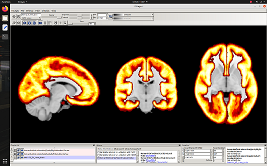
Image 2:
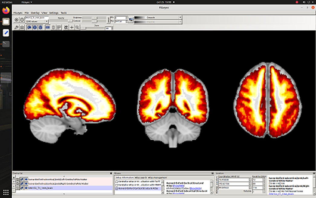
Image 3:
 Conclusions
To our knowledge, the absence of cerebellar myelination disorders in patients at an early-stage schizophrenia is reported for the first time, while the observed decrease in cerebrum myelination in schizophrenia is consistent with the previous findings. The difference in myelination between cerebellum and cerebrum may help to characterize the dynamics of the pathological process and provide additional information for understanding the biological mechanisms of the development of schizophrenia.
Grant RSF 20-15-00299 (partially).
Disclosure of InterestNone Declared


NAA and BOLD dynamics after single short stimulus in motor cortex of schizophrenia patients
-
- Journal:
- European Psychiatry / Volume 66 / Issue S1 / March 2023
- Published online by Cambridge University Press:
- 19 July 2023, pp. S293-S294
-
- Article
-
- You have access
- Open access
- Export citation
-
Introduction
Endogenous psychoses, e.g. schizophrenia, are a pressing problem of modern medicine and biology. Among various neurobiological models of schizophrenia,much attention is paid to disturbances in the brain neural activity and metabolism.
ObjectivesThe aim of this study was to analyze dynamics of motor cortex metabolites in the norm and in early stage of schizophrenia in period of BOLD response to event related single stimulus using MRI methods (fMRI and NMR).
MethodsStudy was performed on clinical Phillips Achieva 3.0 T MRI scanner. Volume of interest in motor cortex was localized on the base of fMRI study (EPI FFE, TR = 3000 ms, TE = 30 ms) as the zone of activation (Fig 1) caused by bottom push with the forefinger in response to single auditory stimuli transmitted with the 18 s periodicity. The BOLD signal was measured each 3 sec. 1Н МR spectra (PRESS, TE = 30 ms TR = 3000 ms) were run; FID signals for time points t = 0, 3, 6, 9, 12, 15, 18 s after stimulus were summarized (Fig 2). Thus, the synchronization of BOLD and metabolic responses to single stimulus was achieved. The same method was applied for spectra accumulation in resting state. For FID processing custom made software was used (with apodization filtering (LB = 20, GB = -5), FT and manual phase correction).
NAA, Cho, Cr signal intensities for each time point were normalized to their values at t = 0 and to the volume of activated cells containing in the voxel (segmented manually). Intergroup difference and time points differences were estimated using Mann-Whitney criterion with the level of significance p<0.05.
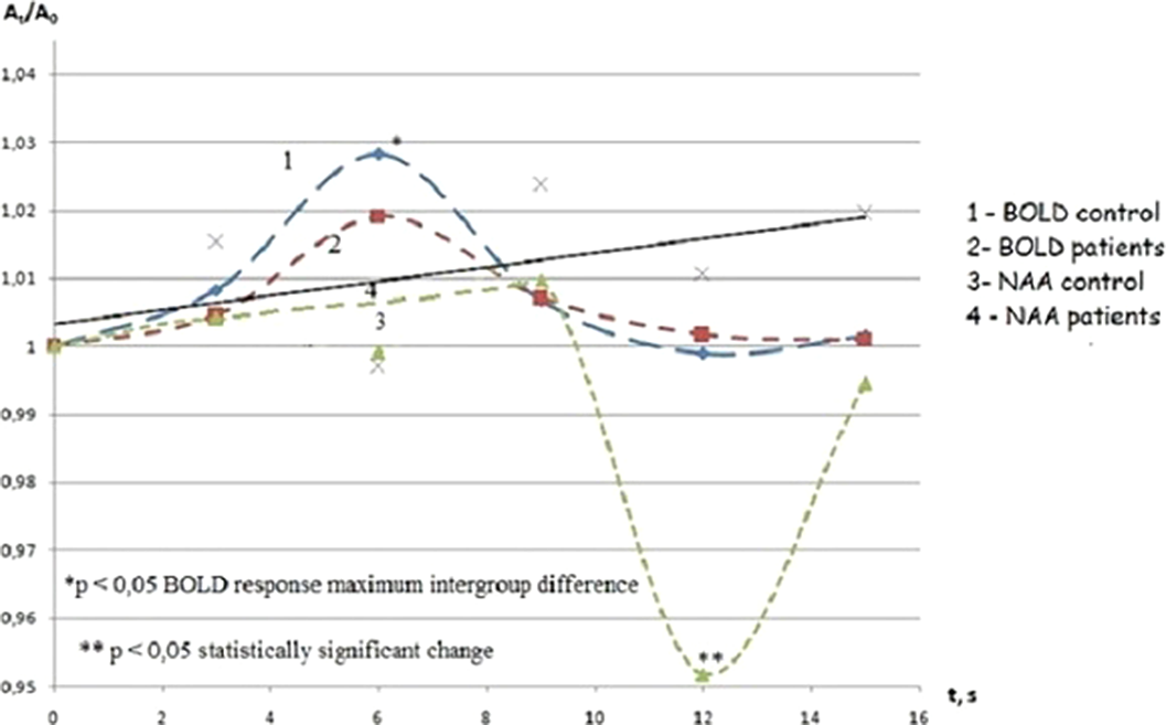
ResultsThe BOLD signal in both groups demonstrated maximum at the 6th s after target stimulus, however its value was reliably lover in schizophrenia in comparison with the control group.
The only [NAA] in normal motor cortex was changed after the stimulation (Fig D). In schizophrenia [NAA], [Cr] and [Cho] were constant. The stable values of [NAA], [Cr] and [Cho] were observed in dynamics in resting state as well. [NAA] in normal cortex statistically significantly decreased at the 12th s after stimulus presentation and returned to initial value at the 15th s (Fig 3). Thus [NAA] minimum delayed relative to maximum of BOLD by 6 s.
Image:
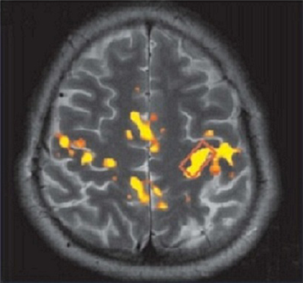
Image 2:
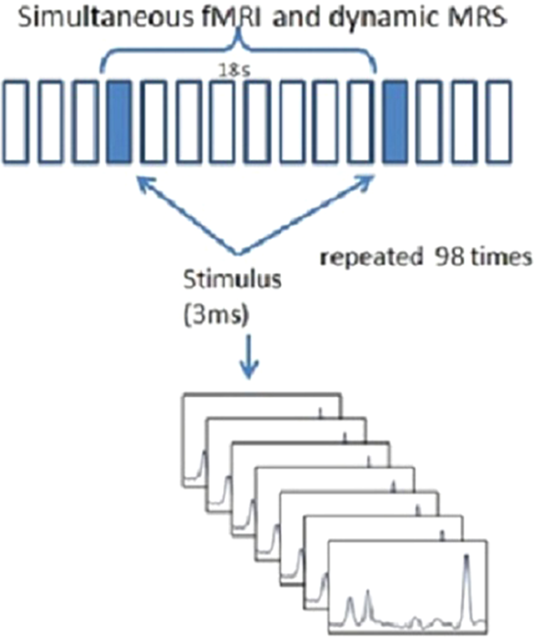
Image 3:
 Conclusions
The reversible decrease of NAA observed for the norm in the study could provide a short-term activation of neuronal Krebs cycle through a synthesis of Ac CoA using acetate obtained in ASPA reaction. Different behavior of [NAA] in the norm and schizophrenia might be related with a difference in location (or activity) of ASPA. Decreased expression of glutamate transporters in schizophrenia could also reduce consumption of NAA as a source of acetate in synthesis of Ac CoA which is used for restoration of ATP.
Disclosure of InterestNone Declared


Alteration in creatine phosphate behavior in excited visual cortex of early-stage schizophrenia patients measured by phosphorus magnetic resonance spectroscopy
-
- Journal:
- European Psychiatry / Volume 33 / Issue S1 / March 2016
- Published online by Cambridge University Press:
- 23 March 2020, p. S88
-
- Article
-
- You have access
- HTML
- Export citation
