
3 results
Decrease in anterior cingulate cortex GABA in schizophrenia at early stage
-
- Journal:
- European Psychiatry / Volume 66 / Issue S1 / March 2023
- Published online by Cambridge University Press:
- 19 July 2023, pp. S609-S610
-
- Article
-
- You have access
- Open access
- Export citation
-
Introduction
There is evidence that the concentrations of the main inhibitory neurotransmitter (GABA) may be altered in schizophrenia. The purpose of this study is to find the changes in the GABA concentration in the area of anterior and posterior cingulate cortex of patients with early-stage schizophrenia using the spectral-edited magnetic resonance spectroscopy.
ObjectivesTo measure the cerebral concentrations of the gamma-aminobutyric acid in schizophrenia patients at early stage.
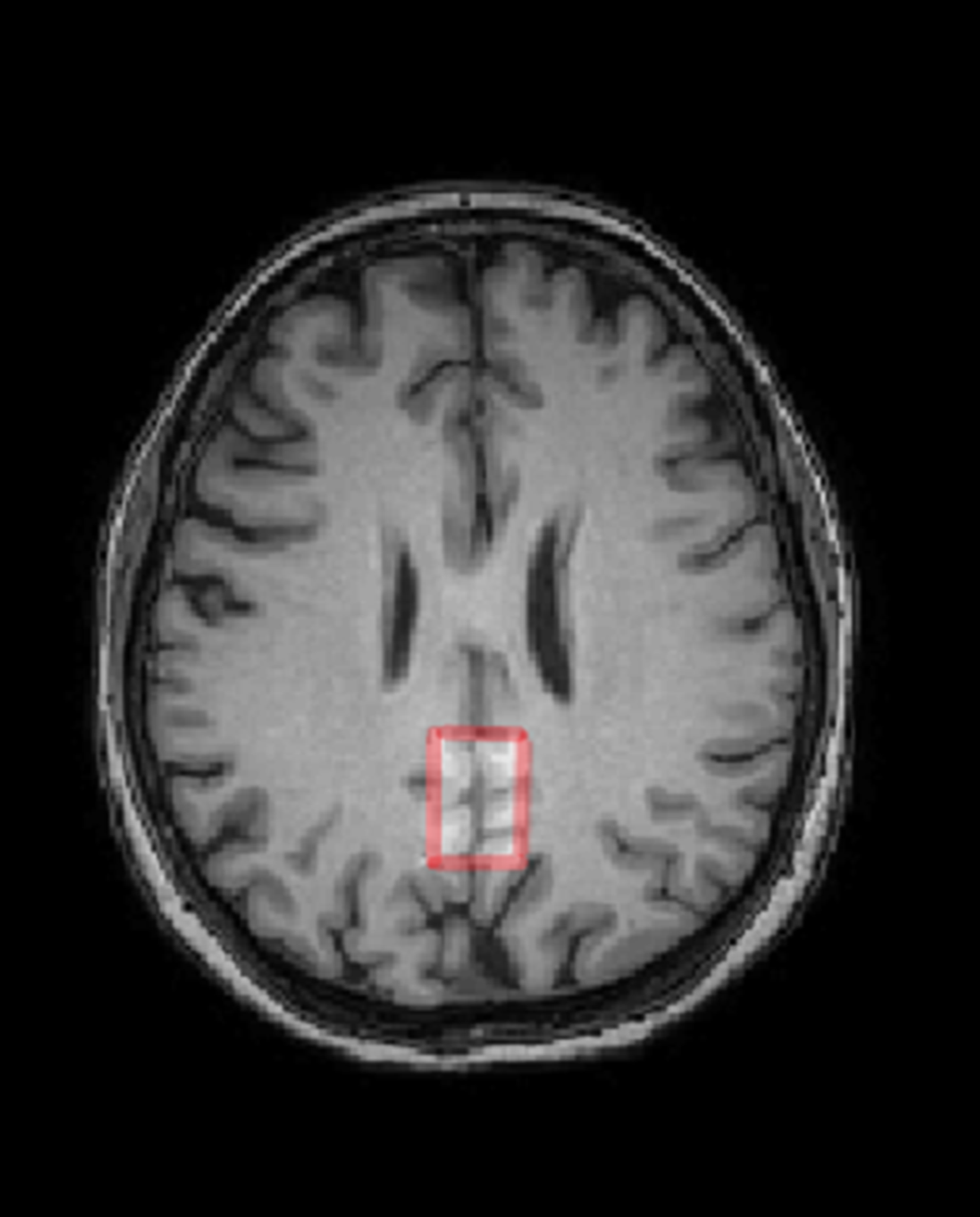
MethodsThirty-one subject, 18 controls (11m+7f, 29.6±5.7 y.o.) and 13 schizophrenia patients (F20.0, 8m+5f, 27.5±3.1 y.o.). Philips Achieva dStream 3T MRI scanner, standard head coil. The 3D T1w head images and MEGA-PRESS GABA spectra in ACC and PCC areas were acquired with the following parameters: 50x25x25 mm, TR = 2 s, TE = 64 ms, 180-editing pulses applied at 1.9 ppm and 7.6 ppm, NSA = 288 (acq.time ~10 min). GABA spectra were processed in Gannet program. The intensities of the GABA, Glutamate+glutamine (Glx), creatine (Cr) and unsuppressed water signals were acquired. T-test was used in search for between-group differences.
ResultsIn ACC region, significant reduction of the GABA/Water was observed (by ~15%, p=0.02) as well as a trend to a decrease in GABA/Cr (by ~10%, p=0.07) in schizophrenia. In PCC, no significant GABA/Water or GABA/Cr differences were observed. Glx/Water and Glx/Cr in both areas were also unchanged.
Image:
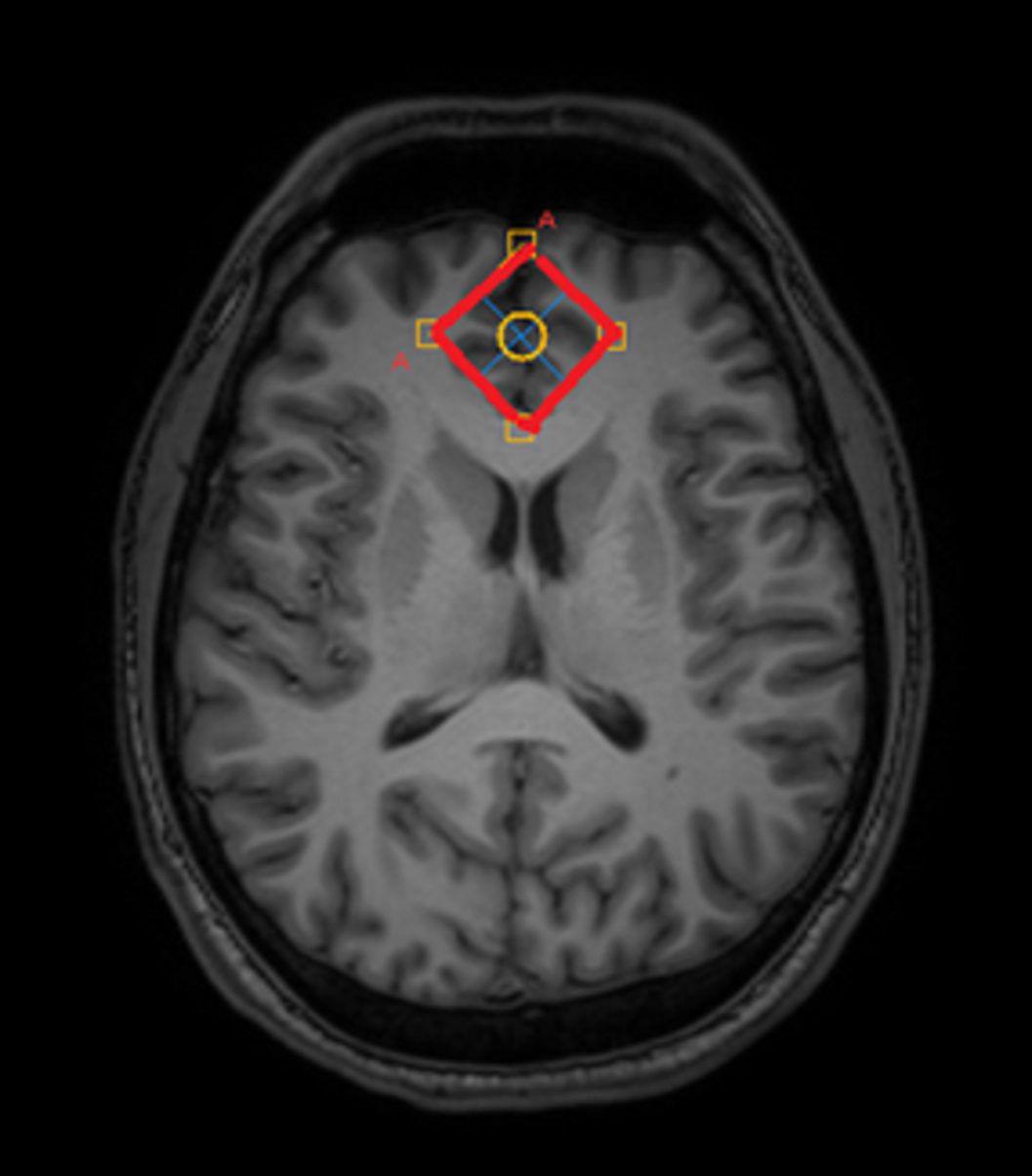
Image 2:
 Conclusions
This study provides insight into neurotransmitter alterations at early-stage schizophrenia. The results demonstrate the region-specific changes in the balance of the main neurotransmitters. Since this balance is crucial for the normal cerebral functioning, the results may facilitate better understanding of the dynamics of the pathological process and provide additional information for understanding the biological mechanisms of the schizophrenia development.
Disclosure of InterestNone Declared


Alterations in brain myelination at early-stage schizophrenia detected by macromolecular proton fraction MRI
-
- Journal:
- European Psychiatry / Volume 66 / Issue S1 / March 2023
- Published online by Cambridge University Press:
- 19 July 2023, pp. S134-S135
-
- Article
-
- You have access
- Open access
- Export citation
-
Introduction
There is evidence that cerebral myelination is impaired in schizophrenia. The purpose of this study is to find the myelin content changes in the brain structures of patients with early-stage schizophrenia using the macromolecular proton fraction (MPF) method, and also to evaluate the differences in the myelination of these structures.
ObjectivesTo measure MPF in the brain structures of schizophrenia patients
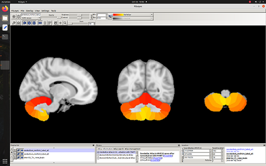
MethodsForty-five subjects, 22 controls (10m+12f, 31.6±9.7 y.o.) and 23 schizophrenia patients (F20.0, 11m+12f, 31.5±5.1 y.o.). Philips Achieva dStream 3T MRI scanner, standard head coil. The magnetization transfer (TR=20 ms, TE=4.60 ms, FA=10°), T1-weighted (TR=20 ms, TE=4.60 ms, FA=20°) and PD-weighted (TR=20 ms, TE=4.60 ms, FA=4°) were acquired. The MPF maps were reconstructed using home-made software. In FSL, non-brain structures were removed and MPF maps were registered to a standard MNI152 1 mm atlas. Harvard Oxford Cortical and Subcortical atlases were used to select areas of interest. T-test was used in search for between-group differences.
ResultsA 3% decrease in myelination in schizophrenia was observed in whole cerebral cortex p = 0.03) and cerebral white matter (p=0.02). Trends to cortical demyelination were found: paracingular cortex (p=0.06), anterior (p=0.1) and posterior cingulate cortex (p=0.07). No myelination disorders were detected in the cerebellum.
Image:
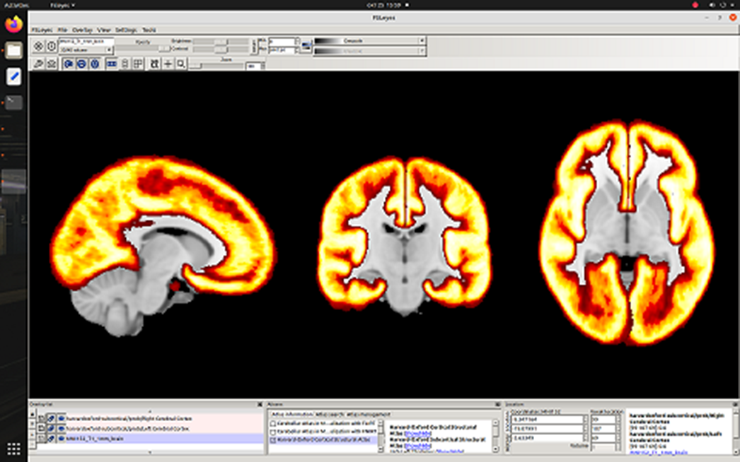
Image 2:
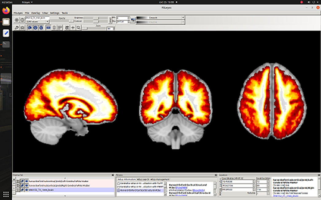
Image 3:
 Conclusions
To our knowledge, the absence of cerebellar myelination disorders in patients at an early-stage schizophrenia is reported for the first time, while the observed decrease in cerebrum myelination in schizophrenia is consistent with the previous findings. The difference in myelination between cerebellum and cerebrum may help to characterize the dynamics of the pathological process and provide additional information for understanding the biological mechanisms of the development of schizophrenia.
Grant RSF 20-15-00299 (partially).
Disclosure of InterestNone Declared


21 - BEC and the Relaxation Explosion in Magnetically Trapped Atomic Hydrogen
-
-
- Book:
- Bose-Einstein Condensation
- Published online:
- 15 December 2009
- Print publication:
- 06 April 1995, pp 472-477
-
- Chapter
- Export citation
