Skip to main content Accessibility help
×
Hostname: page-component-848d4c4894-wg55d Total loading time: 0 Render date: 2024-05-20T20:53:59.681Z Has data issue: false hasContentIssue false

Chapter 3 - Fetal Anatomy

Second and Third Trimester Assessment

Published online by Cambridge University Press:  08 June 2023

Zarko Alfirevic
Affiliation:
Liverpool Women's Hospital
Seshadri Suresh
Affiliation:
Mediscan, Chennai
Jonathan Hyett
Affiliation:
Prince Alfred Hospital, Sydney
Get access

Summary

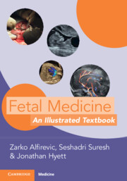
This chapter covers definitions and characteristics, ultrasound assessment, investigations, counselling and management of fetal anomalies encountered in the second and third trimesters. The chapter has seven sections (head and neck, fetal heart, thoracic and pulmonary abnormalities, spine, abdomen, genitourinary tract and skeletal abnormalities). All sections are richly illustrated with ultrasound pictures, X-ray fetograms and pathological specimens.

Type
Chapter
Information
Fetal Medicine
An Illustrated Textbook
, pp. 58 - 261
Publisher: Cambridge University Press
Print publication year: 2023

Access options

Get access to the full version of this content by using one of the access options below. (Log in options will check for institutional or personal access. Content may require purchase if you do not have access.)

References

Primary Sources

Griffiths, PD, Brackley, K, Bradburn, M, et al. Anatomical subgroup analysis of the MERIDIAN cohort: ventriculomegaly. Ultrasound Obstet Gynecol 2017;50(6):736744.CrossRefGoogle ScholarPubMed
Haratz, KK, Shulevitz, SL, Leibovitz, Z, et al. Fourth ventricle index: sonographic marker for severe fetal vermian dysgenesis/agenesis. Ultrasound Obstet Gynecol 2019;53(3):390395.CrossRefGoogle ScholarPubMed
Maduram, A, Farid, N, Rakow-Penner, R, et al. Fetal ultrasound and magnetic resonance imaging findings in suspected septo-optic dysplasia: a diagnostic dilemma. J Ultrasound Med 2020;39(8):16011614.CrossRefGoogle ScholarPubMed
Paladini, D. Fetal micrognathia: almost always an ominous finding. Ultrasound Obstet Gynecol 2010;35(4):377384.CrossRefGoogle ScholarPubMed
Pasquier, L, Marcorelles, P, Loget, P, et al. Rhombencephalosynapsis and related anomalies: a neuropathological study of 40 fetal cases. Acta Neuropathol 2009;117(2):185200.CrossRefGoogle ScholarPubMed
Viñals, F, Correa, F, Gonçalves-Pereira, PM. Anterior and posterior complexes: a step towards improving neurosonographic screening of midline and cortical anomalies. Ultrasound Obstet Gynecol 2015;46(5):585594.CrossRefGoogle ScholarPubMed
Vos, FI, De Jong-Pleij, EA, Bakker, M, et al. Nasal bone length, prenasal thickness, prenasal thickness-to-nasal bone length ratio and prefrontal space ratio in second- and third-trimester fetuses with Down syndrome. Ultrasound Obstet Gynecol 2015;45(2):211216.CrossRefGoogle ScholarPubMed

Secondary Sources

Burns, NS, Iyer, RS, Robinson, AJ, Chapman, T. Diagnostic imaging of fetal and pediatric orbital abnormalities. Am J Roentgenol 2013;201(6):W797W808.CrossRefGoogle ScholarPubMed
Paladini, D, Malinger, G, Birnbaum, R, et al. ISUOG Practice Guidelines (updated): sonographic examination of the fetal central nervous system. Part 2: performance of targeted neurosonography. Ultrasound Obstet Gynecol 2021;57(4):661671.CrossRefGoogle ScholarPubMed
Sacco, A, Pannu, D, Ushakov, F, Dyet, L, Pandya, P. Fetal dural sinus thrombosis: a systematic review. Prenat Diagn 2021;41(2):248257.CrossRefGoogle ScholarPubMed
Donofrio, MT, Moon-Grady, AJ, Hornberger, LK, et al. Diagnosis and treatment of fetal cardiac disease: a scientific statement from the American Heart Association. Circulation 2014;129(21):21832242. Erratum in: Circulation. 2014;129(21):e512.CrossRefGoogle ScholarPubMed
International Society of Ultrasound in Obstetrics and Gynecology, Carvalho, JS, Allan, LD, et al. ISUOG Practice Guidelines (updated): sonographic screening examination of the fetal heart. Ultrasound Obstet Gynecol 2013;41(3):348359.CrossRefGoogle Scholar
Suresh, S, Indrani, S, Shanthi, C, Vijayalakshmi, S. Mediscan Manual of Fetal Echocardiography. Avichal Publishing Company, New Delhi, 2021.Google Scholar
Chaoui, R, Kalache, KD, Heling, KS, et al. Absent or hypoplastic thymus on ultrasound: a marker for deletion 22q11.2 in fetal cardiac defects. Ultrasound Obstet Gynecol 2002;20(6):546552.CrossRefGoogle ScholarPubMed
Deprest, JA, Nicolaides, KH, Benachi, A, et al. Randomized trial of fetal surgery for severe left diaphragmatic hernia. N Engl J Med 2021;385(2):107118.CrossRefGoogle ScholarPubMed
Derderian, SC, Coleman, AM, Jeanty, C, et al. Favorable outcomes in high-risk congenital pulmonary airway malformations treated with multiple courses of maternal betamethasone. J Pediatr Surg 2015;50:515518.CrossRefGoogle ScholarPubMed
Nørgaard, LN, Nygaard, U, Damm, JA, et al. OK-432 treatment of early fetal chylothorax: pregnancy outcome and long-term follow-up of 14 cases. Fetal Diagn Ther 2019;46(2):8187.CrossRefGoogle ScholarPubMed
Abbasi, N, Ryan, G. Fetal primary pleural effusions: prenatal diagnosis and management. Best Pract Res Clin Obstet Gynaecol 2019;58:6677.CrossRefGoogle ScholarPubMed
Basurto, D, Russo FM, Van der Veeken, L, et al. Prenatal diagnosis and management of congenital diaphragmatic hernia. Best Pract Res Clin Obstet Gynaecol 2019;58:93106.CrossRefGoogle ScholarPubMed
Chaoui, R, Kalache, KD, Heling, KS, et al. Absent or hypoplastic thymus on ultrasound: a marker for deletion 22q11.2 in fetal cardiac defects. Ultrasound Obstet Gynecol 2002;20(6):546552.CrossRefGoogle ScholarPubMed
David, M, Lamas-Pinheiro, R, Henriques-Coelho, T. Prenatal and postnatal management of congenital pulmonary airway malformation. Neonatology 2016;110(2):101115.CrossRefGoogle ScholarPubMed
Friszer, S, Dhombres, F, Morel, B, et al. Limited dorsal myeloschisis: a diagnostic pitfall in the prenatal ultrasound of fetal dysraphism. Fetal Diagn Ther 2017;41(2):136144.CrossRefGoogle ScholarPubMed
Al Naimi, A, Baumüller, JE, Spahn, S, Bahlmann, F. Prenatal diagnosis of multicystic dysplastic kidney disease in the second trimester screening. Prenat Diagn 2013;33(8):726731.CrossRefGoogle ScholarPubMed
Fontanella, F, Duin, L, Adama van Scheltema, PN, et al. Fetal megacystis: prediction of spontaneous resolution and outcome. Ultrasound Obstet Gynecol 2017;50(4):458463.CrossRefGoogle ScholarPubMed
Turkyilmaz, G, Cetin, B, Sivrikoz, T, et al. Antenatally detected ureterocele: associated anomalies and postnatal prognosis. Taiwan J Obstet Gynecol 2019;58:531535.CrossRefGoogle ScholarPubMed
Cheung, KW, Morris, RK, Kilby MD. Congenital urinary tract obstruction. Best Pract Res Clin Obstet Gynaecol 2019;58:7892.CrossRefGoogle ScholarPubMed
Dias, T, Sairam, S, Kumarasiri, S. Ultrasound diagnosis of fetal renal abnormalities. Best Pract Res Clin Obstet Gynaecol 2014;28(3):403415.CrossRefGoogle ScholarPubMed
Gimpel, C, Avni, FE, Bergmann, C, et al. Perinatal diagnosis, management, and follow-up of cystic renal diseases: a clinical practice recommendation with systematic literature reviews. JAMA Pediatr 2018;172(1):7486.CrossRefGoogle ScholarPubMed
Huber, C, Shazly, SA, Blumenfeld, YJ, Jelin, E, Ruano, R. Update on the prenatal diagnosis and outcomes of fetal bilateral renal agenesis. Obstet Gynecol Surv 2019;74(5):298302.CrossRefGoogle ScholarPubMed
Liu, DB, Armstrong WR 3rd, Maizels M. Hydronephrosis: prenatal and postnatal evaluation and management. Clin Perinatol 2014;41(3):661678.CrossRefGoogle ScholarPubMed
Nguyen, HT, Herndon, CD, Cooper, C, et al. The Society for Fetal Urology consensus statement on the evaluation and management of antenatal hydronephrosis. J Pediatr Urol 2010;6(3):212231.CrossRefGoogle Scholar
Rosenblum, S, Pal, A, Reidy, K. Renal development in the fetus and premature infant. Semin Fetal Neonatal Med 2017;22(2):5866.CrossRefGoogle ScholarPubMed
Stambough, K, Magistrado, L, Perez-Milicua, G. Evaluation of ambiguous genitalia. Curr Opin Obstet Gynecol 2019;31:303308.CrossRefGoogle ScholarPubMed
Taghavi, K, Sharpe, C, Stringer, MD. Fetal megacystis: a systematic review. J Pediatr Urol 2017;13(1):715.CrossRefGoogle ScholarPubMed
Talati, AN, Webster, CM, Vora, NL. Prenatal genetic considerations of congenital anomalies of the kidney and urinary tract (CAKUT). Prenat Diagn 2019;39(9):679692.CrossRefGoogle ScholarPubMed
Westland, R, Schreuder, MF, Ket, JCF, van Wijk, JAE. Unilateral renal agenesis: a systematic review on associated anomalies and renal injury, Nephrol Dialy Transplant 2013;28(7):18441855.CrossRefGoogle ScholarPubMed
Mathiesen, JM, Aksglaede, L, Skibsted, L, et al. Outcome of fetuses with short femur length detected at second-trimester anomaly scan: a national survey. Ultrasound Obstet Gynecol 2014;44(2):160165.CrossRefGoogle ScholarPubMed
Chitty, LS, Everett, F, Usakov, F. Songoraphic diagnosis of skeletal anomalies. In: Fetal Medicine (Kumar, B, Alfirevic, Z, eds), Cambridge University Press, Cambridge, 2016; 161181.CrossRefGoogle Scholar
Di Mascio, D, Buca, D, Khalil, A, et al. Outcome of isolated fetal talipes: a systematic review and meta-analysis. Acta Obstet Gynecol Scand 2019;98(11):13671377.CrossRefGoogle ScholarPubMed

Save book to Kindle

To save this book to your Kindle, first ensure coreplatform@cambridge.org is added to your Approved Personal Document E-mail List under your Personal Document Settings on the Manage Your Content and Devices page of your Amazon account. Then enter the ‘name’ part of your Kindle email address below. Find out more about saving to your Kindle.

Note you can select to save to either the @free.kindle.com or @kindle.com variations. ‘@free.kindle.com’ emails are free but can only be saved to your device when it is connected to wi-fi. ‘@kindle.com’ emails can be delivered even when you are not connected to wi-fi, but note that service fees apply.

Find out more about the Kindle Personal Document Service.

Available formats
×

Save book to Dropbox

To save content items to your account, please confirm that you agree to abide by our usage policies. If this is the first time you use this feature, you will be asked to authorise Cambridge Core to connect with your account. Find out more about saving content to Dropbox.

Available formats
×

Save book to Google Drive

To save content items to your account, please confirm that you agree to abide by our usage policies. If this is the first time you use this feature, you will be asked to authorise Cambridge Core to connect with your account. Find out more about saving content to Google Drive.

Available formats
×