Botany, the eldest daughter of medicine.

Approximately one-fourth of all current prescription medicines are formulations that are either substances or derivatives of molecules from botanical sources.
Cannabis sativa, also known as “marijuana,” has been used for thousands of years. In 1964 Δ9-tetrahydrocannabinol (THC) was first isolated and identified as the psychoactive constituent in the plant.
In the late 1980s the first cannabinoid receptor that bound THC was discovered in the brain. Molecules referred to as endocannabinoids that bind to the cannabinoid receptors and are produced within the body were subsequently identified.
Two types of cannabinoid receptors have been identified and are named CB1 and CB2. Cannabinoid receptors are G protein-coupled receptors widely distributed within the central and peripheral nervous system and multiple regions of the body.
Phytocannabinoids are three-ring structures synthesized by the cannabis plant. Endocannabinoids are bioactive fatty acids derived from arachidonic acid in the body. Both groups bind to cannabinoid receptors.
Introduction
Plants have been a plentiful source of useful drugs and remedies throughout human history. In the early nineteenth century Friedrich Sertürner isolated morphine from the opium plant. By 1827, morphine was marketed by Merck in Germany and the origins of the modern pharmaceutical industry began. Over the remainder of the nineteenth century further advances in organic chemistry led to identification of other drugs from plant material. Examples of important medicines developed from plants included quinine from the bark of the cinchona tree for the treatment of malaria and salicylic acid from the willow tree that eventually led to the development of aspirin (Reference AndersonAnderson, 2005). Today, the World Health Organization estimates that over 100 plant species are the source of approved pharmaceutical products and thousands of other plants are used for medical and recreational applications (Reference Potter and PertweePotter, 2014).
Progress in the identification of the constituents of cannabis (also known as marijuana) lagged behind the advances in plant chemistry. Although the cannabis plant has been used for fiber, as a medication, as a source of nutrition and as an element of religious ceremonies for over 4000 years, the constituents of the plant were extremely difficult to chemically separate because they were highly lipophilic, water-insoluble and frequently gelatinous. Isolating the substance that induced the psychoactive effect of cannabis was especially resistant to exploration (Reference IversenIversen, 2008). Only in the 1930s was the structure of cannabinol (CBN) partially isolated by the British chemist Robert Cahn and work by Alexander Todd and Roger Adams in the 1940s in Britain and the United States came close to identifying cannabidiol (CBD) (Reference IversenIversen, 2008). Finally, in 1964 Raphael Mechoulam and Yehiel Gaoni at Hebrew University separated (-)Δ9-tetrahydrocannabinol (also known as THC), a three-ring, 21-carbon terpenophenolic structure, from cannabis and demonstrated that the molecule was responsible for the psychoactive effects (Reference Gaoni and MechoulamGaoni and Mechoulam, 1964). From this discovery followed 25 years of research that identified multiple naturally occurring cannabinoids from plants, referred to as phytocannabinoids and new compounds synthesized from chemical manipulation of the phytocannabinoid structure. The cannabinoids that were newly identified from cannabis were found to be extremely lipophilic and it was assumed that the action of THC likely occurred through nonspecific interactions within the neuron membrane (Reference IversenIversen, 2008). The observation that THC activity was stereospecific (Reference MechoulamMechoulam et al., 1988) led to the search for specific targets that cannabinoids were likely to bind. In the mid-1980s Allyn Howlett at St. Louis University discovered the cannabinoid receptor 1 (CB1) in the rat brain (Reference Howlett, Qualy and KhachatrianHowlett, Qualy and Khachatrian, 1986; Reference DevaneDevane et al., 1988).
Obviously, a receptor in the brain is present to bind to an endogenous substance and not to specifically bind to a plant molecule and a search for an endogenous ligand began. A similar search had occurred earlier in the 1970s after the discovery of opiate receptors in the brain. That search ultimately led to the discovery of endogenously produced endorphins functioning within a previously unknown complex system of opiate receptors in the brain (Reference IversenIversen, 2008).
Similar to the discoveries with opiates earlier, within a few years of the identification of the CB1 receptor, the endogenous cannabinoid, N-arachidonoylethanolamide (anandamide; AEA), an ethanolamine of arachidonic acid, was discovered in pig brain using radiolabeled ligands by Mechoulam and Devane (Reference DevaneDevane et al., 1992). Later, a second endogenous cannabinoid, 2-arachidonoylglycerol (2-AG), an arachidonate ester of glycerol, was identified in canine intestines by the same investigators and in rat brain by Sugiura and colleagues (Reference SugiuraSugiura et al., 1995). Both AEA and 2-AG, referred to as endocannabinoids since they are synthesized within the body, bind to the cannabinoid receptor. However, the affinities for the cannabinoid receptors and the chemical structure of endocannabinoids differ significantly from the familiar three-ring structures of phytocannabinoids.
This discovery of the CB1 and the binding of AEA and 2-AG was quickly followed by the identification of a second cannabinoid receptor (CB2), initially located on white blood cells and spleen suggesting a role for cannabinoids in immune function and protection from attack and tissue repair (Reference Munro, Thomas and Abu-ShaarMunro, Thomas and Abu-Shaar, 1993). Although it was originally believed that the CB2 was only a peripheral receptor, subsequent work found that CB2 is also present in microglial cells in the brain.
It is possible that more cannabinoid receptors may be found in the future. GPR55 is a G protein-coupled receptor that binds THC and endocannabinoids (Reference DerocqDerocq et al., 1998; Reference RybergRyberg et al., 2007). CBD acts as an antagonist on this receptor and the CB1 inverse antagonist rimonabant acts as an agonist at this site (Reference PertweePertwee et al., 2005). TRPV1 is another candidate and is a postsynaptic receptor that binds AEA in addition to activation by capsaicin. Both AEA and capsaicin bind to the same site within the cell and appear to mediate pain in sensory neurons (Reference Starowicz and PrzewlockaStarowicz and Przewlocka, 2012).
Brief History of Cannabis
The first historical record describing cannabis occurs in ancient Chinese literature dating from 2727 BC and the Chinese Emperor Shen Nung. Considered the “father of Chinese medicine,” Shen Nung described hundreds of drugs from plants, animals and minerals in the Pen Ts’ao, believed to be the first pharmacopeia in history. The Emperor recommended in his work the use of hemp elixir to treat a wide range of ailments including gout and malaria. Although the original text no longer exists, the oldest surviving text of the Pen Ts’ao is found in the first century AD and refers to cannabis as “Ma,” which is the Chinese word for chaos. We can only conjecture if the meaning of Ma and chaos was intended to describe the medicinal and psychoactive effects of cannabis. There is little doubt, however, that the Chinese regarded cannabis as a valuable medicine (Reference AbelAbel, 1980).
Probably as a result of invasion and migration from central Asia, cannabis arrived later to the subcontinent of India. Although the medicinal uses were also recognized, the Indian culture embraced cannabis as an important social and religious element. Legend states that Prince Siddhartha, later named Buddha, survived for six years exclusively on a diet of hemp seed in his quest for enlightenment. Later in Buddhism, cannabis was reserved for the priests and upper classes. Cannabis eventually became stratified into three classes: bhang, ganga and charas (known in other regions as hashish) and the use of cannabis became even more embedded into Indian culture (Reference BoothBooth, 2004).
Further awareness of cannabis extended beyond India into Persia, Syria and Greece. By 600 BC hemp was brought to the Middle East carried by the Indo-Aryan migration and was used both as a fiber and a medicine. In Syria cannabis was referred to as “qunubu” or the “drug for sadness” (Reference BoothBooth, 2004). The Greeks and Romans were certainly aware of cannabis as hemp and this played an important role in Greco-Roman society. Dioscorides, a Roman physician trained in the arts of Greek and Oriental medicine served in the Roman army during the first century AD and wrote De Materia Medica describing the medical uses of cannabis. De Materia Medica would remain among the most influential pharmacopeias in Western medicine for the next 1700 years (Reference AbelAbel, 1980; Reference BoothBooth, 2004).
In the Muslim era, there was little agreement if cannabis was an intoxicant or a medicine. In a culture where alcohol was prohibited, cannabis presented daunting religious questions. One resolution was to severely punish any recreational use of the plant while in other instances it could be used by physicians to administer treatment (Reference IversenIversen, 2008).
Other uses were also found in the Muslim world. Hemp was used as a source of paper and later exported to Europe where it remained a medium of writing and printing until the introduction of wood pulp in the mid-nineteenth century (Reference AbelAbel, 1980; Reference BoothBooth, 2004)
The first reference to the term “hashish” is found in Egypt during the twelfth century AD and probably derives from the Arabic word for dried foliage. Hashish contains dried leaves and flowers along with the highly potent resin that was consumed originally by eating and not by the more common method today of smoking. The concept of burning dried plants to activate drug release in smoking was unknown until the sixteenth century with the importation of tobacco from the new world. Thus, according to legend popularized by the publication of Marco Polo’s “The Travels of Marco Polo” in the twelfth century, another possible origin of the word hashish was from “assassins” that described the violent followers of an old man in the mountains (Reference BoothBooth, 2004).
Several trends converged in the nineteenth century and contributed to the growing awareness of the medicinal and psychoactive properties of cannabis in Western Europe. In 1839 William Brooke O’Shaughnessy, an Irish physician and professor of chemistry working for the British East India Company in Calcutta, presented the first medical paper on cannabis and hashish in western medicine to medical societies in India. In this paper he described his observations over seven years of the therapeutic effect of cannabis and hashish in the treatment of seizures, pain, fever and malaria and other conditions. O’Shaughnessy returned to London in 1842 where he published The Bengal Dispensatory, and Companion to the Pharmacopoeia that introduced to the British medical societies cannabis and other herbs used in India. He also provided hashish to a London pharmacist to make a medical extract with alcohol that was patented and sold as an analgesic. It was this extract that eventually found its way to North America as Tilden’s Extract. So impactful were these activities that Queen Victoria is reported to have used cannabis for premenstrual pain (Reference BoothBooth, 2004).
O’Shaughnessy had a brilliant and open mind that saw the therapeutic possibilities of cannabis although he is better remembered in history for other work. He is credited with the first use of intravenous fluids for rehydration while a young physician recently out of training. Later, while in India, he was appointed Director-General of Telegraphs and built the first telegraph network in India. For this achievement he was knighted in 1856 (Reference BoothBooth, 2004).
Other events also contributed to the growing awareness that cannabis was more than a source of fiber. In 1796 Napoleon occupied Egypt with the design to disrupt British trade to the Middle East and India. Although he accomplished this goal of interfering with commerce, overall the mission was not a success. On August 1–3, 1798 the British navy under the command of Admiral Horatio Nelson defeated the French fleet at the Battle of the Nile. This effectively quarantined the French army of 35,000 men in Egypt and prevented supplies from France reaching them while the British blockaded the ports. It was during this time as the French were building fortifications to defend their occupation that the Rosetta Stone was discovered in July 1799 by Pierre-François Bouchard, an officer and engineer in the French army. Stranded in a Moslem country without alcohol, the French soldiers became acquainted with hashish much to the dismay of Napoleon. On September 2, 1801 the Capitulation of Alexandria was signed and the French army surrendered and were transported along with their supplies of hashish back to France by the British navy. The Rosetta Stone became the property of Britain and was shipped back to London and has been on continuous display in the British Museum since 1802 (Reference IversenIversen, 2008).
Jacques-Joseph Moreau, a French psychiatrist, also contributed to this raising awareness about cannabis and hashish after studying about medical practices in India and China for several years. Returning to France in the 1830s, Moreau was the first to conduct clinical trials on psychiatric patients and described the effect of cannabis and hashish on the central nervous system (CNS). Moreau concluded that hashish could mimic mental illness but recognized that this action might also hold clues to potential treatment. From these observations the discipline of psychopharmacology arose (Reference BoothBooth, 2004).
Description of the Cannabis Plant
Cannabis is believed by many to have originated in central Asia or in the great river valleys of eastern China. Cannabis is an annual plant propagated by wind, widely distributed in temperate and tropical zones, and reported to grow at altitudes up to 8000 feet. The plant prefers direct sunlight, requires little water, germinates within six days and can reach maturity within five months. Because of its versatility, it is estimated that cannabis can grow in over two-thirds of the landmass of the globe (Reference BoothBooth, 2004).
In 1753 Carolus Linnaeus, the Swedish “father of botany,” was aware of cannabis as hemp and its importance in rope and sail manufacture and named the plant “Cannabis sativa.” Linnaeus chose the word “cannabis” from the Greek “kannabis” for hemp and “sativa” from the Latin word for cultivated. The evolutionary biologist Jean-Baptiste Lamarck in 1783 proposed a second species of the cannabis plant with the name “Cannabis indica” for a smaller, denser plant that grows in India. Lamarck believed that there were important distinctions between the two plants and noted that C. sativa possessed more fiber-like qualities and C. indica had more behavioral effects. Finally, in 1924 Janischewski, a Russian botanist, introduced a third species of cannabis named Cannabis ruderalis. C. ruderalis is found along the banks of the Volga River and in Siberia (Reference AbelAbel, 1980; Reference BoothBooth, 2004).
The appearance and geographic distribution of C. sativa and C. indica differ significantly. C. sativa is the more widespread of the two with the height of up to 15 feet. In contrast, C. indica can grow up to 4 feet in height and has dense branching and foliage (Reference BoothBooth, 2004).
Despite these differences in appearance and properties, controversy remains over classifying cannabis as several species (polytypic), or as one species (monotypic). Central to this argument is the belief that the observed differences may reflect the robust adaptability of the cannabis plant to different climates and soil. This view is based on the observation that seeds from C. sativa, when planted in regions where C. indica thrive, will over several generations display many of the characteristics of C. indica. In addition, the various types of cannabis can easily be interbred, cuttings can be cloned, and hybrids established resulting in myriad versions of the plant (Reference BoothBooth, 2004; Reference IversenIversen, 2008).
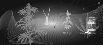
The male plant produces pollen in the anther within a prominent flower while the smaller female flower after pollination produces seeds in the perianth.
The stem of the plant can be up to 5 cm in diameter and is covered with stiff fine hairs. When harvested and dried, the stem serves as the primary source of hemp and contains less than 0.3% tetrahydrocannabinol (THC). The distribution of THC varies in the plant with less than 1% THC in the leaves closer to the ground with increasing content up to 2–3% at the apex of the plant. Unpollinated female flowers do not produce seeds and contain higher THC concentrations up to 20%. When pollinated, seeds are produced and the concentration of THC in the flower is reduced since seeds do not contain cannabinoids. Thus, in the cultivation of cannabis for THC, male plants are usually removed from the growth area and only unpollinated female plants remain (Reference Potter and PertweePotter, 2014).
Both male and female plants produce a yellow colored resin containing high concentrations of THC in glandular structures at the base of the fine hairs. The resin is quite thick and sticky. Glandular secretions adhere to the male anthers and female perianths within the flowers increasing their cannabinoid content. In addition, female plants are prolific producers of resin compared with males. The purpose of the resin is unknown but may provide a natural protection for the plants against sunlight and heat as increased ambient temperature increases resin secretion. Others have speculated that the resin provides both a protective barrier and an impairing intoxicant that discourages insects and other pests (Reference Potter and PertweePotter, 2014).
Cannabis has a complex sexual physiology and usually each plant is one of two sexes (defined as dioecious). However, less commonly, individual plants may bear both male and female flowers and are termed hermaphrodites. Unpollinated female flowers do not produce seeds and are the preferred plant for production of cannabinoids while monoecious plants are cultivated for hemp and paper production (Reference BoothBooth, 2004; Reference Potter and PertweePotter, 2014).
Perhaps cannabis is best known today for the psychoactive properties exhibited when consumed. The psychoactive effect occurs when the phytocannabinoid THC traverses the lipid blood–brain barrier from the circulating bloodstream. Since all phytocannabinoids are extremely lipophilic, THC and the other compounds from the plant enter the brain with the well-known intoxication in addition to other non-psychoactive effects. In the blood, THC is converted into 11-hydroxy-THC and absorbed into fatty tissue. After approximately 30 minutes, THC is distributed back into the bloodstream. The phytocannabinoids exhibit a characteristic C21 terpenophenolic structure and within the plant other compounds including flavonoids and terpenes are to be found. The number of known cannabinoids exceeds 110 today and it is possible that others are yet to be found (Reference IversenIversen, 2008).
THC is classified into three types based upon the concentration of the cannabinoid. Marijuana usually refers to dried leaves, flowers and stems. The THC content is between 5% and 10% and marijuana is the primary source of recreational use in North America. Hashish is made from the resin and glandular trichomes in addition to the flower and leaves of the cannabis plant. The resin contains high concentrations of all the cannabinoids and THC often exceeds 20%. Typically, the female unpollinated flower contains the most trichomes and highest concentration of cannabinoids and is the predominate constituent of Hashish. In Europe, hashish is the preferred form of THC in recreational use. Hashish oil is another version and may have THC content up to 85%. Typically, in most countries where THC is legalized, the hashish oil remains prohibited (Reference IversenIversen, 2008).
The Cannabinoid System: Receptors and Neurotransmitters
Cannabinoids are molecules that alter neurotransmitter release by binding to cannabinoid receptors in the body. Cannabinoid receptors belong to the superfamily of G protein-coupled receptors (GPCR), a ubiquitous, 7-transmembrane family of protein receptors involved in the transduction of extracellular molecular messaging to intracellular response. After cannabinoids bind to the cannabinoid receptor, Gi/o-transduced inhibition of the enzyme adenyl cyclase results and the synthesis of cyclic adenosine monophosphate (cAMP), the second messenger for the intracellular signal transduction, is reduced. Through this second messenger system, binding at the cannabinoid is coupled with the mitogen-activated protein kinases that influence diverse signaling pathways within the cell (Reference MechoulamMechoulam et al., 2014).
The reduction of cAMP by binding at the cannabinoid receptor results in inhibition of calcium influx into the cell and is likely important in the reduction in neurotransmitter release. Most neurotransmitters are stored in vesicles and other mechanisms are involved in release into the synaptic cleft. It is possible these alternative systems may also prove important in understanding how cannabinoids influence neurotransmission.
Although cannabinoids binding at the receptor reduce neurotransmitter release, the overall effect of activation or inhibition is determined by the neurotransmitter that is inhibited. For example, glutamate and gamma-aminobutyric acid (GABA) are major neurotransmitters in the CNS and CB1 receptors are found on these neuronal membranes. Glutamate is a ubiquitous neurotransmitter known to be activating and involved in tissue damage. Inhibition of glutamate by activation of the CB1 receptor would be expected to result in reduced activation and even neuroprotection. In contrast, CB1 receptors are also prominent on GABA neurons. GABA is the primary inhibitory neurotransmitter in the CNS and inhibition of GABA would result in increased activation and anxiety. Other neurotransmitter systems including the noradrenergic and cholinergic systems that are key systems in neurological and psychiatric disease are also known to be influenced by activation of the CB1 receptor (Reference Nestler, Hyman and MalenkaNestler, Hyman and Malenka, 2001).
CB1 receptors were originally found in brain and were initially believed to be limited to the CNS. Later work has found that CB1 receptors are also present in lesser amounts within peripheral tissue including heart, liver, fat tissue, stomach, intestines and testes. Despite this wide distribution throughout the body, the highest concentrations of the CB1 receptor are in the brain and they have the highest density of any GPCRs. In the brain, the CB1 receptors are distributed in the basal ganglia, substantia nigra, globus pallidus, hippocampus and cerebellum with little presence in the brain stem. The receptor is found predominantly on the presynaptic neuron axon terminal and this is an ideal location to influence neurotransmitter activity. The inhibition of synaptic transmission is the most common effect of CB1 agonist activity.
CB1 receptors are also found in the peripheral nervous system and are involved in sensory neurons, postganglionic sympathetic neurons, and in the parasympathetic nervous system. Because of the density of CB1 receptors in the brain and wide distribution in diffuse organs, it is likely that when cannabinoids bind to the receptor significant physiological functions are influenced.
In close proximity to the CB1 binding site, an allosteric site is situated on the membrane and, when activated, influences the primary CB1 site. This modulation of CB1 activity caused by the nearby allosteric binding can have significant effects including reduction or termination of response to the cannabinoid. (Reference PricePrice et al., 2005). In the absence of cannabinoid binding to the CB1 site, molecules that bind to the allosteric site will have no effect on the response.
The presence of endogenous allosteric molecules thus allows enhancement or diminution of the response to the primary binding site.
Just as the CB1 receptor was originally thought to be only in the CNS, the CB2 receptor was believed to be a peripheral cannabinoid receptor and involved in the immune response against protein attack and injury. Later studies confirmed that the CB2 receptor is also present in the CNS especially in microglial tissue. Although the CB2 receptor number is substantially less when compared with the CB1 receptor in the CNS, the wide distribution in the body including cardiovascular, reproductive, gastrointestinal, pulmonary and hepatic systems suggest a broader role for the CB2 receptor. Reference Pacher and MechoulamPacher and Mechoulam (2011) have speculated about a broad protective role of the CB2 receptor by writing, “Are there mechanisms through which our body lowers the damage caused by various types of neuronal as well as non-neuronal insults? The answer is, of course, positive. Through evolution numerous protective mechanisms have evolved to prevent and limit tissue injury. We believe that lipid signaling through CB2 receptors is a part of such a protective machinery and CB2 receptor stimulation leads mostly to sequences of activities of the protective nature.”
Cannabinoids are a major constituent of the cannabis plant. In mammals, two endocannabinoids have been identified that bind with a higher affinity to the cannabinoid receptors and have significant differences from phytocannabinoids in their chemical structure. Phytocannabinoids as previously noted are three-ring structures while endocannabinoids are long-chain fatty acids. A third class of cannabinoids are synthetic molecules manufactured in a laboratory setting and consists of various chemical structures that also bind to the cannabinoid receptor. Synthetic cannabinoids consist of multiple chemical structures including the three-ring structures of phytocannabinoids, eicosanoids, aminoalkylindoles, quinolines and others.
The phytocannabinoids are grouped into 11 types of cannabinoids. THC is the best-known cannabinoid due to its psychoactive properties. CBD is the other major constituent of the phytocannabinoids and has no psychoactive properties. Other cannabinoid types include (-)-Δ8-trans-tetrahydrocannabinol, cannabigerol (CBG), cannabichromene (CBC), cannabinodiol (CBND), cannabielsoin (CBE), cannabicyclol (CBL), cannabinol (CBN), cannabitriol (CBT) and miscellaneous cannabinoids (Reference IversenIversen, 2008).
THC is a partial agonist at both CB1 and CB2 receptors. Compared with endocannabinoids, the THC has a lower receptor affinity.
CBD, although non-psychoactive, may have important physiological properties. Research suggests CBD may work as a negative allosteric modulator on the CB1 and CB2 receptors. CBD may eventually demonstrate important benefits in the treatment of diverse diseases including epilepsy, psychiatric and sleep disorders, pain and inflammation. CBD may also have antioxidant effects and potentially provide neuroprotective benefits in neurodegenerative diseases such as the dementias including Alzheimer’s disease.
The endocannabinoids consist of AEA and 2-AG. AEA is a N-derivative and 2-AG an O-derivative long-chain fatty acid. Unlike neurotransmitters that are stored in vesicles and wait for release, endocannabinoids are synthesized on demand and released immediately into the synaptic cleft to bind on presynaptic receptors. AEA is synthesized by phospholipase D and 2-AG by another enzyme named diacylglycerol lipase (DAG) (Reference Di Marzo, De Petrocellis and BisognoDi Marzo, De Petrocellis and Bisogno, 2005).
Both endocannabinoids are rapidly removed by a membrane transport process and inactivated. AEA is transported through this mechanism across the postsynaptic membrane and rapidly degraded by intracellular fatty acid amide hydrolase (FAAH) to arachidonic acid and ethanolamine. In contrast, 2-AG is transported across the presynaptic membrane and enzymatically deactivated by the enzyme monoacylglycerol lipase (MAGL) (Reference Dinh, Kathuria and PiomelliDinh, Kathuria and Piomelli, 2004).
Similar to the situation with cannabinoid receptors, other endocannabinoids are likely to be discovered since other lipid molecules interact with the cannabinoid receptors or share some common metabolic pathways or targets.
Fatty acids are widely distributed throughout the body but have significant differences in the amount and distribution of the lipid and their degradative enzymes. Both AEA and 2-AG are derivatives of arachidonic acid which is a polyunsaturated fatty acid commonly found in cell membranes and is especially prominent in the brain. Arachidonic acid is known to be involved in cellular signaling and as an inflammatory intermediate and differences in the distribution of degradative enzymes may lead to different roles for the two endocannabinoids.
AEA has been found throughout the brain and peripheral tissue and can be present where the CB1 receptor is highly concentrated or has limited distribution suggesting that the molecule may be important in other non-cannabinoid processes. AEA is a partial agonist at the CB1 receptor and can act as a weak partial agonist/antagonist at the CB2 site.
2-AG is more prominent than AEA and has been proposed as the primary endocannabinoid for the known cannabinoid receptors (Reference SugiuraSugiura et al., 2002). Unlike AEA, 2-AG is a full agonist of both CB1 and CB2 receptors. Because of the differences in concentration and binding affinity to the cannabinoid receptors, it has been suggested that these two endocannabinoids have different roles to play within the cell (Reference Maccarrone, Dainese and OddiMaccarrone, Dainese and Oddi, 2010; Reference Alger and KinAlger and Kim, 2011).
Other arachidonic acid derivatives are also known to act on cannabinoid receptors: 2-arachidonoyl glyceryl ether (noladin) (Reference HanusHanus et al., 2001), virodhamine (Reference PorterPorter et al., 2002), and N-arachidonoyldopamine (NADA) (Reference HuangHuang et al., 2002).
The Pharmacokinetics of Phytocannabinoids
Although the cannabis plant contains multiple cannabinoids, terpenes and flavonoids, THC and CBD have been the most important and studied due to their prevalence and pharmacodynamic effects. Smoking is the most common route of administration although oral ingestion is a frequent option.
Smoking results in rapid absorption in the lungs and distribution of cannabinoids to the brain. The euphoriant effects from smoking cannabis are the result of the rapid uptake into the CNS of THC. As might be expected with smoking, there are large differences in dose-dependent individual choices of depth and length of inhalation. Peak plasma concentrations (Cmax) of THC absorbed from the lungs occur within 6–10 minutes and peak concentrations are only slightly less from smoking when compared with intravenous administration. Bioavailability is defined as the percentage of unchanged drug that reaches the systemic circulation and is 25% of plasma concentrations by 10 minutes. Because smoking allows the user to “titrate” the dose, there is considerable variation in the mean concentrations after 15 and 30 minutes.
It is estimated that between 2 and 22 mg of THC must be administered to experience the psychoactive effects of THC. With a bioavailability of approximately 25%, this suggests that between 0.2 and 4.4 mg is the dose that must be smoked (Reference ParkerParker, 2017).
THC is highly fat-soluble and can also be ingested as a food or liquid. Under these conditions the absorption is considerably delayed and there is a reduced peak level in comparison with smoking. Time to peak value occurs after 2–6 hours (compared with less than 10 minutes by smoking) with bioavailability lower (6%) compared with smoking (25%) (Reference HuestisHuestis, 2005).
THC is metabolized by the CYP 450 system to the equipotent 11-hydroxy-THC and the inactive 11-nor-9-carboxy-THC metabolites and glucuronide conjugates. Cytochromes 2C9, 2C19 and 3A4 are involved in the metabolism to the 11-hydroxy-THC and the plasma concentration of this metabolite is about 10% of THC after smoking and approximately equivalent after oral ingestion. As discussed earlier, more than 100 other metabolites have been identified from the metabolism of THC and there is no effect by gender (Reference Huestis, Smith and Pertwee.Huestis and Smith, 2014).
Within five days the majority of THC and its metabolites are cleared from the bloodstream and eliminated primarily in stool (65%) and urine (25%). Hydroxylated and carboxylated metabolites are the most common with the carboxylated glucuronide primary in urine and the 11-hydroxy-THC in stool.
THC is rapidly removed from the bloodstream due to its high lipophilicity and is absorbed in organs richly supplied by the vasculature including the heart, lungs, brain and liver. While the blood flow to fat tissue is more limited, THC is more slowly absorbed into adipose tissue but sequestered for a longer duration. For this reason, inactive THC metabolites may be detected in plasma for up to a month after conception although the intoxicant effects of THC have long subsided (Reference Huestis, Smith and Pertwee.Huestis and Smith, 2014).
Endocannabinoids structurally are much different from the phytocannabinoids and the synthesis, distribution and elimination differ dramatically from the phytocannabinoids just discussed. Endocannabinoids will be discussed in detail in Chapter 3.
Regulation of Cannabis in the Twentieth Century
In 1909 the USA and China convened the first opium conference to address the growing concerns regarding opium and other drugs of potential abuse. From this meeting, international restrictions were imposed on the exportation of opium, cocaine and cannabis.
Neither the USA nor China, however, were signatories of the international treaty signed at the Hague in 1912. Both countries were dissatisfied with simple restrictions on exportation agreed at the conference and advocated for prohibition of cannabis and opium cultivation and sale. Later in 1925, a second opium conference was held and sponsored by the League of Nations in Geneva. This time the participants agreed to the prohibition of cultivation and production of hashish. One exception to this was the Indian government which held the position that hashish represented an important social and religious role in the Indian culture and refused to accept this decision.
For the next 30 years this agreement was in force and survived the world war and the development of synthetic opiates by the pharmaceutical industry. However, by 1960 it was clear that the agreement needed to be updated to reflect the scientific advances in the pharmaceutical industry.
The International Single Convention on Narcotic Drugs was convened in 1961 with the objective of updating existing international agreements and providing flexibility to accommodate future scientific advances. An outcome of the conference was the establishment of a classification of potentially addictive drugs into five schedules. At the time, the cannabinoid responsible for the psychoactive effects had yet to be identified by Mechoulam and his group in Israel. Perhaps for this limited knowledge, the cannabis plant and all its substances and derivatives were classified in the most restrictive schedule along with opium and cocaine.
One objective of the convention was to establish a format that member nations could follow and develop their individual internal regulations consistent with international agreements. In 1969 the US Congress began consideration of the Controlled Substances Act to align US drug law to international agreements. President Nixon signed the act into law in 1970 and cannabis was classified as a Schedule I drug. With this designation, prohibition of use in humans became law with substantial penalties and access to cannabis for preclinical and clinical trials became severely limited. This designation was determined by Congress during the drafting of the bill and included provisions that future additions or deletions from scheduling would reside under the authority of the US Drug Enforcement Agency (DEA) in the Department of Justice and the US Food and Drug Administration (FDA) (Reference Mead and PertweeMead, 2014).
Shortly after the US passage of the Controlled Substances Act, the United Nations in 1971 held the UN Psychotropic Convention and recognized that THC was the psychoactive component of cannabis and would remain as a Schedule I drug. The other constituents in marijuana including non-psychoactive cannabinoids, terpenes and flavonoids would subsequently be placed in less restrictive schedules. Many countries have recognized this decision and revised their drug schedules to reflect this change. The USA and Canada, however, were exceptions to this trend and maintained all cannabis products in the most restrictive classification until very recently.


