Skip to main content Accessibility help
×
Hostname: page-component-76fb5796d-2lccl Total loading time: 0 Render date: 2024-04-27T00:41:00.632Z Has data issue: false hasContentIssue false

Chapter 8 - Cerebral Mass Lesions

Published online by Cambridge University Press:  01 July 2017

Murat Gokden
Affiliation:
University of Arkansas for Medical Sciences, Little Rock
Manoj Kumar
Affiliation:
University of Arkansas for Medical Sciences, Little Rock
Get access
Type
Chapter
Information
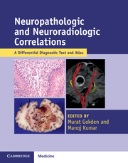
Neuropathologic and Neuroradiologic Correlations
A Differential Diagnostic Text and Atlas
, pp. 151 - 221
Publisher: Cambridge University Press
Print publication year: 2000

Access options

Get access to the full version of this content by using one of the access options below. (Log in options will check for institutional or personal access. Content may require purchase if you do not have access.)

References

Yung, WA, Horten, BC, Shapiro, WR. Meningeal gliomatosis: A review of 12 cases. Ann Neurol 1980;8:605–8.CrossRefGoogle ScholarPubMed
Nishio, S, Korosue, K, Tateishi, J, Fukui, M, Kitamura, K. Ventricular and subarachnoid seeding of intracranial tumors of neuroectodermal origin: A study of 26 consecutive autopsy cases with reference to focal ependymal defect. Clin Neuropathol 1982;1:8391.Google ScholarPubMed
Pezeshkpour, GH, Henry, JM, Armbrustmacher, VW. Spinal metastases. A rare mode of presentation of brain tumors. Cancer 1984;54:353–6.3.0.CO;2-R>CrossRefGoogle ScholarPubMed
Gajjar, A, Bhargava, R, Jenkins, JJ, et al. Low-grade astrocytoma with neuraxis dissemination at diagnosis. J Neurosurg 1995;83:6771.CrossRefGoogle ScholarPubMed
Hukin, J, Siffert, J, Velaszquez, L, Zagzag, D, Allen, J. Leptomeningeal dissemination in children with progressive low-grade neuroepithelial tumors. Neuro-Oncology 2002;4:253–60.Google Scholar
Knopp, EA, Cha, S, Johnson, G, et al. Glial neoplasms: Dynamic contrast-enhanced T2*-weighted MR imaging. Radiology 1999;211:791–8.CrossRefGoogle ScholarPubMed
Bian, W, Khayal, IS, Lupo, JM, et al. Multiparametric characterization of grade 2 glioma subtypes using magnetic resonance spectroscopic, perfusion, and diffusion imaging. Translat Oncol 2009;2:271–80.Google ScholarPubMed
Saito, T, Yamasaki, F, Kajiwara, Y, et al. Role of perfusion-weighted imaging at 3 T in the histopathologic differentiation between astrocytic and oligodendroglial tumors. Eur J Radiol 2012;81:1863–9.CrossRefGoogle ScholarPubMed
Daumas-Duport, C, Tucker, M-L, Kolles, H, et al. Oligodendrogliomas, Part II: A new grading system based on morphological and imaging criteria. J Neuro oncol 1997;34:6178.CrossRefGoogle Scholar
Miller, DC, Lang, FF, Epstein, FJ. Central nervous system gangliogliomas, I: Pathology. J Neurosurg 1993;79:859–66.Google Scholar
Nishio, S, Takeshita, I, Fukui, M. Primary cerebral ganglioneurocytoma in an adult. Cancer 1990;66:358–62.3.0.CO;2-8>CrossRefGoogle ScholarPubMed
Giangaspero, F, Cenacci, G, Losi, L, et al. Extraventricular neoplasms with neurocytoma features: A clinicopathologic study of 11 cases. Am J Surg Pathol 1997;21:206–12.Google Scholar
Ng, H-K, Ko, HCW, Tse, CCH. Immunohistochemical and ultrastructural studies of oligodendrogliomas revealed features of neuronal differentiation. Int J Surg Pathol 1994;2:4756.CrossRefGoogle Scholar
Vyberg, M, Uhhøi, BP, Tegelbjærg, PS. Neuronal features of oligodendrogliomas: An ultrastructural and immunohistochemical study. Histopathol 2007;50:887–96.Google Scholar
Sharma, MC, Deb, P, Sharma, S, Sarkar, C. Neurocytoma: A comprehensive review. Neurosurg Rev 2006;29:270–85.Google Scholar
Myung, JK, Cho, HJ, Park, C-K, et al. Clinicopathologic and genetic characteristics of extraventricular neurocytomas. Neuropathol 2013;33:111–21.Google Scholar
Miller, DC. Modern Surgical Neuropathology. Cambridge: Cambridge University Press, 2009, 118–22. (Hereafter Miller, Mod Surg NP)Google Scholar
Miller, DC, Koslow, MK, Budzilovich, GN, Burstein, DE. Synaptophysin: A sensitive and specific marker for ganglion cells in central nervous system neoplasms. Hum Pathol 1990;21:271–6.Google Scholar
Kros, JM, de Jong, AAW, van der Kwast, TH. Ultrastructural characterization of transitional cells in oligodendrogliomas. J Neuropathol Exp Neurol 1992;51:186–93.Google Scholar
Kros, JM, Eden, CG Van, Stefanko, SZ, Batenburg, M Waayer-Van, van der Kwast, TH. Prognostic implications of glial fibrillary acidic protein containing cell types in oligodendrogliomas. Cancer 1990;66:120412.Google Scholar
Miller, , Mod Surg NP, 4–77.Google Scholar
Miller, , Mod Surg NP, 1921.Google Scholar
Barker 2nd, FG, Davis, RL, Chang, SM, Prados, MD. Necrosis as a prognostic factor in glioblastoma multiforme. Cancer 1996;77:161–6.Google ScholarPubMed
Kim, S-D, Jung, T-Y, Kim, I-Y, et al. The prognosis of anaplastic astrocytoma with radiologic necrosis mimicking glioblastoma. Br J Neurosurg 2013;27:74–9.CrossRefGoogle ScholarPubMed
Lacroix, M, Abi-Said, D, Fourney, DR, et al. A multivariate analysis of 416 patients with glioblastoma multiforme: Prognosis, extent of resection, and survival. J Neurosurg 2001;95:190–8.Google Scholar
Ecici, MA, Bulut, T, Tucer, B, Kurtsoy, A. Analysis of the mortality probability of preoperative MRI features in malignant astrocytomas. Turkish Neurosurg 2011;21:271–9.Google Scholar
Kim, H, Choi, SH, Kim, J-H, et al. Gliomas: Application of cumulative histogram analysis of normalized cerebral blood volume on 3T MRI to tumor grading. PLoS One 2013;8:e63462.Google Scholar
Alexiou, GA, Zikou, A, Tsiouris, S, et al. Correlation of diffusion tensor, dynamic susceptibility contrast MRI and 99mTc-tetrofosmin brain SPECT with tumour grade and Ki-67 immunohistochemistry in glioma. Clin Neurol Neurosurg 2014;116:41–5.Google Scholar
Bobek-Billewicz, B, Staski-Pres, G, Hebda, A, et al. Anaplastic transformation of low-grade gliomas (WHO II) on magnetic resonance imaging. Folia Neuropathol 2014;52:128–40.Google Scholar
Cairncross, JG, Ueki, K, Zlatescu, MC, et al. Specific genetic predictors of chemotherapeutic response and survival in patients with anaplastic oligodendrogliomas. J Natl Cancer Inst 1998;90:1473–9.Google Scholar
Chinot, O-L, Honore, S, Dufour, H, et al. Safety and efficacy of temozolomide in patients with recurrent anaplastic oligodendrogliomas after standard radiotherapy and chemotherapy. J Clin Oncol 2001;19:2449–55.Google ScholarPubMed
Jiang, H, Zhang, Z, Ren, X, et al. 1p/19q-driven prognostic molecular classification for high-grade oligodendroglial tumors. J Neurooncol 2014;120:607.CrossRefGoogle ScholarPubMed
Minniti, G, Arcella, A, Scaringi, C, et al. Chemoradiation for anaplastic oligodendrogliomas: Clinical outcomes and prognostic value of molecular markers. J Neurooncol 2014;116:275–82.CrossRefGoogle ScholarPubMed
Law, M, Brodsky, JE, Babb, J, et al. High cerebral blood volume in human gliomas predicts deletion of chromosome 1p: Preliminary results of molecular studies in gliomas with elevated perfusion. J Magn Reson Imaging 2007;25:1113–19.CrossRefGoogle ScholarPubMed
Chawla, S, Vossough, KA, Zhang, Y, et al. Differentiation between oligodendroglioma genotypes using dynamic susceptibility contrast perfusion-weighted imaging and proton MR spectroscopy. Am J Neuroradiol 2013;34:1542–9.Google ScholarPubMed
Rodriguez, FJ, Mota, RA, Scheithauer, BW, et al. Interphase cytogenetics for 1p19q and t(1;19)(q10;p10) may distinguish prognostically relevant subgroups in extra-ventricular neurocytomas. Brain Pathol 2009;19:623–9.Google Scholar
Sabha, N, Knobbe, CB, Maganti, M, et al. Analysis of IDH mutation, 1p/19q deletion, and PTEN loss delineates prognosis in clinical low-grade diffuse gliomas. Neuro-Oncology 2014;16:914–23.Google Scholar
Brandner, S, von Deimling, A. Review: Diagnostic, prognostic, and predictive relevance of molecular markers in gliomas. Neuropathol Appl Neurobiol 2015;41:694720.Google Scholar
Mur, P, Mollejo, M, Hernandez-Iglesias, T, et al. Molecular classification defines 4 prognostically distinct glioma groups irrespective of diagnosis and grade. J Neuropathol Exp Neurol 2015; 74:241–9.CrossRefGoogle ScholarPubMed
Sahm, F, Reuss, D, Koelsche, C, et al. Farewell to oligoastrocytoma: In situ molecular genetics favor classification as either oligodendroglioma or astrocytoma. Acta Neuropathol 2014;128:551–9.CrossRefGoogle ScholarPubMed
Kelly, PJ, Daumas-Duport, C, Scheithauer, BW, Kall, BA, Kispert, DB. Stereotactic histologic correlations of computed tomography- and magnetic resonance imaging-defined abnormalities in patients with glial neoplasms. Mayo Clin Proc 1987;62:450–9.Google Scholar
Miller, , Mod Surg NP, 129.Google Scholar
Law, M, Cha, S, Knopp, EA, et al. High-grade gliomas and solitary metastases: Differentiation using perfusion and proton spectroscopic MR imaging. Radiology 2002;222:715–21.Google Scholar
Neiman, OH, Sadetzki, S, Chetrit, A, et al. Perfusion-weighted imaging of peritumoral edema can aid in the differential diagnosis of glioblastoma multiforme versus brain metastasis. Israel Med Assoc J 2013;15:103–5.Google Scholar
Lee, EJ, Ahn, KJ, Lee, EK, Lee, YS, Kim, DB. Potential role of advanced MRI techniques for the peritumoural region in differentiating glioblastoma multiforme and solitary metastatic lesions. Clin Radiol 2013;68:e689–97.Google Scholar
Rath, TJ, Hughes, M, Arabi, M, Shah, GV. Imaging of cerebritis, encephalitis, and brain abscess. Neuroimaging Clin N Am 2012;22:585607.Google Scholar
Lerner, A, Shiroishi, MA, Zee, C-S, Law, M, Go, JL. Imaging of neurocysticercosis. Neuroimaging Clin N Am 2012;22:659–76.Google Scholar
Peeraully, T, Landolfi, JC. Herpes encephalitis masquerading as tumor. ISRN Neurol 2011; doi:10.5402/2011/474672.Google Scholar
Kepes, JJ. Large focal tumor-like demyelinating lesions of the brain: Intermediate entity between multiple sclerosis and acute disseminated encephalomyelitis? A study of 31 patients. Ann Neurol 1993;33:1827.Google Scholar
Zagzag, D, Miller, DC, Kleinman, GM, et al. Demyelinating disease versus tumor in surgical neuropathology. Clues to a correct pathologic diagnosis. Am J Surg Pathol 1993;17:537–45.Google Scholar
Tan, HM, Chan, LL, Chuah, KL, Goh, NSS, Tang, KK. Monophasic, solitary tumefactive demyelinating lesion: Neuroimaging features and neuropathologic diagnosis. Br J Radiol 2004;77:153–6.Google Scholar
Law, M, Meltzer, DE, Cha, S. Spectroscopic magnetic resonance imaging of a tumefactive demyelinating lesion. Neuroradiology 2002;44:986–9.Google Scholar
Saindane, AM, Cha, S, Law, M, et al. Proton MR spectroscopy of tumefactive demyelinating lesions. Am J Neuroradiol 2002;23:1378–86.Google Scholar
Masdeu, JC, Moreira, J, Trasi, S, et al. The open ring. A new neuroimaging sign in demyelinating disease. J Neuroimaging 1996;6:104–7.Google Scholar
Masdeu, JC, Quinto, C, Olivera, C, et al. Open-ring imaging sign: Highly specific for atypical brain demyelination. Neurology 2000;54:1427–33.Google Scholar
Mitha, AP, Scott, JN, George, D, et al. Neuroimaging highlight: Tumefactive demyelinating lesions. Can J Neurol Sci 2007;34:362–4.Google Scholar
Given, CA IInd, Stevens, BS, Lee, C. The MRI appearance of tumefactive demyelinating lesions. Am J Radiol 2004;182:195–9.Google ScholarPubMed
Miller, , Mod Surg NP, 363–72.Google Scholar
Miller, , Mod Surg NP, 363–4.Google Scholar
Truong, M-T, Golfinos, JG, Rush, SC, et al. Results of surgical resection for progression of brain metastases previously treated by gamma knife radiosurgery. Neurosurgery 2006;59:8697.Google Scholar
Brat, DJ, Ryken, TC, Kalkanis, SN, Olson, JJ. The role of neuropathology in the management of progressive glioblastoma. A systematic review and evidence-based clinical practice guideline. J Neurooncol 2014;118:461–78.Google Scholar
Raimbault, A, Cazals, X, Lauvin, M-A, et al. Radionecrosis of malignant glioma and cerebral metastasis: A diagnostic challenge in MRI. Diagn Interven Imag 2014; http://doi.org/10.1016/j.diii.2014.06.013Google Scholar
Powell, SZ, Yachnis, AT, Rorke, LB, Roijiani, AM, Eskin, TA. Divergent differentiation in pleomorphic xanthoastrocytoma: Evidence for a neuronal element and possible relationship to ganglion cell tumors. Am J Surg Pathol 1996;20:80–5.CrossRefGoogle ScholarPubMed
Leung, SY, Gwi, E, Ng, HK, Yam, KY. Dysembryoplastic neuroepithelial tumor. A tumor with small neuronal cells resembling oligodendroglioma. Am J Surg Pathol 1994;18:604–14.Google Scholar
Hirose, T, Scheithauer, BW, Lopes, MBS, VandenBerg, SR. Dysembryoplastic neuroepithelial tumor (DNT): An immunohistochemical and ultrastructural study. J Neuropathol Exp Neurol 1994;53:184–95.Google Scholar
Brat, DJ, Scheithauer, BW, Fuller, GN, Tihan, T. Newly codified glial neoplasms of the 2007 WHO Classification of Tumours of the Central Nervous System: Angiocentric glioma, pilomyxoid astrocytoma, and pituicytoma. Brain Pathol 2007;17:319–24.CrossRefGoogle ScholarPubMed
Koral, K, Koral, KM, Sklar, F. Angiocentric glioma in a 4-year old boy: Imaging characteristics and review of the literature. Clin Imag 2012; 36: 61–4.Google Scholar
Lellouch-Tubiana, A, Boddaert, N, Bourgeois, M, et al. Angiocentric neuroepithelial tumor (ANET): A new epilepsy-related clinicopathologic entity with distinctive MRI. Brain Pathol 2005;15:281–6.Google Scholar
Wang, M, Tihan, T, Rojiani, AM, et al. Monomorphous angiocentric glioma: A distinctive epileptogenic neoplasm with features of infiltrating astrocytoma and ependymoma. J Neuropathol Exp Neurol 2005;64:875–81.Google Scholar
Cerda-Nicolas, M, Kepes, JJ. Gliofibromas (including malignant forms) and gliosarcomas: Comparative study and review of the literature. Acta Neuropathol 1993;85:349–61.Google ScholarPubMed
Vazquez, M, Miller, DC, Epstein, F, Allen, JC, Budzilovich, GN. Glioneurofibroma: Renaming the pediatric “gliofibroma.” Modern Pathol 1991;4:519–23.Google Scholar
Blümcke, I, Thorn, M, Aronica, E, et al. The clinicopathologic spectrum of focal cortical dysplasias: A consensus classification proposed by an ad hoc Task Force of the ILAE Diagnostic Methods Commission. Epilepsia 2011;52:158–74.Google Scholar
Perry, A, Kurtkaya-Yapicier, O, Scheithauer, BW, et al. Insights into meningioangiomatosis with and without meningioma: A clinicopathologic and genetic series of 24 cases with review of the literature. Brain Pathol 2005;15:5565.Google ScholarPubMed
Squires, LA, Constantini, S, Allen, JC et al. Hypothalamic hamartoma and the Pallister–Hall syndrome. Pediatr Neurosurg 1995; 22: 303–8.Google Scholar
Tihan, T, Fisher, PG, Kepner, JL, et al. Pediatric astrocytomas with monomorphous pilomyxoid features and a less favorable outcome. J Neuropathol Exp Neurol 1999;58:1061–8.Google Scholar
Brat, DJ, Scheithauer, BW, Staugaitis, SM, et al. Pituicytoma: A distinctive low-grade tumor of the neurohypophysis. Am J Surg Pathol 2000;24:362–8.Google Scholar
Secci, F, Merciadri, P, Rossi, DC, D'Andrea, A, Zona, G. Pituicytomas: Radiologic findings, clinical behavior, and surgical management. Acta Neurochir, 2012;154:649–57.Google Scholar
Kamil, ZS, Sinson, G, Gucer, H, Asa, SL, Mete, O. TTF-1 expressing sellar neoplasm with ependymal rosettes and oncocytic change: Mixed ependymal and oncocytic variant pituicytoma. Endocr Pathol 2013; doi 10.1007/s12022–013–9279–2Google Scholar
Miller, , Mod Surg NP, 188194.Google Scholar

Save book to Kindle

To save this book to your Kindle, first ensure coreplatform@cambridge.org is added to your Approved Personal Document E-mail List under your Personal Document Settings on the Manage Your Content and Devices page of your Amazon account. Then enter the ‘name’ part of your Kindle email address below. Find out more about saving to your Kindle.

Note you can select to save to either the @free.kindle.com or @kindle.com variations. ‘@free.kindle.com’ emails are free but can only be saved to your device when it is connected to wi-fi. ‘@kindle.com’ emails can be delivered even when you are not connected to wi-fi, but note that service fees apply.

Find out more about the Kindle Personal Document Service.

Available formats
×

Save book to Dropbox

To save content items to your account, please confirm that you agree to abide by our usage policies. If this is the first time you use this feature, you will be asked to authorise Cambridge Core to connect with your account. Find out more about saving content to Dropbox.

Available formats
×

Save book to Google Drive

To save content items to your account, please confirm that you agree to abide by our usage policies. If this is the first time you use this feature, you will be asked to authorise Cambridge Core to connect with your account. Find out more about saving content to Google Drive.

Available formats
×