Granulocyte vs. oncosphere – who’s calling the shots?

11 February 2021
Last update: 11/02/21 14:23
The latest Paper of the Month for Parasitology is Agranulocytosis leads to intestinal Echinococcus multilocularis oncosphere invasion and hepatic metacestode development in naturally resistant Wistar rats
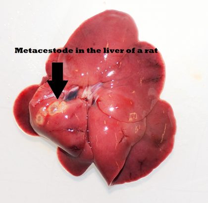
Let me introduce you to a tiny tapeworm that is widely distributed in the northern hemisphere: Echinococcus multilocularis. At just 4-6 millimeters in length, the adult worm parasitizes the small intestines of foxes and other wild canids, where it produces eggs that are excreted with the fox feces. For parasite transmission, the egg containing an infectious larva (oncosphere) needs to be ingested by intermediate hosts, such as rodents. After ingestion, the hatched oncosphere migrates through the intestinal wall and via blood vessels to the intermediate host’s liver, where it develops within days into the larval stage known as a metacestode. From this moment on, the metacestode starts its invasive growth into the liver parenchyma with differentiation into thousands of small worm heads called protoscoleces. The parasite lifecycle is completed when a fox eats infected prey and adult tapeworms develop from the protoscoleces in the fox intestine. So far, so good.
But why should this tiny fox tapeworm or the relevant Paper of the Month be of interest for you? Alveolar echinococcosis (AE) is a serious zoonotic disease caused by the above mentioned metacestode of the fox tapeworm and the incidence in humans is increasing annually in many parts of Central Europe and Asia. Susceptibility to AE varies considerably among intermediate hosts (rodents) and dead-end host species (e.g., humans and other mammals). Wistar rats, for example, are, in contrast to mice, highly resistant to infection and subsequent metacestode development after oral inoculation with E. multilocularis eggs, but become susceptible after immunosuppressive treatment with dexamethasone. This led us to the hypothesis that innate immune cells may primarily be responsible for defense against E. multilocularis oncosphere invasion and subsequent metacestode development in rats. Interestingly, there are parallels in susceptibility between rats and humans, as humans are also considered resistant rather than susceptible to AE, whereas severe progression of disease occurs in patients with compromised immunological status (e.g., caused by disease or immunosuppressive treatment).
But which population of the immune cell orchestra should we choose? We examined the role of three innate immune cell populations that are abundant in the intestinal mucosa and/or part of the intestinal immunological barrier in this process: macrophages (MΦ), granulocytes (i.e. neutrophils, eosinophils and basophils) and natural killer (NK) cells. We investigated whether these innate immune cells are involved in the natural resistance against intestinal E. multilocularis oncosphere invasion, by experimental egg inoculation of Wistar rats that were depleted of one or several cell types.
And how lucky we were with our findings! Although NK cell and MΦ depletion did not alter the resistance status of rats, the majority of granulocyte-depleted animals developed liver metacestodes, indicating that granulocytes are key players in preventing oncosphere migration and/or development in Wistar rats. Although the granulocyte population and detailed mechanism of resistance remain unknown to date, we hope that the discovery of relevant mechanisms inhibiting E. multilocularis larval invasion and establishment in animal species considered as resistant may contribute to the understanding of the hitherto unknown mechanisms of susceptibility in humans.
The paper Agranulocytosis leads to intestinal Echinococcus multilocularis oncosphere invasion and hepatic metacestode development in naturally resistant Wistar rats, by Deborah E. Joekel, Selim Nur, Josep Monné Rodriguez, Philipp A. Kronenberg, Anja Kipar, Salomé LeibundGut-Landmann and Peter Deplazes, published in Parasitology, is available free for a month.
Images and video © 2018 Institute of Parasitology, Vetsuisse Faculty Zurich, University of Zurich, Switzerland
Related Posts

Why is it dangerous to walk dogs where foxes live? A parasite on the rise
The latest Paper of the Month for Parasitology is Conquering Switzerland: emergence of Angiostrongylus vasorum over three decades and rapid regional increase in the fox population contrasts with the stable prevalence of lungworms Did you know that foxes can harbour a heartworm which can cause respiratory problems or bleeding in your dog? Foxes are widely distributed in the […]

Schistosomiasis then and now; what has changed in the last 100 years?
Centennial Reflections – a distinguished parasitologist reflects on a paper published in their field in Parasitology 100 years ago A paper entitled “Bilharziasis in Natal”, published in Parasitology in 1918 by Dr F. G. Cawston, provides a window on the state of knowledge at the time (Cawston, 1918). He was a British physician and zoologist […]

Coccidiosis in humans – the past 100 years: A Revision of the Coccidia Parasitic in Man
“Centennial Reflections – a distinguished parasitologist reflects on a paper published in their field in Parasitology 100 years ago” Coccidiosis in humans – the past 100 years: A Revision of the Coccidia Parasitic in Man BY: J. P. Dubey, United States Department of Agriculture, Agricultural Research Service, Beltsville Agricultural Research Service, Animal Parasitic Disease Laboratory, […]

Watching the 2016 presidential debates
Watching the 2016 presidential debates Patrick A. Stewart, Ph.D. (pastewar@uark.edu) If a picture is worth a thousand words, volumes will be spoken during the upcoming televised U.S. presidential debates. At thirty frames per second, the facial displays, vocalizations, body movements, and, yes, even the words themselves, will enlighten TV viewers concerning Hillary Clinton and […]


