Introduction
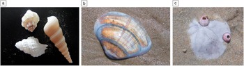
Many of us probably recollect walking along a sandy shore and marveling at the beautiful, ornate shells that are revealed and left behind by a retreating tide (Figure 1). The intricacy of the patterns on these shells are eye-catching, their texture and robustness a product of several million years of evolutionary engineering. They represent some of the marvels of materials conjured up by Mother Nature.
Figure 1. Mantles of (a) mollusks, (b) bivalve, and (c) sand dollar, collectively referred to as seashells. The stiff shell of the sand dollar is composed of plates of calcium carbonate arranged in an intricate radial pattern.
The primary constituent of the protective armor (mantle) that encases the soft body of mollusks and some bivalves is calcium carbonate. The mantle of a mollusk differs from that secreted by bivalves in that it is additionally composed of chitin, a biopolymer commonly associated with the exoskeleton of shrimps and lobsters. Biogenic calcite along with amorphous silica represent the two most common biominerals on the planet.
Biomineralization is the assembly of these highly organized mineral phases by and in living systems and may be viewed as an accumulation of metal by an organism through a carefully orchestrated biological process, which, in some instances, has remained unchanged over millennia. Interestingly, biomineralization also provides a record of the environmental history of an organism over its lifetime.
The motivation to understand and shed light on biomineralization processes is multifaceted. The assembly of mineral phases achieved by living organisms is yet to be rivaled by any synthetic effort by scientists to date. Biominerals, due to their unique hierarchical organization, exhibit very interesting properties that have potential applications in all walks of life. An interesting aspect of biomineralization is that the formation of the mineral phase occurs from conditions that are not thermodynamically favorable for the mineral phase’s nucleation and growth. Therefore, deciphering the assembly algorithm and components that initiate and promote hierarchical deposition of cations has significant implications for the development of nanocomposites and nanotechnology as a whole.
There are numerous examples of biomineralization and classes of biominerals in nature. This diversity provides a unique opportunity for creative and defining collaboration between materials scientists, chemists, biophysicists, and biologists for harnessing the structure–property relationships in these naturally occurring minerals. One can envisage applications for biominerals in several arenas ranging from military hardware, nanocontainers and nanosystems for medical and diagnostic applications, novel materials for photonics with hierarchical structures, and tunable photonic properties (bioinspired optics).Reference Yu, Bagaev, Bukin, Voznesenski, Drozdov, Zinin, Nagornyĭ, Pestryakov and Trunov1,Reference Fuhrmann-Leiker and Karthaus2
The objective of this MRS Bulletin issue on biomineralization is to highlight some of the challenges in characterizing and replicating biomineralization processes, specifically using biogenic calcite and magnetosomes as representative examples. The articles in the issue do not cover all aspects of biomineralization. For a more comprehensive overview on biomineralization and synthetic systems, refer to the reviews by Palmer et al.Reference Palmer, Newcomb, Kaltz, Spoerke and Stupp3 and Nudelman and Sommerdijk.Reference Nudelman and Sommerdijk4
Role of macromolecules in biomineralization
Molecular systems recognized over the years as being important for biomineralization are well-defined highly charged proteins (biomineralization promoter proteins [BPPs])Reference Wang and Nilsen-Hamilton5 and polysaccharides. Knowing the structure and chemical makeup of these macromolecules and the mechanism by which these molecules drive biomineralization can aid in the design of synthetic mimics.
Two common aspects have emerged with regard to macromolecules in biomineralization: (1) These macromolecules have a high degree of negative charge along their backbone, and (2) in the case of proteins, the presence of a β-sheet motif appears to be important.Reference Addadi and Weiner6,Reference He, Dahl, Veis and George7 Both features are thought to aid in the initiation of crystal growth and possibly function as a molecular template for the organization of the mineral phase. In view of the compelling evidence for the part of proteins and polysaccharides in biomineralization, proteomics and glycomics can be expected to play an important role in furthering our knowledge base in the coming years.
Biogenic calcite
Biogenic calcium carbonate occurs in two polymorphs: aragonite and biogenic calcite. Aragonite, which is a thermodynamically unstable form, will transform to calcite at elevated temperatures, although this transformation in nature is on the timescale of a few million years. Biogenic calcite is the primary constituent material of the mantles of mollusks and bivalves.
The formation of calcite occurs from a poorly organized precursor called amorphous calcium carbonate (ACC).Reference Addadi, Raz and Weiner8 Aizenberg et al. reported that calcareous sponges and ascidians, which are invertebrate marine filter feeders and commonly referred to as sea squirts, synthesize skeletal elements composed of both ACC and calcite, with calcite covered in an ACC layer followed by a thinner calcite in the case of the former, and an ACC core enveloped in calcite in the case of the latter.Reference Aizenberg, Weiner and Addadi9 This is interesting because calcareous sponges and ascidians are wide apart in the phylogenetic tree, in that calcareous sponges belong to the phylum Porifera, and, ascidians belong to the phylum Cordata. In both instances, the growth of these discrete phases was found be under the control of macromolecules. In the biomineralization of abalone shells, cooperative interactions between polyanionic proteins by themselves have been shown to promote the abrupt transition between calcite and c-axis, oriented aragonite.Reference Belcher, Wu, Christensen, Hansma, Stucky and Morse10
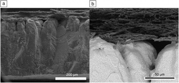
Biominerals also play an important role in vertebrates. In avians, the eggshell is composed of calcium carbonate; two families of proteins, ovocleidins and ovocalyxins, are responsible for shell’s biomineralization.Reference Panheleux, Bain, Fernandez, Morales, Gautron, Arias, Solomon, Hincke and Nys11 The robust mechanical properties of the eggshell are due to the organized deposition of calcite spherulites on an organic framework. The eggshell is composed of six layers, with the calcified zone sandwiched between organic layers and organized in irregular cones (Figure 2).Reference Nys, Gautron, Garcia-Ruiz and Hincke12

Figure 2. Scanning electron micrograph of the shell of a hen egg. (a) Image showing the calcite layer and the eggshell membrane above. (b) Higher magnification image showing the juxtaposition of the dense proteinaceous membrane layer with the calcite phase. Images courtesy of Melika Sarem, Ralf Thomann, and Prasad Shastri.
Hydroxyapatite
Hydroxyapatite (HAP), a naturally occurring form of calcium phosphate (CP), is the mineral phase in bones that is responsible for conferring strength to the bony skeletal framework in vertebrates.Reference Fratzl and Weinkamer13 Very often the ions within the HAP in bone are replaced by other ions, with the substitution of phosphate with carbonate being the most prevalent, resulting in the formation of carbonated HAP. A calcium deficient form of HAP is also found in tooth dentin and enamel. In bone, HAP is assembled within a fibrillar collagen network, with the triple helical collagen bundles (tropocollagen) forming a template of sorts for the nucleation and growth of the platelet-like HAP phase. While the origins of the platelet-like HAP phase in bone remain a work in progress, it is thought to occur through an octacalcium phosphate intermediate phase.Reference Brown, Eidelman and Tomoazic14 Interestingly, the staggered arrangement (assembly) of adjacent and adjoining collagen bundles results in a 67 nm offset (or gap) overall.Reference Fratzl and Weinkamer13 This gap is very critical for HAP formation and provides the vacancy in the organic framework for the interpenetrating deposition of collagen by the osteoblasts in bone and is also thought to be the nucleating site for HAP formation.
In human bone formation, collagen is thought to have a role in the nucleation and growth of the HAP phase; however, up until now, there is no direct evidence to directly implicate a role for its amino acid composition or the helical structure in HAP formation. In human bone formation, collagen is thought to have a role in the nucleation and growth of the HAP phase. Although a recent study has demonstrated that the orientation and growth of carbonated HAP and its atomic and structural characteristics can be influenced by collagen in vitro,Reference Wang, Azais, Robin, Vallee, Catania, Legriel, Pehau-Arnaudet, Babonneau, Giraud-Guille and Nassif15 thus far there is no direct evidence to implicate a role for collagen, its amino acid composition, or the helical structure in initiating HAP formation in vivo. Nevertheless, it is clear that the collagen framework serves as a medium for the presentation of BPPs.Reference Yee16 In the formation of a tooth’s dentin structure, a family of dentin matrix proteins (DMPs), most notably DMP-1, is thought to play a critical role in driving the supersaturation of Ca2+ ions necessary for initiating the nucleation process. In bone tissue, analogous proteins, namely bone sialoprotein and osteonectin, which are secreted by osteoblast, have been identified. However, whether these proteins are directly responsible or have a critical role in the nucleation and growth of HAP in bone is still debatable. Likewise, phosvitin, the predominant protein found in egg yolk, is a highly phosphorylated protein and has very high affinity for bivalent cations such as calcium and iron. Strong evidence supporting a physiological role for phosvitin, analogous to DMP-1 and BSP in mammalian physiology, as a BPP in avian osteogenesis is beginning to emerge.Reference Liu, Czernick, Lin, Alasmari, Serge and Salih17
In addition to the collagen network and BPPs, the formation of HAP precursors can be influenced by other local variables. In a recent study, Wang et al. characterized the structure and organization within crystalline and biomimetic HAP using various characterization techniques, including cryo-transmission electron microscopy (TEM), and made a surprising find: water molecules can orient the formation of the amorphous apatite phase (similar to ACC), which covers the crystalline core in bone.Reference Wang, Von Euw, Fernandes, Cassaignon, Selmane, Laurent, Pehau-Arnaudet, Coelho, Bonhomme-Coury, Giraud-Guille, Babonneau, Azais and Nassif18 This example not only emphasizes the need for robust characterization tools, but also highlights how much more still needs to be understood about biomineralization processes.
Bone is a spectacular example of an inorganic/organic nanocomposite; however, such nanocomposites are not a monopoly of vertebrates and inorganic–organic composites involving HAP are also common in invertebrates. The exceptional toughness of the dactyl clubbing appendage of the peacock mantis shrimp (Odontodactylus scyllarus) can be attributed to the presence of highly organized crystalline HAP, which in this case is present as a layered composite with chitin.Reference Weaver, Milliron, Miserez, Evans-Lutterodt, Herrera, Gallana, Mershon, Swanson, Zavattieri, DiMasi and Kisailus19 Therefore, understanding the role of protein mediators in biogenic HAP formation within the bone environment has big implications for not only understanding pathologies that involve bone loss, such as osteoporosis, but also to enable the development of new coatings for improving bone conduction and induction and open up new treatment options for improving bone mineral density. Toward this larger objective, synthetic systems that can replicate the bone mineralization microenvironment could be important.
Magnetosomes
In addition to invertebrates and mammals, bacteria are also capable of processing minerals into highly ordered structures. Certain strains of marine bacteria, called magnetotactic bacteria, are capable of processing iron salts into magnetite. Devouard et al. studied the size distribution and twinning of magnetite crystals from various strains of helical and rod-shaped marine magnetotactic bacteria (Magnetospirillum magnetotacticum, MC-1, MC-2, etc.) and observed that not only were the magnetite crystals produced by these magnetotactic bacteria of a much narrower size distribution than their abiotic cousins, they also exhibited an asymmetry in size distribution that is not observed in synthetic samples.Reference Devouard, Posfai, Hua, Bazylinksi, Frankel and Buseck20 TEM analysis of magnetosomes synthesized by magnetotactic bacterium MC-1 has revealed that control over crystal structure occurs at a very early stage of crystal growth, and the medium of the culture impacts both the morphology and number of magnetite crystals, with an acetate-containing medium yielding more crystals than a sulfide-containing medium. Such exquisite control over magnetite structure is of extreme relevance in many applications, including the development of nanotechnology-based therapeutics.Reference Meldrum, Mann, Heywood, Frankel and Bazylinksi21
Biosilica
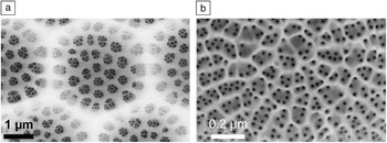
Biominerals composed of silica constitute the second most abundant mineral composition after biogenic calcium carbonate in nature. It is now well known that many plant species, such as rice, accumulate silica from the soil, and this accumulation is thought to have a beneficial effect for the survival of the plant. Diatoms constitute the most prevalent structure composed of amorphous silica found in nature. Diatoms are found in every body of water on the planet; each body of water has a unique population of diatoms, and this is frequently used in forensics to pinpoint the geographical location of the body of water.Reference Krstica, Dumab, Janevskab, Levkova, Nikolovab and Noveskaa22,Reference Peabody23 Diatoms are actually living systems: unicellular microalgae with well-defined exoskeletons of silica. The process of deposition of silica (i.e., silicification) occurs via silica deposition vesicles that are secreted by the microalgae. The factors and biological molecules involved in the nanofabrication and patterning of silica into intricate structures (Figure 3) are just beginning to be understood.

Figure 3. Scanning electron microscopy images of diatom cell walls. (a) Coscinodiscus asteromphalus. (b) Thalassiosira pseudonana. Reproduced with permission from Reference 21. © 1993 Royal Society.
As mentioned earlier, the role of organic molecules in the biomineralization processes is now a well-understood phenomenon. In 1999, Kröger and Sumper identified and isolated a family of cationic polypeptides called silaffins from the diatom species Cylindrotheca fusiformis. Reference Kroger, Deutzmann and Sumper24 Silaffins are tightly associated with the biosilica synthesized by the diatom, and it has been shown that silaffins and other polyamines play a direct role in biosynthesis of silica.Reference Sumper and Kroger25 The deposition of the silica skeleton is believed to occur through precipitation within the water phase, and the repeated phase separation that ensues to the sol-gel transition of the growing silica phase is thought to be responsible for the observed hierarchy in the diatom silica exoskeleton.Reference Sumper26
Diatoms offer an interesting platform to make highly functionalized silica structures. Lang et al. have demonstrated that chemical modification of the diatom can be carried out without compromising its structure, via the incorporation of thiol moieties into the diatom during frustule synthesis.Reference Lang, del Monte, Collins, Rodriguez, Thompson, Dockery, Finn and Pandit27 One can imagine such modified diatoms being used in drug delivery and catalysis. For more details on biogenic silica, refer to the in-depth review by Coradin and Lopez.Reference Coradin and Lopez28
Synthetic systems
In spite of these recent advances in understanding the biomineralization process in bacteria, bivalves, and vertebrates, there are very few examples of synthetic systems that can replicate biomineralization processes. In fact, most efforts to date rely on protein extracts from the biomineralization system of interest to replicate the phase and structure of the biominerals. A notable example of a novel synthetic system that offers templated biomineralization is self-assembled peptide ampiphiles containing phosphorylated-serine residues capable of directing the mineralization of HAP such that its c-axis is aligned with the long axis of the self-assembled peptide fibers.Reference Hartgerink, Beniash and Stupp29
As mentioned previously, polyanionic proteins are known to mediate the formation of calcite and aragonite in sponges and abalone shells.Reference Aizenberg, Weiner and Addadi9,Reference Belcher, Wu, Christensen, Hansma, Stucky and Morse10 However, in a recent study, Smeets et al. showed that control over the calcium carbonate phase can also be achieved using synthetic polymers. They studied the effect of poly(styrene sulfonate) (PSS) as a polyanionic mimic of biomineralization promoting proteins, in the formation of ACC.Reference Smeets, Cho, Kempen, Sommerdijk and De Yoreo30 They found that the binding of calcium to PSS is a key step in the nucleation and growth of metastable ACC, thus providing more evidence that one of the primary roles of proteins and macromolecules in biomineralization might be in the nucleation and growth of metastable ACC, which is the precursor to calcite.
One direction of research could be the development of nanocomposites via direct biomineralization of nanoscale synthetic templates. In this context, Znidarsic et al. studied the formation of CP on silica nanoparticles (SNPs) bearing positive charges, collagen, and phosvitin.Reference Znidarsic, Chen and Shastri31 They found that generally while the formation of CP on SNPs was heterogeneous, negatively charged surfaces promoted a more uniform deposition of CP nodules, even in comparison to collagen-coated SNPs. One important conclusion of this study was that protein-mediated biomineralization might be governed by surface energetics as much as biology.
Together, these findings provide the foundation for developing synthetic systems wherein biomineralization can be realized in a controlled three-dimensional metal or polymer framework. In this context, synthetic biologyReference Purnick and Weiss32 offers tools and concepts to manipulate living systems at the genetic and protein expression level. Combined with optogenetics,Reference Deisseroth33 this can provide a unique set of tools to controllably perturb the biomineralization environment within living systems and assess its impact on the structure of the mineral phase. Gaining control over the biomineralization process can foster the development of new materials for photonics and regenerative medicine, where hierarchical structures are critical for function.
In this issue
In this issue of MRS Bulletin, the article by Hendley et al., provides a comprehensive overview of the microscopic tools that can be used to elucidate the biomineralization process, with a particular focus on the organic molecules that control biomineralization. As discussed earlier, protein mediators are critical for the nucleation and growth of the biomineral.
While microscopic techniques are useful for the visualization of the mineral phase, understanding dynamic changes to the protein structure in the presence of cations can provide valuable information on the nucleation loci. Toward this objective, Sarem and Lüdeke make the case for incorporating circular dichroism as a tool in interrogating biomineralization.
In the first of two articles focused on specific biominerals, Schenk and Kim present the current understanding of biogenic calcite formation and discuss how this feeds into design strategies for artificial materials. In doing so, they present analogies between biogenic and synthetic calcite single crystals. Finally, the article by Faivre discusses the biosynthesis of highly ordered magnetite nanoparticles in magnetosomes, which are essentially nano-reactors for the production of magnetite.
It is hoped that the diverse topics covered in this issue will prompt the readership to engage in and explore the topic of biomineralization.
Acknowledgments
This work was funded by the excellence initiative of the German Federal and State Governments Grant EXC 294 (Centre for Biological Signalling Studies) and the Swiss National Foundation-Sinergia grant (Nr. CRSII3_136179). V.P.S thanks all of the contributors to this issue of MRS Bulletin for their time and commitment, without which this issue would not have materialized.