

Introduction
Babunaite-(Nd) NdAsO4 was discovered in a dark pink muscovite-quartz schist near the Nežilovo village in the northern Macedonia, where metasomatic rocks of the Mixed Series of the Pelagonian Massif are distributed. These Mixed Series rocks are a source of new Pb-Zn-bearing oxide minerals, including nežilovite, PbZn2Mn4+2Fe3+8O19 (Bermanec et al., Reference Bermanec, Holstam, Sturman, Criddle, Back and Šćavničar1996), zincohögbomite-2N6S, (Zn,Al,Fe)3(Al,Fe,Ti)8O15(OH) (Armbruster et al., Reference Armbruster, Bermanec, Zebec and Oberhansli1998, Armbruster, Reference Armbruster2002), ferricoronadite, Pb(Mn4+6Fe3+2O16 (Chukanov et al., Reference Chukanov, Aksenov, Jančev, Pekov, Göttlicher, Polekhovsky, Rusakov, Nelyubina and Van2016), zincovelesite-6N6S, Zn3(Fe3+,Mn3+,Al,Ti)8O15(OH) (Chukanov et al., Reference Chukanov, Krzhizhanovskaya, Jančev, Pekov, Varlamov, Göttlicher, Rusakov, Polekhovsky, Chervonnyi and Ermolaeva2018), zincorinmanite-(Zn), ZnSb5+(Fe3+2Zn)O7(OH) (Chukanov et al., Reference Chukanov, Gridchina, Rastsvetaeva, Varlamov, Kasatkin, Pekov, Vigasina, Virus, Jančev and Britvin2025), and minerals of the epidote group, such as piemontite-(Pb), CaPb(Al2Mn3+)(Si2O7)(SiO4)O(OH) (Chukanov et al., Reference Chukanov, Varlamov, Nestola, Belakovskiy, Goettlicher, Britvin, Lanza and Jančev2012).
It is widely accepted that the uniqueness of Nežilovo’s rocks is related to the presence of chalcophile elements, which are usually chemically bound in sulfides, but here form oxides and oxysalts. This is explained by the oxidising conditions during formation, the high chemical activity of barium and the binding of sulfur in baryte, which is distributed widely in these rocks (Chukanov et al., Reference Chukanov, Varlamov, Ermolaeva and Jančev2020, Bermanec et al., Reference Bermanec, Chukanov, Varlamov, Rajačić, Jančev and Ermolaeva2023). Endogenic ores free of sulfur with chalcophile elements are relatively rare. These have a metasomatic origin and are found in the Fe-Zn Franklin and Sterling mines in New Jersey, USA (Tarr, Reference Tarr1929; Palache, Reference Palache1929a, Reference Palache1937; Wilkerson, Reference Wilkerson1962), the Fe-Mn Långban mine, Nordmark (including Jacobsberg) and Pajsberg (including Harstigen) in the Bergslagen mining district, Värmland County, Sweden (Palache, Reference Palache1929b; Holtstam and Langhof, Reference Holtstam and Langhof1999), and the Kombat mine in Namibia (Innes and Chaplin, Reference Innes, Chaplin, Anheusser and Maske1986; Dunn, Reference Dunn1991). All of the above localities are well known thanks to the wide variety of minerals found here, many of which are rare and only occur in this type of ore.
Babunaite-(Nd), NdAsO4, is the seventh new mineral discovered in the Nežilovo area. Initially, we thought we had found a potentially new mineral, ‘gasparite-(Nd)’, which belongs to the monazite supergroup and has previously been reported in Lugau, Salzburg, Austria, but was not approved by the Commission on New Minerals, Nomenclature and Classification of the International Mineralogical Association (Kolitsch et al., Reference Kolitsch, Schachinger and Auer2025). It turned out, however, that this mineral has a scheelite-type structure. The known synthetic NdAsO4 with a scheelite-type structure [tetragonal, I41/a; a = 5.1046(4) Å and c = 11.6032(11) Å]. It can be obtained from the monoclinic phase of NdAsO4 [‘gasparite-(Nd)’] under high-pressure and high-temperature conditions of 11 GPa and 1100–1300°C respectively (Metzger et al., Reference Metzger, Ledderboge, Heymann, Huppertz and Schleid2016; Kolitsch et al., Reference Kolitsch and Holtstam2004). Furthermore, the scheelite-type NdAsO4 has been synthesised from component oxides at 550°C in an evacuated silica ampoule (Mazhenov et al., Reference Mazhenov, Nurgaliev and Muldakhmetov1988).
In this paper, we provide a description of a new mineral, babunaite-(Nd) (IMA2025-032, symbol Bbu-Nd), that was approved by the CNMNC-IMA in 2025. It is named after the Babuna River, near an outcrop of muscovite-quartz schist containing the mineral was found. This paper presents data on the composition and structure of babunaite-(Nd) and discusses questions concerning its genesis.
Methods of investigations
The composition and morphology of babunaite-(Nd) and associated minerals were studied using a Phenom XL scanning electron microscope with an EDS detector, as well as a Quanta scanning electron microscope with a Thermo Fisher Scientific EDS UltraDry X-ray microanalyser. The composition of babunaite-(Nd) was measured using a Cameca SX100 with an accelerate voltage of 15kV, a beam current of 40 nA and a spot size of 2 μm, using the following analytical lines and standards: CaKα = wollastonite, MnKα = rhodonite, WMα = scheelite, AsLβ = skutterudite; VKα = vanadinite; ThMα = Th-Glass; YLα = xenotime; LaLα = La-Glass; CeLα = Ce-Glass; PrLβ = Pr-glass; NdLβ = Nd-Glass; SmLβ = Sm-glass; EuLβ = Eu-Glass; GdLβ = Gd-Glass; TbLα = Tb-Glass; and DyLα = Dy-glass.
Raman spectra of babunaite-(Nd) were recorded using a WITec alpha 300R confocal Raman microscope equipped with an air-cooled solid-state laser (488 and 532 nm) and a CCD camera operating at –61°C. An air Zeiss LD EC Epiplan-Neofluar DIC-100/0.55NA objective was used. The Raman scattered light was focused onto a multimode fibre and a monochromator with a 600 or 1800 mm–1 grating. The laser power at the sample position was ∼25 mW. 15 scans with an integration time of 3 s were collected and averaged, with a resolution of 3.5 cm–1 (600 nm) and 2 cm–1 (1800) being set. The monochromator of the spectrometer was calibrated using the Raman scattering line of a silicon plate (520.7 cm–1).
Single-crystal X-ray studies were carried out using a SuperNova diffractometer (Agilent Technologies) with an Atlas detector (Institute of Physics, Faculty of Science and Technology, University of Silesia, Poland). Measurements were performed under ambient conditions (290 K) using MoKα radiation (λ = 0.71073 Å). The structure solution and refinement were performed using the SHELXL-2018/3 programme (Sheldrick, Reference Sheldrick2015).
Occurrence
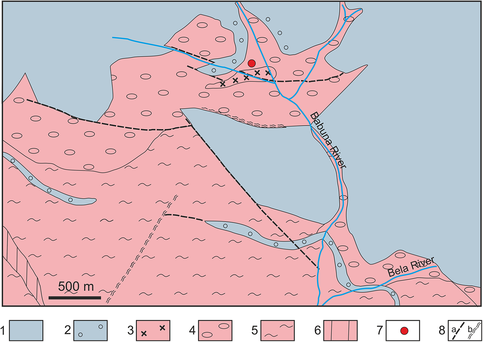
Babunaite-(Nd) was discovered in muscovite-quartz schist ∼5 km from the village of Nežilovo in Northern Macedonia. The locality is situated alongside the macadam road to the Čeples campus, near the footpath leading to the source of the Babuna River (N41°41’06.2”, E021°25’31.2”). This area belongs to the high-grade metamorphic region of the Upper Precambrian Pelagonian Massif. According to Arsovski (Reference Arsovski1959, Reference Arsovski1961) and Stojanov (Reference Stojanov1960), the region is divided into two parts. The lower complex comprises gneiss and mica schists that have been intruded by younger granitic and granodioritic bodies. The upper part consists of the so-called ‘Mixed Series’ (an irregular intercalation of gneiss, schist, calciphyre and marble with metariolite relics) and a series of massive marble (Fig. 1). During geological mapping occurrences of Pb-Zn mineralisation were found in the vicinity of the field of massive marble within the Mixed Series rocks through exploratory trenching (Stoyanov, Reference Stojanov1960; Ivanov, Reference Ivanov1961; Jančev, Reference Jančev1975a, Reference Jančev1975b; Reference Jančev1979).
Geological scheme of the Nežilovo area (modified after Jančev, Reference Jančev1979). Blue-grey colour – massive marble series, pink colour – Mixed Series. 1 – marble, 2 – calciphyre, 3 – metariolite, 4 – albite schist and augen gneiss, 5 – banded feldspar schist, 6 – gneiss, 7 – type locality of babunaite-(Nd), 8: a – fault, b – fault zone.

Figure 1 Long description
The geological map of the Nežilovo area illustrates various geological formations and features. The map includes a legend with symbols and colors representing different geological units and structures. The blue-grey color indicates the massive marble series, while the pink color represents the Mixed Series. The legend identifies several geological components: 1) marble, 2) calciphyre, 3) metariolite, 4) albite schist and augen gneiss, 5) banded feldspar schist, 6) gneiss, 7) type locality of babunaite-(Nd) and 8) fault and fault zone. The map shows the Babuna River and Bela River flowing through the area, with fault lines marked by dashed lines. The type locality of babunaite-(Nd) is marked with a red dot. Fault zones are indicated with a specific symbol and the map scale is 500 meters. The spatial distribution of these geological features is concentrated around the rivers and extends across the mapped area, highlighting the complex geological structure of the region.
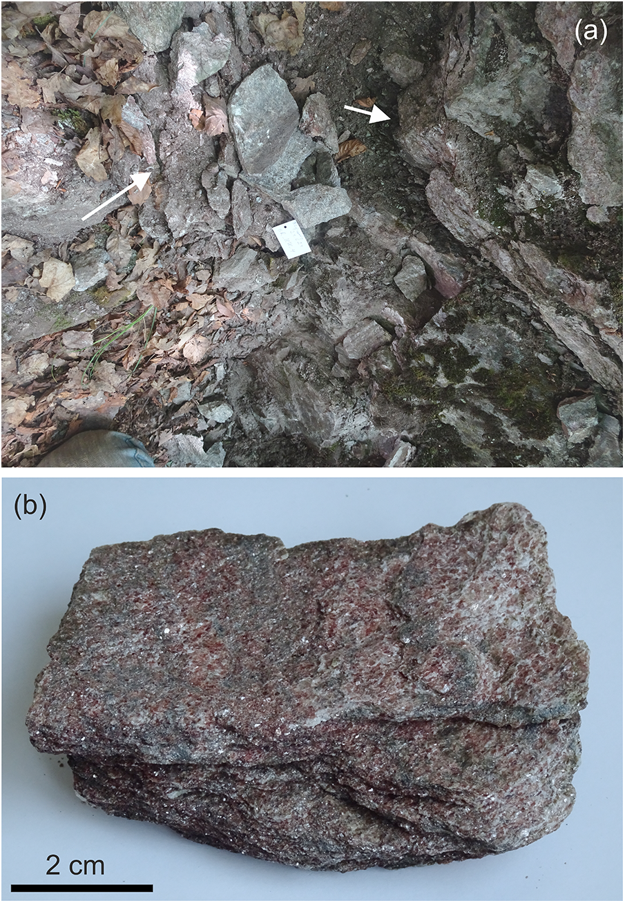
In one such trench, which contains metasomatically altered multicoloured schists and marble-like rocks with piemontite, dolomite, baryte and Pb-Zn mineralisation, a layer of pink mica schist up to 0.5 m thick was found containing babunaite-(Nd) (Fig. 2). A few dozen metres from the babunite-(Nd) type locality, there is a small body of metariolites (Fig. 1), which was first reported by Stojanov (Reference Stojanov1961).
(a) Old inspection pit, where samples for investigation were collected. Pink muscovite-quartz schists are clearly visible (white arrows). Muscovite schists are intercalated with gneiss. (b) Sample of muscovite-quartz schist from which thin-sections were prepared.
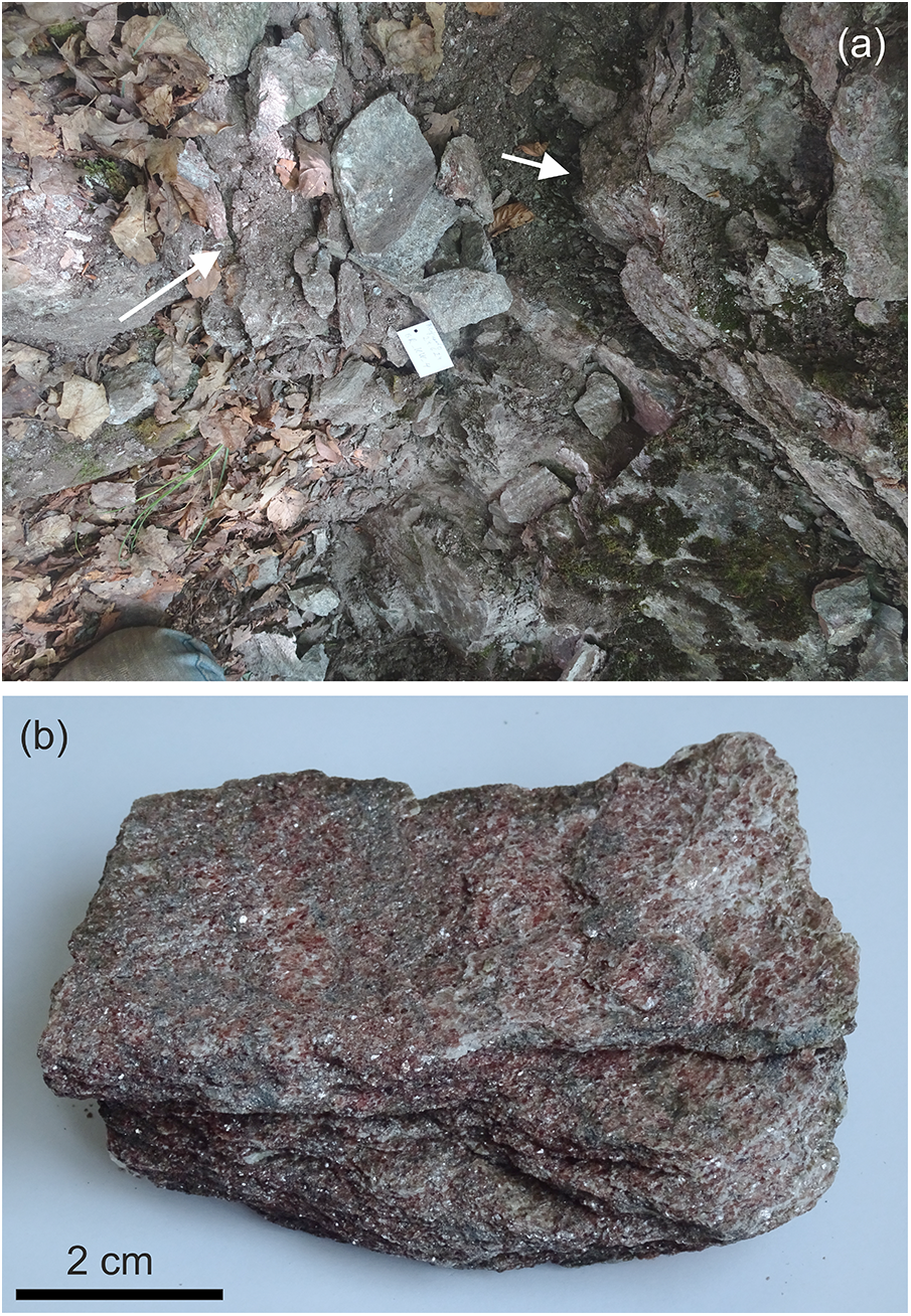
Figure 2 Long description
Image A shows an old inspection pit with various rock formations. White arrows point to pink muscovite-quartz schists intercalated with gneiss. Image B displays a sample of muscovite-quartz schist, with a scale indicating 2 cm for reference. The schist has a layered texture and a reddish-brown appearance.
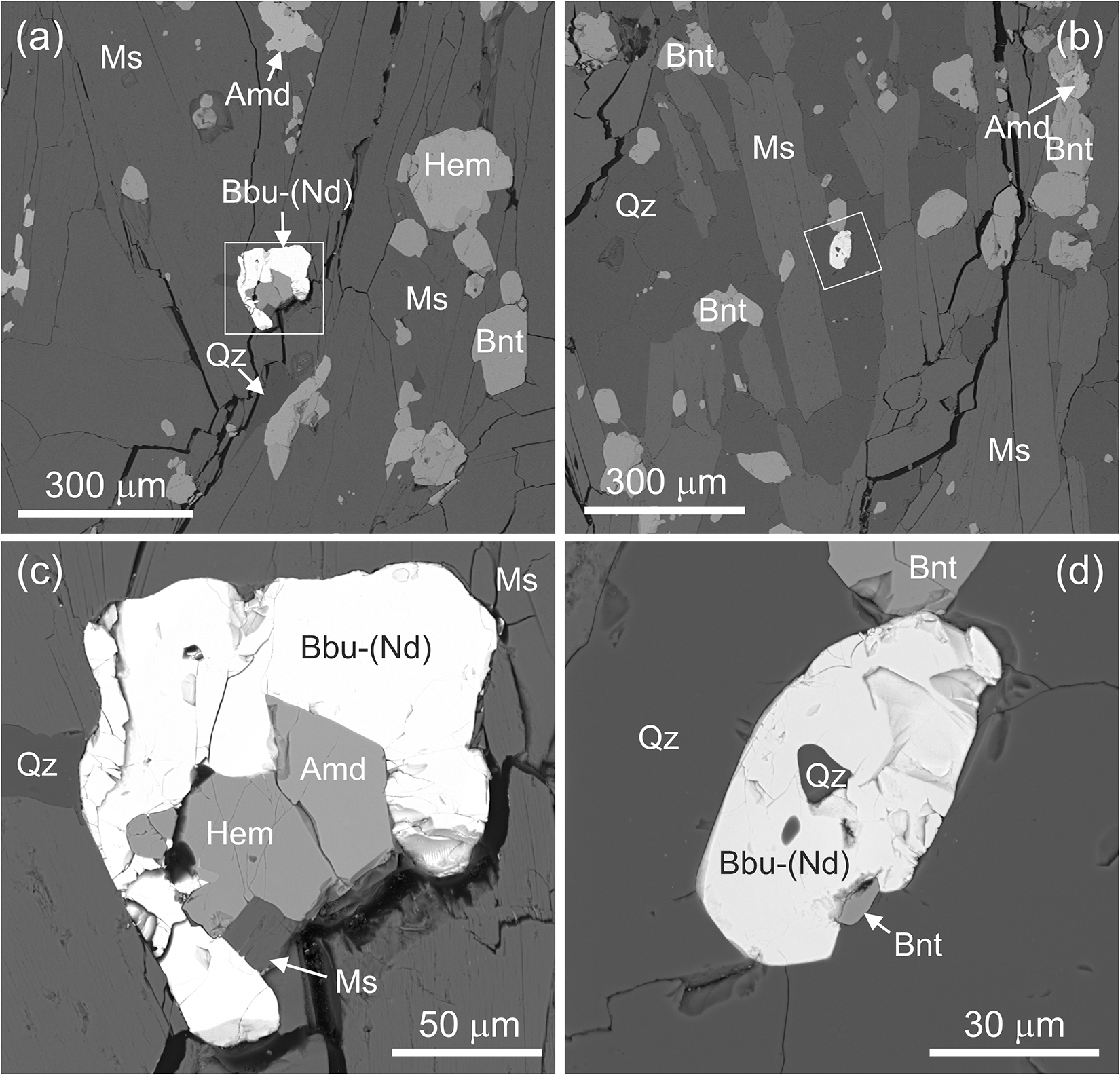
The dark pink mica schists are composed of Mn-bearing muscovite and quartz, with minor braunite (Fig. 3). The diverse accessory minerals include: hematite, gahnite, almeidaite, långbanite, zircon, piemontite, piemontite-(Pb), nežilovite, Sb-bearing rutile, fluorapatite and As-bearing fluorapatite, gasparite-(La), chernovite-(Y) and arsenoflorencite-(La). Single crystals of babunaite-(Nd) measure up to 70 μm and often have rounded edges. The transparent, pale-yellow crystals have a strong lustre (adamantine). The crystals exhibit good visible yellow luminescence at 638 nm. The microhardness of babunaite-(Nd) is VHN25 = 578(21) kg/mm2 (mean 16) and a range of 503–579 kg/mm2, which correspond to a hardness of 5 on the Mohs scale. The mineral is brittle and does not show cleavage. The calculated density based on the empirical formula and single-crystal X-ray diffraction (SC-XRD) data is 5.918 g·cm–3. Unfortunately, we were unable to determine the refractive indices of babunaite-(Nd), estimated to be ∼1.9, as our capabilities are limited to measuring the optical properties of minerals with refractive indices of less than 1.8. The optical sign of babunaite-(Nd) is uniaxial (+) and its birefringence, measured by maximum interference colouration, is Δ = 0.035. The calculated mean refractive index is 1.914. In reflected light babunaite-(Nd) is light grey with light yellow internal reflections. The reflectivity is in the range of 10–11.5%.
Back-scattered electron image showing the mineral associations and babunaite-(Nd) occurrence in muscovite-quartz schist. Rock fragments with babunaite-(Nd) marked with a square in (a) and (b) are enlarged in (c) and (d), respectively. Amd – almeidaite, Bbu-(Nd) – babunaite-(Nd), Bnt – braunite, Hem – hematite, Ms – muscovite, Qz – quartz.
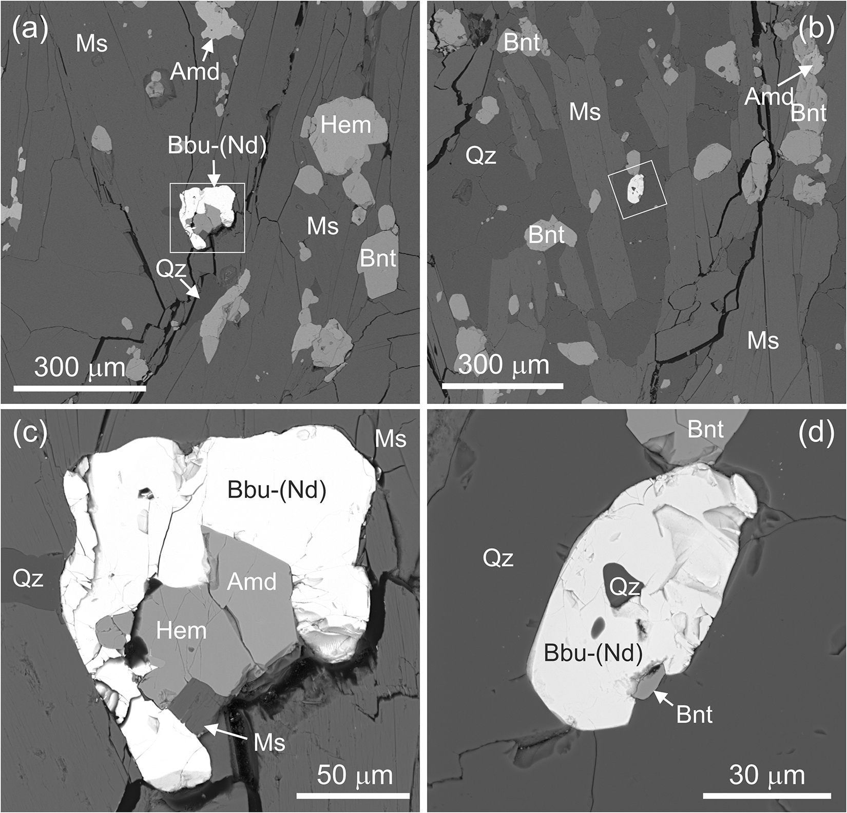
Figure 3 Long description
The image A showing a grayscale micrograph labeled “(a)”. Text labels include “Ms”, “Amd”, “Bbu-(Nd)”, “Hem”, “Qz” and “Bnt”. A white rectangular box encloses a bright, irregular area. Two arrows point toward the boxed area. A scale bar at the bottom left reads “300 µm”. The image B showing a grayscale micrograph labeled “(b)”. Text labels include “Bnt”, “Ms”, “Qz” and “Amd”. A white rectangular box encloses a small bright area. A scale bar at the bottom left reads “300 µm”. The image C showing a grayscale micrograph labeled “(c)”. Text labels include “Qz”, “Hem”, “Amd”, “Bbu-(Nd)” and “Ms”. A bright, irregular central region is bordered by darker regions. Two arrows point toward a darker area near the lower edge. A scale bar at the bottom right reads “50 µm”. The image D showing a grayscale micrograph labeled “(d)”. Text labels include “Qz”, “Bbu-(Nd)” and “Bnt”. A bright, irregular central region contains internal tonal variations. Two arrows point toward “Bnt” near the lower right side. A scale bar at the bottom right reads “30 µm”.
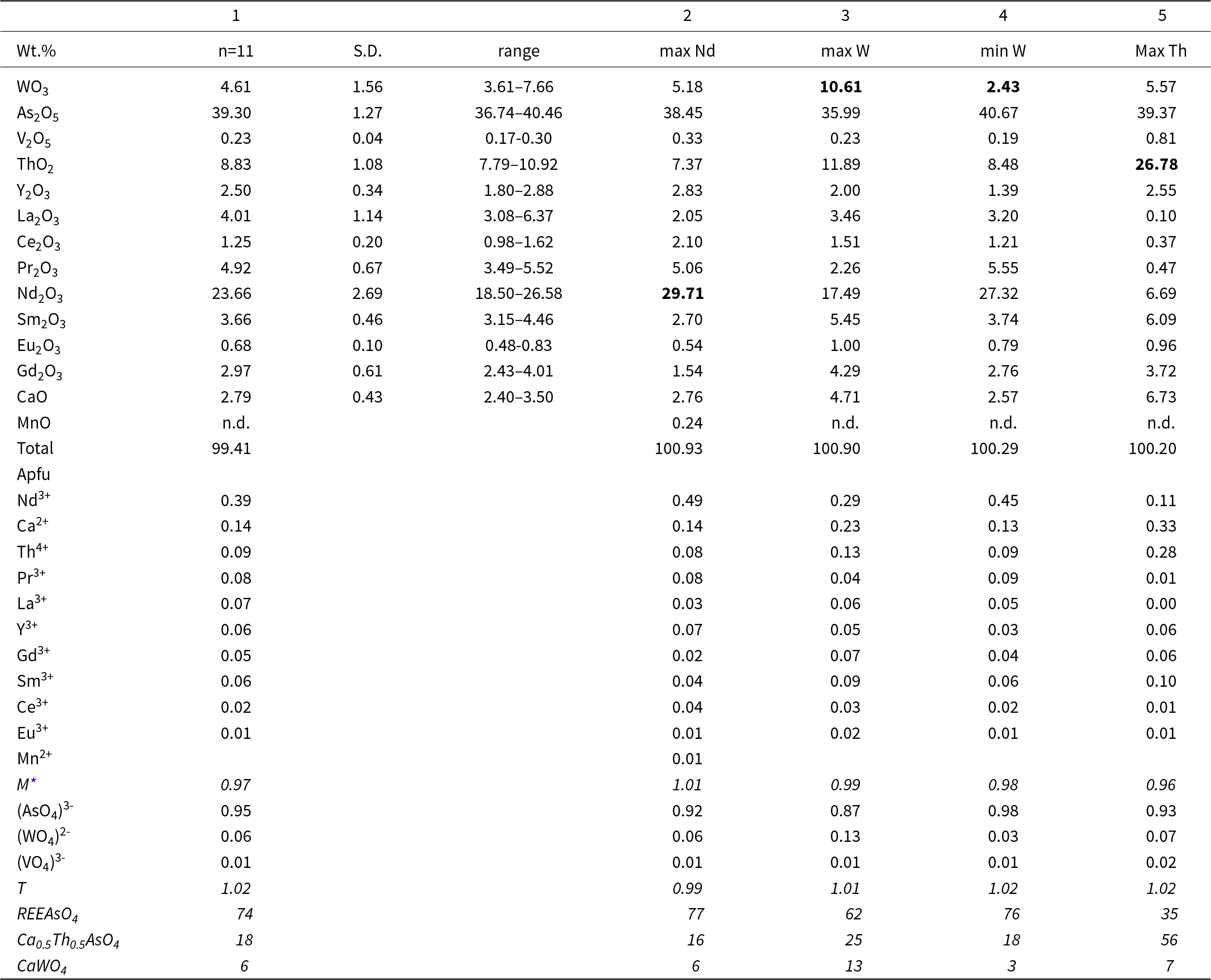
The composition of babunaite-(Nd) varies from grain to grain. We plan to conduct a systematic study of REE arsenates and phosphates in the metasomatic rocks of Nežilovo in the future. The data on the holotype babunaite-(Nd), that was used for the structural investigation, are presented in this article (Table 1). Data on associated grains with the maximum Nd, W and Th and minimum W contents found are also provided (Table 1). Significant fluctuations in composition are observed even within a single grain of babunaite-(Nd). The mean empirical crystal chemical formula of the holotype babunaite-(Nd) crystal is: (Nd3+0.39Ca0.14Th0.09Pr3+0.08La0.07Sm3+0.06Y0.06Gd0.05Ce3+0.02Eu3+0.01)Σ0.97(As5+0.95W6+0.05V5+0.01)Σ1.02O4, which contains the following end-members: REEAsO4 = 74% (including 39% NdAsO4), (Ca0.5Th0.5)AsO4 = 18%, CaWO4 = 6% (Table 1, analysis 1). The maximum content of Nd2O3 in babunaite-(Nd) is 29.7 wt.%, or 49% NdAsO4 end-member (Table 1, analysis 2). The maximum content of WO4 is 10.7 wt.%, and minimum content is 2.4 wt.% (Table 1, analyses 3, 4). In gasparite-(La) and chernovite-(Y), which are associated with babunaite-(Nd), the W content is lower than the limit of detection in microprobe analysis. Rare babunaite-(Nd) grains containing zones with high Th and Ca content could indicate a potentially new mineral with the ideal formula (Th0.5Ca0.5)AsO4 and scheelite structure (Table 1, analysis 5).
Chemical composition of (1) the holotype crystal of babunaite-(Nd) and (2–5) crystals of babunaite-(Nd) in the sample showing various compositions. As these crystals have a zonal structure we have selected analyses with the highest Nd content, the highest and lowest W content, and the highest Th content (marked in bold)

Table 1 Long description
The table reports oxide weight percent chemistry and calculated atoms per formula unit for one averaged crystal analysis and four spot analyses chosen for extreme compositions (highest Nd, highest W, lowest W, and highest Th). In the averaged crystal, major components are As2O5 about 39.30 wt.% and Nd2O3 about 23.66 wt.%, with ThO2 about 8.83 wt.% and WO3 about 4.61 wt.% (WO3 range 3.61 to 7.66 wt.% across the averaged set). Across selected spots, Nd2O3 is highest in the max Nd analysis at 29.71 wt.% and lowest in the max Th analysis at 6.69 wt.%. Tungsten varies widely, from 2.43 wt.% in the min W spot to 10.61 wt.% in the max W spot, while As2O5 stays comparatively stable near 36 to 41 wt.%. Thorium shows the largest enrichment in the max Th spot at 26.78 wt.% compared with 7.37 to 11.89 wt.% in the other selected spots. Several rare earth oxides shift with zoning, for example La2O3 drops to 0.10 wt.% and Pr2O3 to 0.47 wt.% in the max Th spot, while Sm2O3 and CaO rise there to 6.09 wt.% and 6.73 wt.% respectively. In atoms per formula unit, Nd is highest in the max Nd spot at 0.49 and Th is highest in the max Th spot at 0.28, consistent with the oxide trends. Endmember proportions reflect these changes: REEAsO4 decreases from about 74 percent in the average to 35 percent in the max Th spot, while Ca0.5Th0.5AsO4 increases to 56 percent; CaWO4 is highest in the max W spot at 13 percent. Manganese is not detected in most analyses and totals are near 100 wt.% for all spots, supporting overall analytical completeness.
Notes: S.D. – standard deviation, n.d. – not detected.
* Dy, Tb are below the detection limit.
Raman spectroscopy
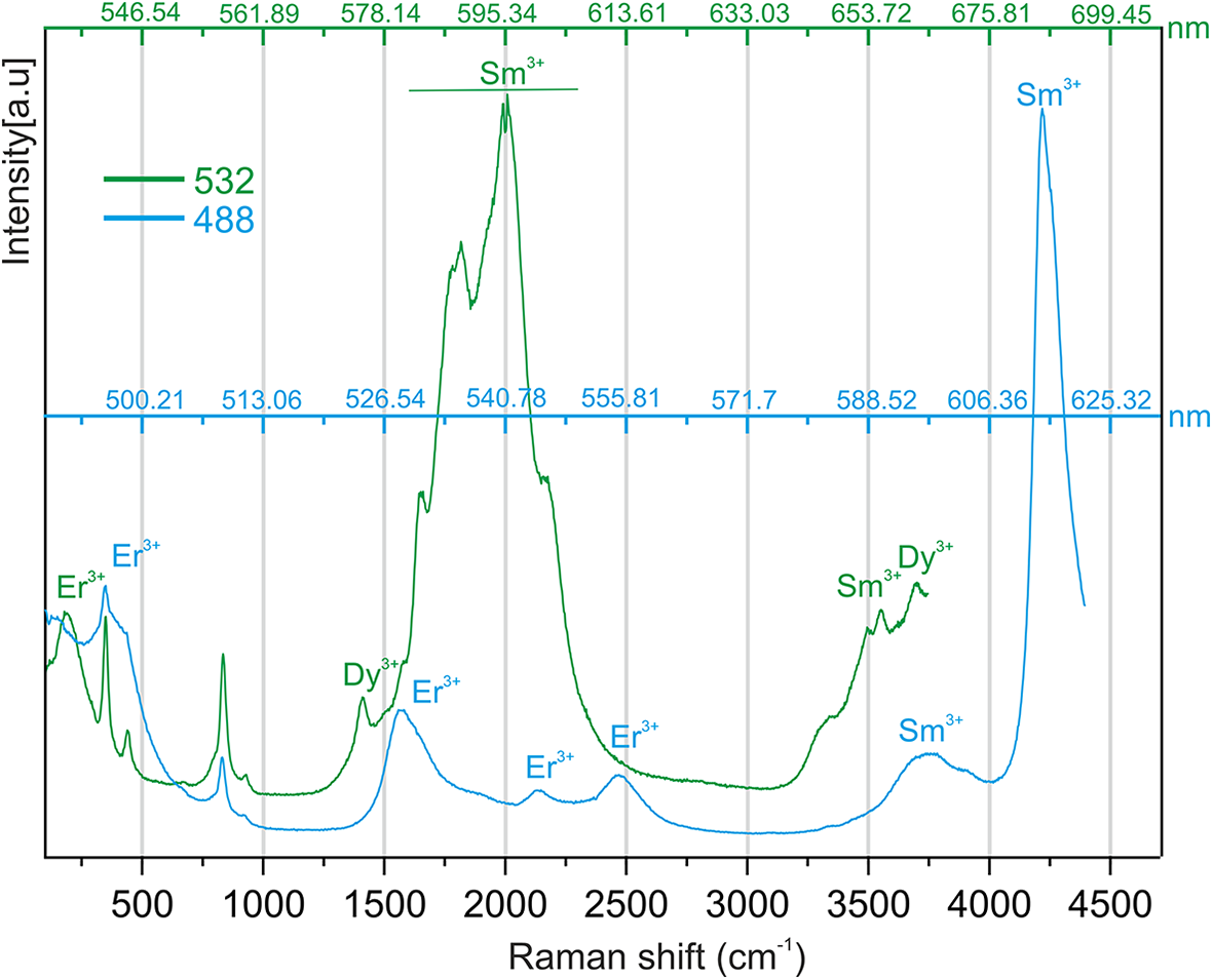
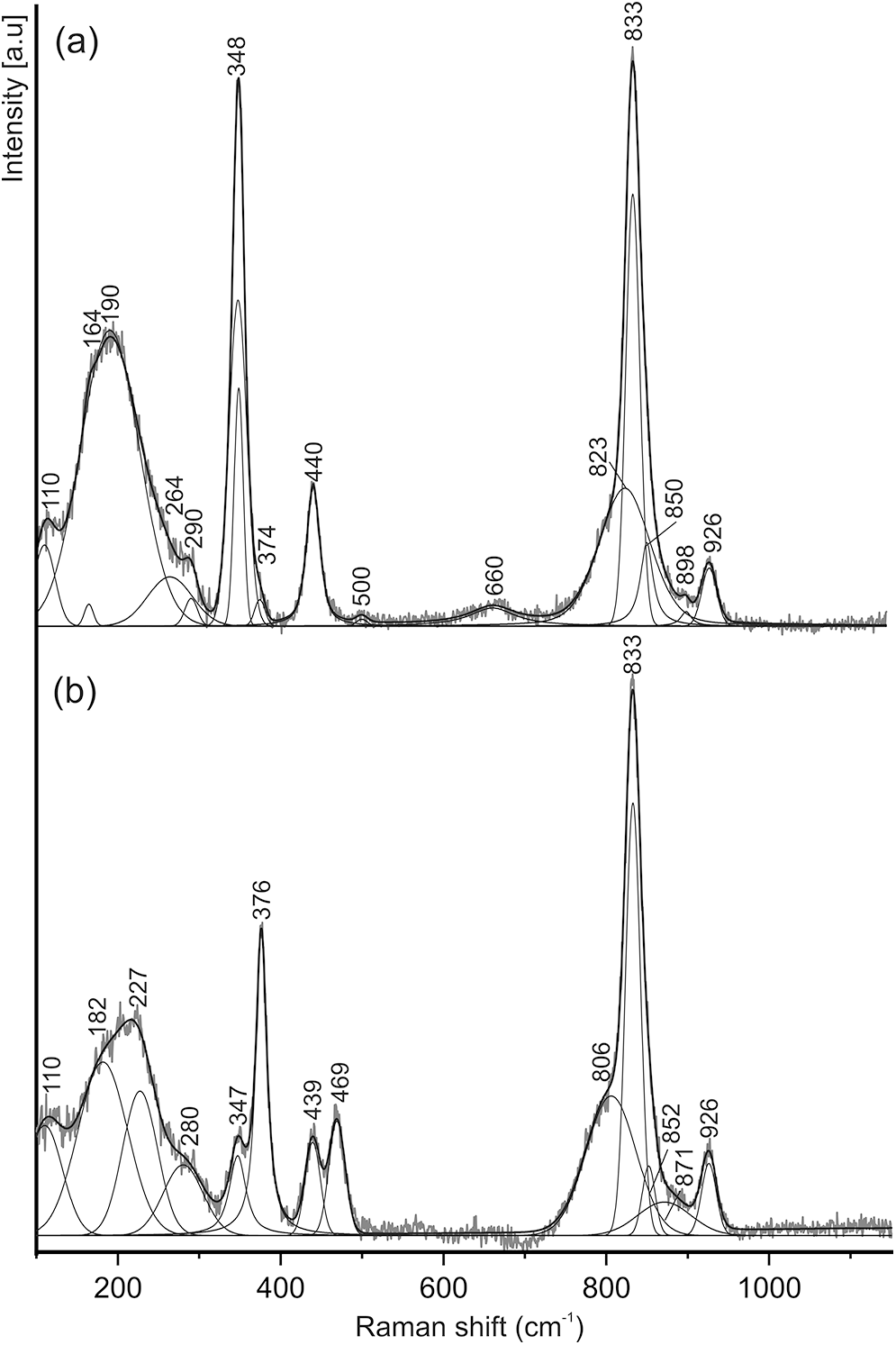
The Raman spectra of babunaite-(Nd) show a quantity of artefacts in the form of broad bands with luminescence from Sm3+, Er3+ and Dy3+ (Fig. 4; MacRae and Wilson, Reference MacRae and Wilson2008). A spectrum obtained using a blue laser (488 nm) indicates the absence of OH groups, which is also confirmed by the structural study of babunaite-(Nd). Raman spectra showing minimal artefact activity were obtained using a green laser (532 nm) in the 100–1150 cm–1 range in the zones with relatively low Th and Ca content (Fig. 5a) and with high Th and Ca of the holotype babunaite-(Nd) (Fig. 5b). Three ranges can be distinguished in the babunaite-(Nd) spectra: (1) 100–300 cm–1 are attributed to the torsional and translational motion of (TO4) group and the lattice vibrations of the (Nd, Ca, Th)-polyhedron. These vibrations are masked by broad bands related to Er3+-luminescence. (2) 300–500 cm–1 corresponds to the bending vibrations ν2 and ν4 in the (ТО4) group with T = As5+, W6+; and (3) 700–950 cm–1 corresponds to the stretching vibrations of ν1 and ν3 in the (ТО4) group (Pradhan and Choudhary, Reference Pradhan and Choudhary1987; Hardcastle and Wachs, Reference Hardcastle and Wachs1995; Rezgui et al., Reference Rezgui, Ouerfelli, Gavinho, Carvalho, Graça and Teixeira2023). The main bands from the stretching vibrations ν1(As–O) and ν1(W–O) occur at 833 cm–1 and 926 cm–1, respectively. In the holotype babunaite-(Nd) spectrum, the band from the bending vibrations ν4(As–O) in (AsO4) is near 440 cm–1, configuration Nb–O–As (Fig. 5a). In the spectrum of babunaite-(Nd) with high Ca and Th contents, an additional band appears at ∼469 cm–1. This band is related to the vibrations ν4(As–O) and the Th–O–As configuration.
Raman spectra of babunaite-(Nd) obtained using green (532 nm) and blue (488 nm) lasers and a monochromator with 600 mm–1 grating.
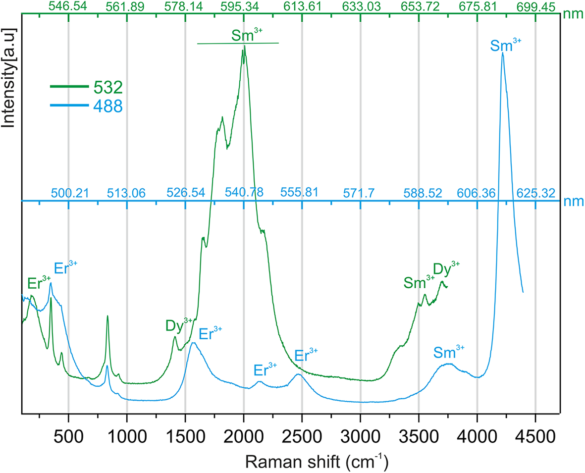
Figure 4 Long description
Raman spectra of babunaite-(Nd) obtained using green (532 nm) and blue (488 nm) lasers and a monochromator with 600 mm grating.–1 A line graph with two overlaid spectra. The x-axis label is Raman shift (cm superscript minus 1). The y-axis label is Intensity (a.u.). Two traces are shown: one labeled 532 and one labeled 488.
Raman spectra of (a) holotype babunaite-(Nd) and (b) babunaite-(Nd) with high Th content obtained using a green laser (532 nm) and a monochromator with 1800 mm–1 grating.
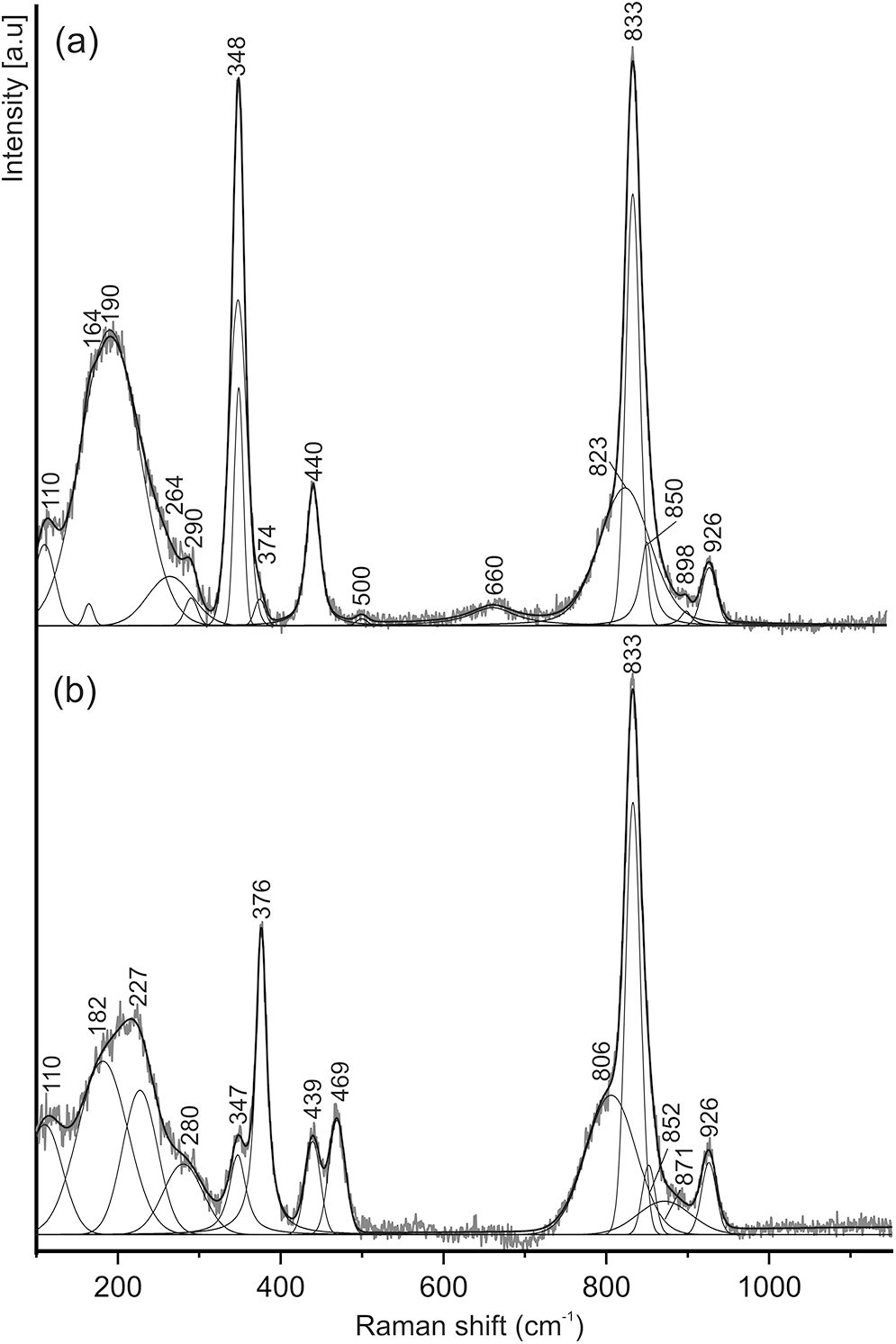
Figure 5 Long description
The Raman spectra line graphs show intensity versus Raman shift in cm superscript -1 for two samples: (a) holotype babunaite-(Nd) and (b) babunaite-(Nd) with high Th content. The x-axis is labeled 'Raman shift (cm superscript -1)' ranging from 100 to 1150. The y-axis is labeled 'Intensity (arbitrary units)'.
Structure of babunaite-(Nd)
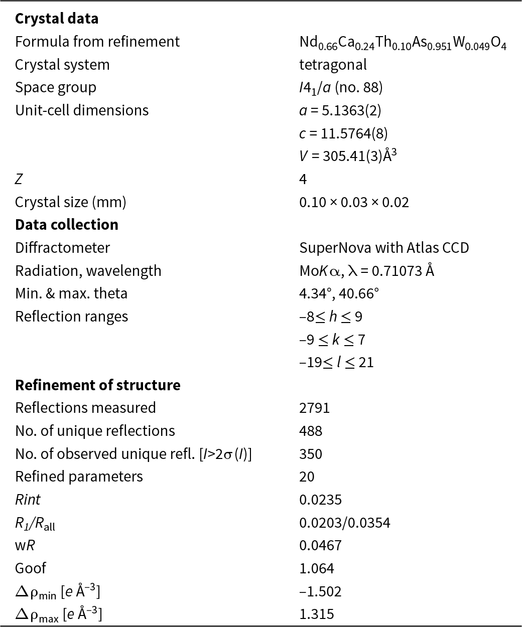
Single-crystal X-ray diffraction data were collected from a 0.10 × 0.03 × 0.02 mm babunaite-(Nd) crystal using a SuperNova diffractometer. The experimental details and refinement data are summarised in Tables 2, 3 and 4.
Crystal data and structure refinement details for babunaite-(Nd)
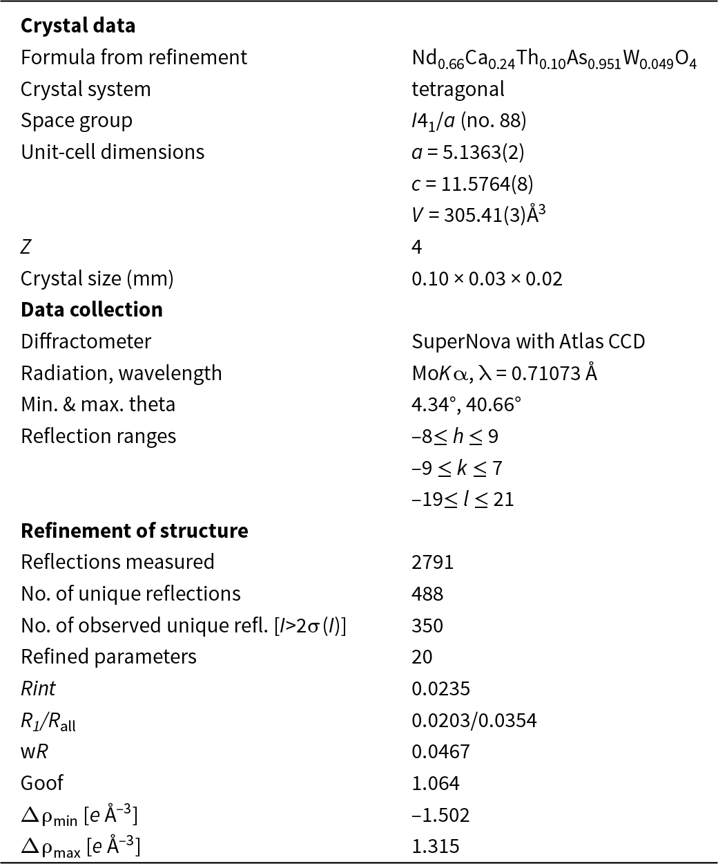
Table 2 Long description
The table summarizes single-crystal diffraction and refinement results for babunaite-(Nd), including composition, lattice metrics, data collection settings, and refinement quality indicators. The refined composition is Nd 0.66, Ca 0.24, Th 0.10, As 0.951, W 0.049, and O 4. The crystal is tetragonal in space group I41/a, with unit-cell parameters a 5.1363 angstroms and c 11.5764 angstroms, giving a unit-cell volume of 305.41 cubic angstroms and Z 4. The measured crystal fragment is 0.10 by 0.03 by 0.02 millimeters. Data were collected on a SuperNova diffractometer with an Atlas CCD using molybdenum K alpha radiation at wavelength 0.71073 angstroms, over theta from 4.34 to 40.66 degrees, with index ranges h minus 8 to 9, k minus 9 to 7, and l minus 19 to 21. A total of 2791 reflections were measured, yielding 488 unique reflections, of which 350 were considered observed using an intensity threshold greater than two times sigma. Refinement used 20 parameters and shows low disagreement factors, with Rint 0.0235, R1 0.0203 for observed and 0.0354 for all data, weighted R 0.0467, and goodness of fit 1.064. Residual electron density ranges from minus 1.502 to 1.315 electrons per cubic angstrom, indicating modest remaining features after refinement.
Atomic coordinates, equivalent-isotropic displacement parameters (Å2) and site occupancy for babunaite-(Nd)
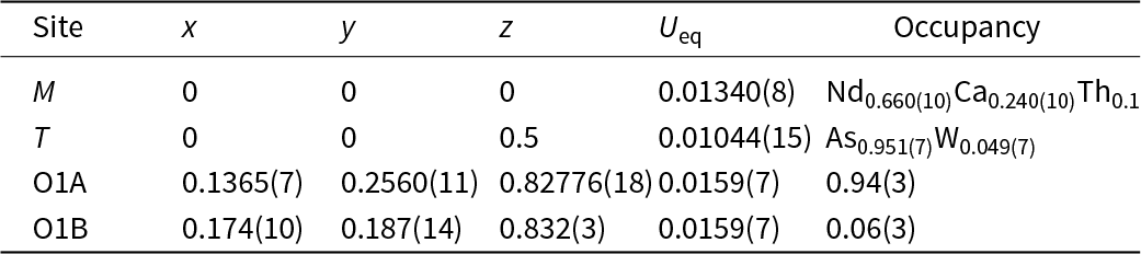
Table 3 Long description
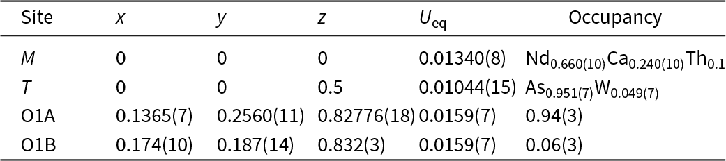
The table lists fractional atomic coordinates (x, y, z), equivalent isotropic displacement values, and site occupancies for four crystallographic sites. The M site at x 0, y 0, z 0 has a displacement value of 0.01340 with mixed occupancy dominated by neodymium about 0.660, plus calcium about 0.240 and thorium about 0.1. The T site at x 0, y 0, z 0.5 has a lower displacement value of 0.01044 and is mainly arsenic about 0.951 with minor tungsten about 0.049. Oxygen site O1A has coordinates near x 0.1365, y 0.2560, z 0.82776 with displacement 0.0159 and high occupancy about 0.94. Oxygen site O1B is close in position to O1A (x about 0.174, y about 0.187, z about 0.832) and shares the same displacement value, but has low occupancy about 0.06. The oxygen positions indicate a split site where most occupancy resides on O1A and a small fraction on O1B. Values in parentheses indicate measurement uncertainty, so small differences should be interpreted with that uncertainty in mind.
Anisotropic displacement parameters (Å2)
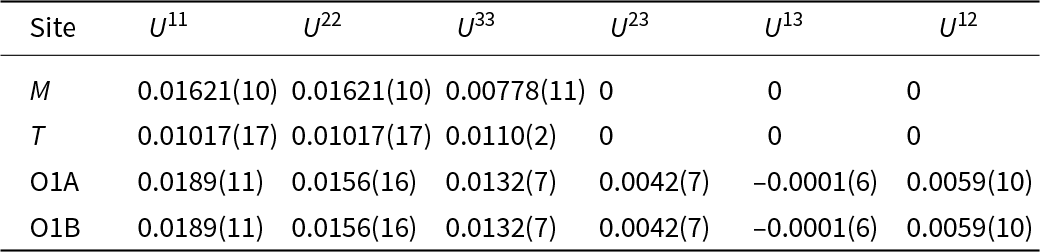
Table 4 Long description
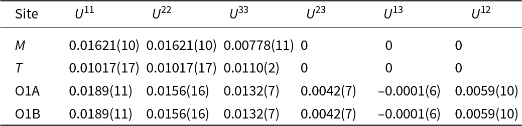
The table lists anisotropic displacement parameters for four crystallographic sites, reporting U11, U22, U33, U23, U13, and U12 with uncertainties in parentheses. Site M has U11 and U22 of 0.01621 and the lowest U33 at 0.00778, with U23, U13, and U12 all zero. Site T has lower U11 and U22 at 0.01017 and a U33 of 0.0110, and its off diagonal terms are also zero. O1A and O1B have identical parameter sets: U11 0.0189, U22 0.0156, U33 0.0132, with nonzero off diagonal terms U23 0.0042, U13 minus 0.0001, and U12 0.0059. Comparing sites, O1A and O1B show the largest U11 and the only nonzero coupling terms, while M and T are constrained to zero for those terms. Values should be interpreted with their reported uncertainties, and the identical O1A and O1B rows may reflect symmetry or shared refinement constraints rather than independent measurements.
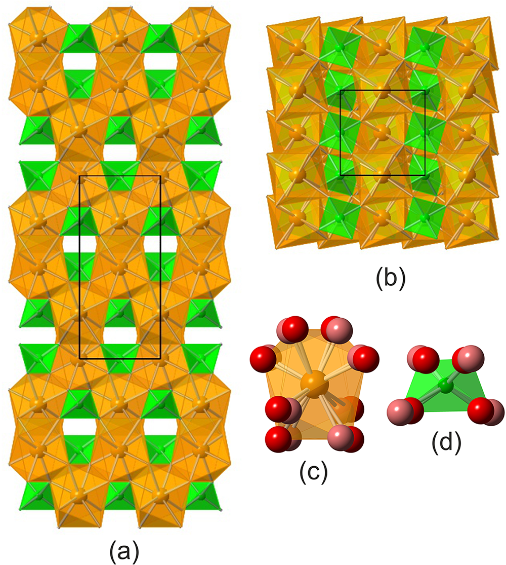
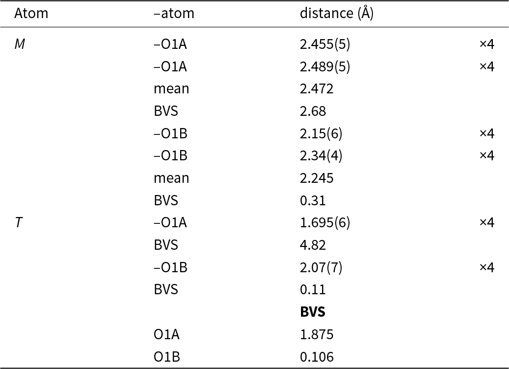
Babunaite-(Nd) has the general crystal chemical formula ABO4 and a scheelite-type structure. It crystallises in the tetragonal I41/a space group (a = 5.1363(2) Å, c = 11.5764(8) Å, V = 305.41(3) Å3, with Z = 4). The babunaite-(Nd) structure can be generally described as a framework comprising a system of intersecting, perpendicular, zigzag columns of edge-linking Nd-polyhedra, orientated along [100] and [010], which are interconnected by As-tetrahedra (Fig. 6). In this case, each top of a Nd polyhedron is shared with an As-tetrahedron. Nd3+ is bonded in an 8-coordinate geometry with eight equivalent O2⁻ atoms. A splitting of the oxygen position is observed: О1А (94%) and О1В (6%), with the distance O1A–O1B of the length 0.425 Å. There are four shorter bonds: Nd1–O1A = 2.455(5) Å or Nd–O1B = 2.15(6) Å, and four longer bonds: Nd1–O1A = 2.489(5) Å and Nd–O1B = 2.34(4) Å (Table 5). As5+ is bonded to four equivalent O atoms in a tetrahedral geometry. The As–O1 bond lengths are 1.695(6) Å to O1A and 2.07(7) Å to O1B. O1 is bonded in a three-coordinate geometry to two equivalent Nd3+ and one As5+ atoms. The disordering of the O1 position is probably due to the entry of the relatively larger W6+ ion (0.42 Å) into the tetrahedral position instead of the As5+ ion (0.335 Å) (Shannon, Reference Shannon1976). The splitting of the oxygen position may also be connected with the differences of ionic radius between Nd3+ (1.109 Å), Th (1.05 Å) and Ca (1.12 Å) (Shannon, Reference Shannon1976), as well as processes caused by the radioactive decay of Th.
The structure of babunaite-(Nd): (a) projection on (101); and (b) projection on (110). O1B is not shown. (c) Yellow polyhedra are occupied by Nd3+, Th, Ca and coordinated by 8 oxygens, which are disordered on O1A (94%, red) and O1B (6%, pink). (d) Green tetrahedra are occupied by As5+, W6+, taking into account O1A (red) and O1B (pink). Drawn using CrystalMaker for Windows, version 2.7.7.

Figure 6 Long description
The scatter plot displays the relationship between r subscript M in angstrom on the horizontal axis, ranging from 0.8 to 1.6 and r subscript T in angstrom on the vertical axis, ranging from 0.0 to 0.65. The plot categorizes minerals into groups based on anion types, represented by different colors: scheelite, zircon or xenotime, monazite, baryte and anhydrite. The data points form horizontal bands, indicating separation by mineral groups such as tantalates, niobates, tungstates, molybdates, vanadates, arsenates, silicates, chromates, phosphates and sulphates. The plot reveals a pattern where minerals are grouped horizontally according to their anion type, with notable outliers like Frm-Y at the highest r subscript T value of approximately 0.64. Dense clusters are observed around r subscript M values from 1.0 to 1.2 and r subscript T values from 0.10 to 0.35, highlighting the concentration of certain mineral types. The purpose of the plot is to illustrate the separation of mineral groups into distinct horizontal bands corresponding to different anion groups, providing insight into the structural classification of minerals based on ionic radii.
Selected bond lengths (Å) and weighted bond valences* (BVS, in valence units) from the empirical formula for babunaite-(Nd)
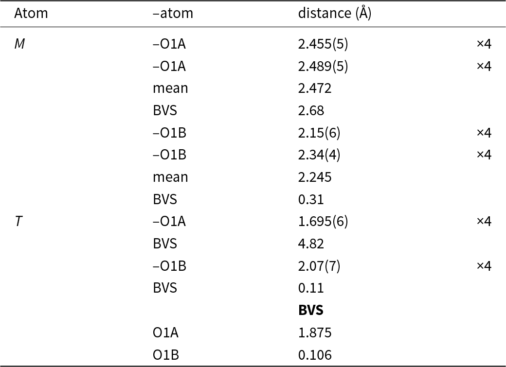
Table 5 Long description
The table lists bond lengths in angstroms and weighted bond valences for two sites, M and T, bonded to oxygen atoms labeled O1A and O1B, with each listed distance occurring four times. For M to O1A, distances are 2.455 and 2.489 angstroms with a mean of 2.472 angstroms and a bond valence of 2.68. For M to O1B, distances are 2.15 and 2.34 angstroms with a mean of 2.245 angstroms and a much lower bond valence of 0.31. For T to O1A, the distance is 1.695 angstroms and the bond valence is 4.82, the largest value in the table. For T to O1B, the distance is 2.07 angstroms and the bond valence is 0.11, indicating a very weak contribution compared with T to O1A. The oxygen totals reported are 1.875 for O1A and 0.106 for O1B, reinforcing that O1A carries most of the bond valence in this dataset. Bond valences are derived from a crystallographic file using a specific calculation program, so values depend on that method and input structure.
* BVS calculated on the basis our crystallographic information file (CIF) using the ECoN21 program (Ilinca, Reference Ilinca2022)
The powder X-ray diffraction data were calculated from the result of the single-crystal refinement. These results are presented in Supplementary Table S1.
Discussion
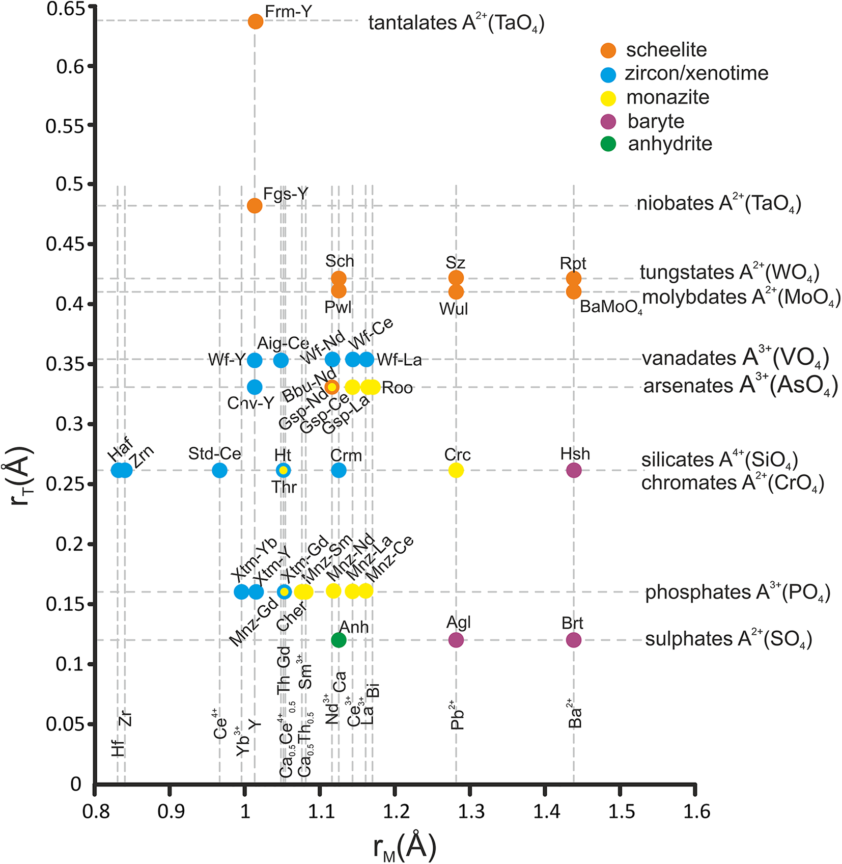
In the pink schists, alongside babunaite-(Nd), other REEAsO4 minerals such as gasparite-(La) and chernovite-(Y) were found. These arsenates belong to the scheelite, monazite and xenotime types, respectively. The structure of pure compounds with stoichiometry VIII–IXM(IVTO4) is determined by the cation radii at the M and T sites. This is clearly reflected in the rM(Å)–rT(Å) diagram of the Bastide type, in which isostructural compounds occupy orthogonal fields, giving the diagram the appearance of a settlement plan with orthogonal zoning (Clavier et al., Reference Clavier, Podor and Dacheux2011; Errandonea, Reference Errandonea2017). However, the diagram of the Bastide type for M(TO4) compounds, which are used in physics and chemistry of the solid state, is not informative for minerals. Minerals in databases are usually represented by the ideal formula, but the natural phase as a rule occurs as a solid solution. For example, there are two polymorphic modifications of xenotime-(Gd) (tetragonal) and monazite-(Gd) (monoclinic), both with the ideal formula Gd(PO4). However, holotype xenotime-(Gd) contains only 38% and holotype monazite-(Gd) contains only 30% of Gd(PO4) end-member, respectively (Ondrejka et al., Reference Ondrejka, Uher, Ferenc, Majzlan, Pollok, Mikuš, Milovská, Molnárová, Škoda, Kopáčik, Kurylo and Bačík2023, Reference Ondrejka, Bačík, Majzlan, Uher, Ferenc, Mikuš, Števko, Čaplovičová, Milovská, Molnárová, Rößler and Matthes2024). In the rM(Å)–rT(Å) diagram, ideal compositions with stoichiometry M(TO4) are shown (Fig. 7). There are only three compounds for which stable dimorphs exist under geological conditions: ThSiO4, GdPO4 and NdAsO4. These are huttonite and thorite (Finch et al., Reference Finch, Harris and Clark1964); monazite-Gd and xenotime-Gd (Ondrejka et al., Reference Ondrejka, Uher, Ferenc, Majzlan, Pollok, Mikuš, Milovská, Molnárová, Škoda, Kopáčik, Kurylo and Bačík2023, Reference Ondrejka, Bačík, Majzlan, Uher, Ferenc, Mikuš, Števko, Čaplovičová, Milovská, Molnárová, Rößler and Matthes2024); babunaite-(Nd) and ‘gasparite-(Nd)’ (Kolitsch et al., Reference Kolitsch, Schachinger and Auer2025), respectively. It is well known that there is a discontinuity in the composition of the phosphates of the xenotime group, which contain heavy rare earth elements (HREE, i.e. Gd–Lu) and Y with relatively small ionic radii, and the phosphates of the monazite group, which contain light rare earth elements (LREE, i.e. La–Eu) with relatively big ionic radii (Fig. 7). The limit of LREE and HREE phosphate miscibility decreases with increasing temperature (Gratz and Heinrich, Reference Gratz and Heinrich1997; Heinrich et al., Reference Heinrich, Andrehs and Franz1997; Pyle et al., Reference Pyle, Spear, Rudnick and McDonough2001; Mogilevsky, Reference Mogilevsky2007). A similar situation is observed for minerals in the REE(AsO4) series, where an increase in temperature is accompanied by an increase in Y content in gasparite (Pagliaro et al., Reference Pagliaro, Comboni, Battiston, Krüger, Hejny, Kahlenberg, Gigli, Glazyrin, Liermann, Garbarino, Gatta and Lotti2022). Interestingly, the ionic radius of the M cation in the pairs huttonite–thorite and monazite-(Gd)–xenotime-(Gd) is very close: Th4+ = 1.05 Å and Gd3+ = 1.053 Å, corresponding to the boundary between LREE and HREE. Therefore, these dimorphic minerals are formed under pressure/temperature (P/T) conditions typical for geological processes. A mineral with the formula NdAsO4 (with an ionic radius of Nd = 1.109 Å, LREE), using the crystal chemical criteria, should have a monazite-type structure, as confirmed by the finding of ‘gasparite-(Nd)’ (Kolitsch et al., Reference Kolitsch, Schachinger and Auer2025). Known minerals, such as the REE and Y arsenates, have a monoclinic structure of the monazite type in the gasparite group [gasparite-(Ce), Ce(AsO4) 21/n, a = 6. 937(3) Å, b = 7.137(4) Å, c = 6.738(6) Å, and β = 104.69(5)° (Graeser and Schwander, Reference Graeser and Schwander1987); gasparite-(La), La(AsO4), 21/n, a = 6. 7646(4) Å, b = 7.2184(9) Å, c = 6.0070(4) Å and β = 104.51(1)° (Vereschagin et al., Reference Vereshchagin, Britvin, Perova, Brusnitsyn, Polekhovsky, Shilovskikh, Bocharov, van der Burgt, Cuchet and Meisser2019)] and a tetragonal structure of the xenotime-type [chernovite-(Y), Y(AsO4), I41/amd, a = 7.039(11) Å and c = 6.272(22) Å (Goldin et al., Reference Goldin, Yushkin and Fishman Fishman1967)]. Arsenates with a scheelite-type structure are not found in Nature. The known synthetic phase with a scheelite-type structure is NdAsO4 [tetragonal, I41/a; a = 5.1046(4) Å and c = 11.6032(11)], which is considered a high-pressure phase. It can be obtained from the monoclinic phase of NdAsO4 under high-pressure and high-temperature conditions of 11 GPa and 1100–1300°C (Metzger et al., Reference Metzger, Ledderboge, Heymann, Huppertz and Schleid2016). Such conditions could not be realised in the formation processes of the Mixed Series rocks. The genesis of the complex polymineral paragenesis of the Mixed Series remains to be fully resolved and merits further study. All petrological elements and ore occurrences of the Mixed Series as well as massive marbles, underwent metamorphism by complex polyphase processes (Stojanov, Reference Stojanov1960; Bermanec et al., Reference Bermanec, Chukanov, Varlamov, Rajačić, Jančev and Ermolaeva2023), reaching the conditions of the kyanite–graphite subfacies (Jančev, Reference Jančev1985). We believe that the formation of babunaite-(Nd) is associated with the stabilisation of its structure by the impurity of tungsten (W). Phases of stoichiometry VIIIM IVTO4 with tetrahedrally coordinated anions and an ionic radius greater than 0.4 Å (W, Mo) have a scheelite-type structure (Fig. 7; Errandonea, Reference Errandonea2017). Babunaite-(Nd) is a unique mineral, it is both the first arsenate and the first REE phase in the scheelite group.
All known minerals M(TO4), M= R2+, R3+, R4+; T = R4+, R5+, R6+ in the diagram rM(Å) – rT(Å).
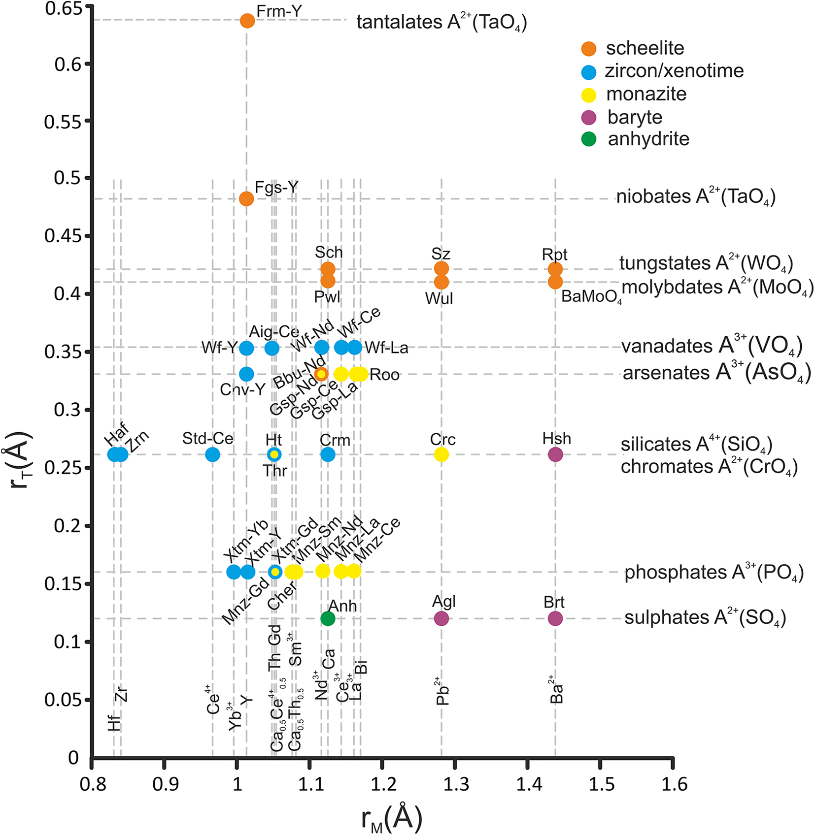
Figure 7 Long description
The scatter plot displays the relationship between r subscript M in angstrom on the horizontal axis, ranging from 0.8 to 1.6 and r subscript T in angstrom on the vertical axis, ranging from 0.0 to 0.65. The plot categorizes minerals into groups based on anion types, represented by different colors: scheelite, zircon or xenotime, monazite, baryte and anhydrite. The data points form horizontal bands, indicating separation by mineral groups such as tantalates, niobates, tungstates, molybdates, vanadates, arsenates, silicates, chromates, phosphates and sulphates. The plot reveals a pattern where minerals are grouped horizontally according to their anion type, with notable outliers like Frm-Y at the highest r subscript T value of approximately 0.64. Dense clusters are observed around r subscript M values from 1.0 to 1.2 and r subscript T values from 0.10 to 0.35, highlighting the concentration of certain mineral types.
Supplementary material
The supplementary material for this article can be found at https://doi.org/10.1180/mgm.2026.10222
Acknowledgements
The authors would like to thank the reviewers and editors for their valuable feedback, which helped to improve the manuscript.
Competing interests
The authors declare none.


 Open access

