


I. INTRODUCTION
Over the past two decades, the triple perovskite with the general formula AA’2BB’2O9 (A,A’ = alkaline and rare-earth metals; B,B’ = metal transition elements) has been one of the most studied by the scientific community because of its wide variety of interesting physical properties, including magnetic, magnetocaloric, multiferroics, metal–insulator transition, relaxor ferroelectrics, dielectric, photocatalytic, superparamagnetic, and SOFCs (Dong and Ye, Reference Dong and Ye2000; Ting et al., Reference Ting, Liu, Withers and Noren2004; Singh et al., Reference Singh, Kumar, Bharat Singh, Kaushik, Gaur, Rayaprol and Simon2011; El Hachmi et al., Reference El Hachmi, El Ouahbi, Manoun and Lassri2022; El Hachmi and Manoun, Reference El Hachmi and Manoun2023; El Hachmi et al., Reference El Hachmi, El Ouahbi, Ounza, Biswas, Lassri, Manoun and Sadoune2024; El Hachmi et al., Reference El Hachmi, Biswas, Sen, Manoun, Draoui and Sadoune2025a).
The BaLn 2CuTi2O9 (Ln = La, Pr, and Nd) triple perovskite compounds have been reported using X-ray and neutron powder diffraction techniques (Iturbe-Zabalo et al., Reference Iturbe-Zabalo, Igartua, Aatiq and Pomjakushin2013). At room temperature, all these structures crystallize in the tetragonal symmetry of space group I4/mcm. The BaLa2CuTi2O9 composition has been reported with unit-cell parameters (a = 5.6032 and b = 7.9192 Å) and refinement factors Rp = 14.2%, R wp = 22.6%, R Bragg = 3.65%, and χ 2 = 1.99. It has been found that the A and B site cations fully occupy the 4c (0, 0, 0) and 4b (0, 1/2, 1/4) positions, respectively.
On the other side, the CaLa2ZnTi2O9 structure has been described as crystallizing in an orthorhombic system (space group Pbnm) with lattice parameters a = 5.5171, b = 5.5444, and c = 7.8109 Å (Aatiq and Boukhari, Reference Aatiq and Boukhari2004). It has also been reported that the double perovskite La2CuTiO6 crystallizes in an orthorhombic Pbnm space group with lattice parameters a = 5.560, b = 5.610, and c = 7.840 Å, which is related to the GdFeO3 structure (Gomez-Romero et al., Reference Gomez-Romero, lacin, Casan, Fuertes and Martinez1993). A study of the electric and magnetic polarizabilities of Ln 2CuTiO6 (Ln = Y, Dy, Ho, Er, and Yb) hexagonal compositions has been reported, revealing unusual combinations of large and extremely stable dielectric responses (Choudhury et al., Reference Choudhury, Hazarika, Venimadhav, Kakarla, Delaney, Sujatha Devi and Mondal2010). Furthermore, the Ln 2CuTiO6 (Ln = La, Y, Pr, and Nd) compositions have been reported to exhibit dielectric relaxation in both hexagonal and orthorhombic compounds (Singh et al., Reference Singh, Kumar, Bharat Singh, Kaushik, Gaur, Rayaprol and Simon2011).
In our recent study, we examined the structural evolution of the perovskite system CaLa2Zn1−x Ca xTi2O9 (x = 0.00, 0.15, 0.30, 0.45, 0.90, and 1.00) employing X-ray powder diffraction (XRPD) combined with Rietveld refinement analysis (El Hachmi et al., Reference El Hachmi, Leila, Brahim El, Brahim, El Hossain and Bouchaib2025b). The crystallographic investigation conducted at room temperature revealed a composition-induced phase transition within the series: samples with x = 0.00, 0.15, 0.30, and 0.45 crystallize in an orthorhombic structure with space group Pbnm, whereas compositions with x = 0.90 and 1.00 adopt a monoclinic structure belonging to space group P21/n. This structural transformation underscores the influence of increasing Ca2+ substitution at the Zn2+ site on the symmetry and lattice parameters of the perovskite framework.
The quadruple perovskites ACu3Ti4O12 (A = Ba, Ca, and Sr) have been characterized by a negative thermal resistance coefficient. The crystalline structure of these compounds at room temperature has been investigated using an X-ray diffraction (XRD) technique, and has been identified as cubic with space group Im-3 (Choudhary and Bhunia, Reference Choudhary and Bhunia2002). The CaCu3Ti4O12 material has been reported to exhibit a large optical nonlinearity (Ning et al., Reference Ning, Chen, Zhou, Heng, Zhang, Ming and Yang2009). The compounds Sr4−x La xFe3ReO12 (x = 0.0 and 1.0) have been described using Neutron Powder Diffraction (NPD) at room temperature as adopting simple perovskite structures without cation ordering on the B-sites, and their structures crystallize with I4/mcm tetragonal symmetry and a tilt system a 0a 0c − (Retuerto et al., Reference Retuerto, Li, Go, Ignatov, Croft, Ramanujachary and Herber2012).
In this work, we describe the synthesis procedure and crystal structure analysis of the triple perovskite materials BaLa2Cu1−x Ba xTi2O9 (x = 0.00, 0.15, and 0.30) using room temperature XRPD and the Rietveld refinement method.
II. EXPERIMENTAL DETAILS
A. Synthesis
Polycrystalline powders of the BaLa2Cu1−x Ba xTi2O9 (x = 0.00, 0.15, and 0.30) ceramics were prepared using a solid-state reaction route in air, and the raw materials BaCO3 (99.98%), La2O3 (99.999%), CuO (99.99%), and TiO2 (99.8%) were used as starting reagents (all received from Sigma-Aldrich). The quantity of each reagent was weighed according to its stoichiometric coefficient to obtain the appropriate metal ratios of the final products, then mixed and ground in an agate mortar to form a homogeneous powder. The resulting mixtures were placed in alumina crucibles and then increasingly heated in air at different temperature stages with intermittent grinding; these three samples were calcined at 1,000 °C for 20 h and 1,200 °C for 12 h, respectively. During the heat treatment process, the powders were cooled to room temperature, reground, and sintered several times to improve homogeneity. The chemical reaction is as follows:
 $$ \begin{array}{c}\left(1+x\right)\;{\mathrm{Ba}\mathrm{C}\mathrm{O}}_3+{\mathrm{La}}_2{\mathrm{O}}_3+\left(1\hbox{--} x\right)\;\mathrm{C}\mathrm{uO}+2\;{\mathrm{Ti}\mathrm{O}}_2\\ {}\frac{\Delta \left(1200\;{}^{\circ}\mathrm{C}\right)}{\mathrm{AIR}}\to {\mathrm{Ba}\mathrm{La}}_2{\mathrm{Cu}}_{1\hbox{--} x}{\mathrm{Ba}}_x{\mathrm{Ti}}_2{\mathrm{O}}_9+\left(1+x\right)\;{\mathrm{CO}}_2\\ {}\left(x=0.00,\hskip0.3em ,0.15,\hskip0.3em \mathrm{and}\hskip0.35em 0.30\right)\end{array} $$
B. X-ray powder diffraction
Diffraction data of the BaLa2Cu1−x Ba xTi2O9 (x = 0.00, 0.15, and 0.30) ceramics were collected at room temperature on a D2 PHASER diffractometer, with the Bragg–Brentano geometry, using a copper anti-cathode tube as the radiation source (K α1, K α2) of the wavelengths λ(K α1) = 1.54056 Å and λ(K α2) = 1.54439 Å with 30 kV and 10 mA, Soller slits of 0.02 rad on incident and diffracted beams; divergence slit of 0.5°; antiscatter slit of 1°; receiving slit of 0.1 mm; with sample spinner, and a Lynxeye detector type with a maximum global count rate > 1000,000,000 cps. The XRD patterns were scanned in steps of 0.020287° (2θ), between 15° and 105° (2θ) with a fixed-time counting of 2 s per step. The WinPlotr software (Roisnel and Rodríguez-Carvajal, Reference Roisnel and Rodríguez-Carvajal2001), integrated into the FullProf program (Rodríguez-Carvajal, Reference Rodríguez-Carvajal1990), was used to analyze the crystal structures by means of the Rietveld method (Rietveld, Reference Rietveld1969). The peak shape was described by a pseudo-Voigt function using linear interpolation between set background points with refinable heights.
The following structural and instrumental parameters – scale factor, unit-cell parameters, pseudo-Voigt corrected for asymmetry parameters, full width at half maximum (FWHM) parameters (u, v, and w), preferred orientation, atomic positions, and isotropic displacement parameters – were included in the refinement of the investigated XRD patterns.
III. RESULTS AND DISCUSSION
A. Crystallite size and strain
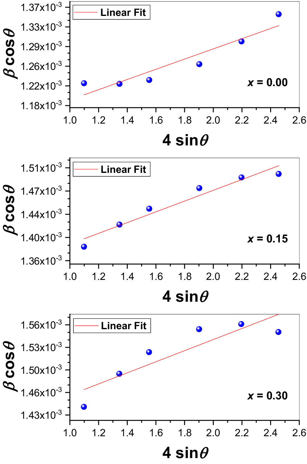
The crystallite size of the three studied compositions (x = 0.00, 0.15, and 0.30) was determined using X-ray line broadening analysis via the Scherrer equation and the Williamson–Hall (W-H) method. X-ray peak broadening can result from crystallite size, microstrain, instrumental effects, temperature factors, and solid solution inhomogeneity. The Scherrer equation is expressed as follows (Scherrer, Reference Scherrer1918):
 $$ D=\frac{K\lambda}{\beta_{(s)}\cos \theta }, $$
where D is the average crystallite size (nm), K is the Scherrer constant equal to 0.9, λ is the X-ray wavelength (Cu-K α average ≈ 1.54178 Å), θ is the Bragg angle corresponding to the most intense XRD peak, and β (s) is the FWHM of the highest peak. In this study, the crystallite size was deduced from the XRD patterns using the strongest peak located at approximately 2θ ≈ 31.89°. The average crystallite size calculated using the Debye–Scherrer equation was found to be 113 nm (x = 0.00), 100 nm (x = 0.15), and 96 nm (x = 0.30).
The broadening caused by microstrain, which arises from lattice distortions and imperfections, was analyzed based on the relation originally introduced by Stokes and Wilson (Reference Stokes and Wilson1944), who described the strain-induced peak broadening as
 $$ \varepsilon =\frac{\beta_{(D)}}{4\tan \theta}, $$
where ε ≈ Δd/d represents the lattice strain and β (D) is the broadening due to deformation. Williamson and Hall (Reference Williamson and Hall1953) combined this strain broadening equation with the Scherrer equation under the assumption that both size and strain broadening contributions are independent and follow Lorentzian (Cauchy) profiles. This combination leads to the following expression:
 $$ {\beta}_{hkl}={\beta}_{(D)}+{\beta}_{(s)}, $$
and from Eqs. (1) and (2), we get
 $$ {\beta}_{hkl}\cos \theta =\varepsilon\;\left(4\sin \theta \right)+\frac{K\lambda}{D}. $$
Plotting β hkl cosθ versus 4sinθ (as shown in Figure 1) allows direct determination of the crystallite size from the y-intercept (Kλ/D) and the microstrain (ε) from the slope of the linear fit. The crystallite size and strain for the three compositions are summarized in Table I. It can be noted that the crystallite size values obtained from the W-H method are larger than those derived from the Scherrer equation alone.
Williamson–Hall plots of the BaLa2Cu1−x Ba xTi2O9 phases.

Rietveld refinement details, crystallite size, and strain for the BaLa2Cu1−x Ba xTi2O9 phases.

Abbreviation: FWHM, full width at half maximum.
B. Crystal structure analysis and structure description
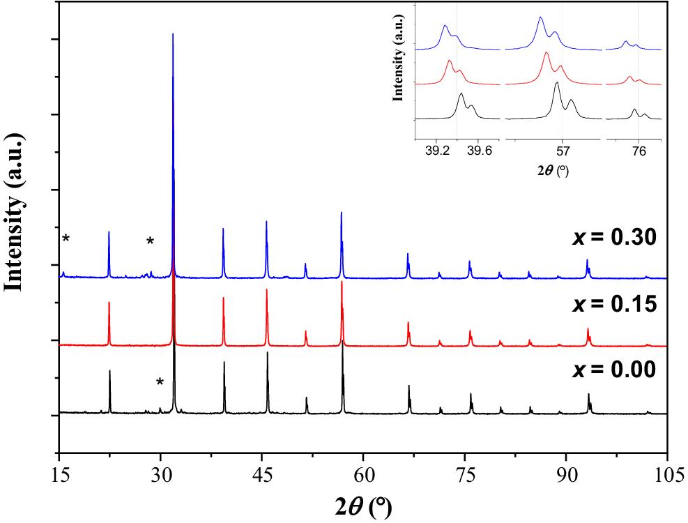
At room temperature, the PXRD patterns of the compositions BaLa2Cu1−x Ba xTi2O9 (x = 0.00, 0.15, and 0.30) are successfully identified by the PDF2 database (Gates-Rector and Blanton, Reference Gates-Rector and Blanton2019) integrated into the HighScore plus software (Degen et al., Reference Degen, Sadki, Bron, König and Nénert2014) and analyzed using the DICVOL program (Louër and Boultif, Reference Louër and Boultif2014) to adopt a phase related to a simple perovskite structure.
A minor presence of secondary phases was detected in the XRD patterns of the as-synthesized samples with compositions x = 0.00 and x = 0.30. For x = 0.00, a small impurity phase was identified as La2Ti2O7 (PDF: 00-027-1182), which crystallizes in the monoclinic system (space group P21/m) with lattice parameters a = 13.01 Å, b = 5.54 Å, c = 7.80 Å, and β = 98.37°. For x = 0.30, the detected secondary phases included La2BaO x (PDF: 00-042-0337), BaLa2O4 (PDF: 00-042-1500), and CuLa2O4 (PDF: 01-081-2446).
Figure 2 presents the powder XRD patterns of BaLa2Cu1−x Ba xTi2O9 (x = 0.00, 0.15, and 0.30) ceramics collected at 298 K. From the enlarged peaks in the inset of Figure 2, the XRD patterns of the powdered samples show an obvious shift of the peaks toward the lower angles (2θ, Bragg position), indicating that a partial substitution of Cu2+ by Ba2+ can have an influence on the crystal structure parameters. The unchanged profile of the broadened peaks implies structural stability without evidence of a phase transition. The crystal structures of all studied compositions were analyzed using the Rietveld refinement method (Rietveld, Reference Rietveld1969). Previously, the BaLa2CuTi2O9 structure (Iturbe-Zabalo et al., Reference Iturbe-Zabalo, Igartua, Aatiq and Pomjakushin2013) has been reported as crystallizing in the tetragonal symmetry and space group I4/mcm (No. 140); its atomic coordinates and lattice parameters were used as starting data for refinement.
X-ray diffraction patterns of BaLa2Cu1−x Ba xTi2O9 perovskites collected at room temperature. The inset shows an enlarged view of the peaks. The stars (*) for x = 0.00 and 0.30 indicate the presence of impurities.

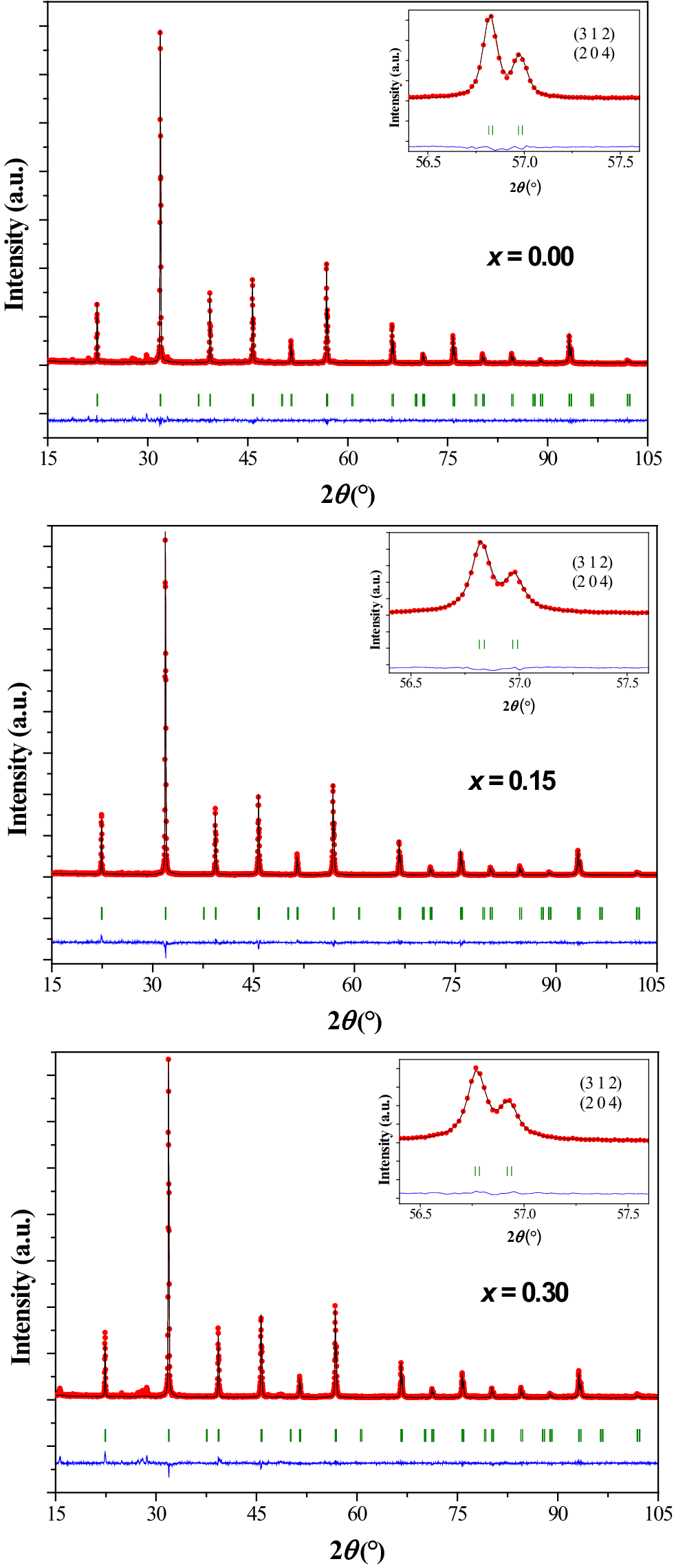
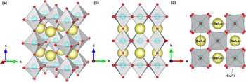
Table I gives the crystal data, data collection, and details of the Rietveld refinements, including lattice parameters, unit-cell volume, crystal system, space group, and various statistical agreement factors. The values of the reliability factors (%), RB, RF, Rp, R wp, and R exp, as well as the goodness-of-fit (χ 2), are small and indicate that we have obtained a good quality of the refinement. The quality of the fits is also confirmed by Figure 3, which presents excellent agreement between the experimental (dots) and theoretical (solid line) patterns. The criteria for refined structures are met, ensuring that they are both chemically plausible and statistically reliable. The structural illustration of the tetragonal phase with space group I4/mcm is shown in Figure 4; it was drawn using the Vesta software (Momma and Izumi, Reference Momma and Izumi2011).
Final Rietveld refinement plots for the tetragonal BaLa2Cu1−x Ba xTi2O9 phases. The three experimental datasets are represented by red dots, and the calculated patterns are shown as black solid lines. The green vertical ticks indicate the Bragg positions of the main phase. The blue line at the bottom represents the difference between the observed and calculated patterns.

Structural view of the tetragonal phase Ba1/3La2/3Cu1/3Ti2/3O3 (x = 0.00) with space group I4/mcm. Panel (a) displays a three-dimensional perspective of the crystal lattice illustrating the arrangement of the Ba/La atoms (large yellow spheres), Cu/Ti atoms (blue spheres), and oxygen atoms (small red spheres) within corner-sharing octahedra. Panel (b) presents the projecti on of the structure along the a-axis, highlighting the layered arrangement and connectivity of the octahedra and the positioning of Ba/La cations in the lattice. Panel (c) illustrates the octahedral tilting consistent with Glazer’s notation a 0a 0c −.

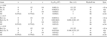
Rietveld refinement analysis reveals that the BaLa2Cu1−x Ba xTi2O9 (x = 0.00, 0.15, and 0.30) structures adopt the tetragonal symmetry of space group I4/mcm (No. 140) with lattice parameters: for x = 0.00, a = 5.6093(1) Å and c = 7.9277(2) Å; for x = 0.15, a = 5.6093(1) Å and c = 7.9269(2) Å; and for x = 0.30, a = 5.6139(2) Å and c = 7.9341(3) Å. The unit-cell volumes were calculated to be 249.43 Å3 (for x = 0.00), 249.42 Å3 (for x = 0.15), and 250.05 Å3 (for x = 0.30). The number of unit formulas within a cell is Z = 4. Reflections from the tetragonal structure with space group I4/mcm (No. 140) are correctly indexed on the studied XRD patterns; where the reflection conditions can be generally expressed as follows: hkl: h + k + l = 2n; hk0: h + k = 2n; 0kl: k, l = 2n; hhl: l = 2n; 00l: l = 2n; h00: h = 2n (International Tables for Crystallography – Volume A). The fractional atomic coordinates and isotropic displacement parameters are listed in Table II. The occupancy factors are given according to the stoichiometry of the formula: (Ba2+1/3La3+2/3)4b (Cu2+(1-x)/3Ba2+ x/3Ti4+2/3)4c O3, which is electrically neutral. The Cu2+, Ba2+(2), and Ti4+ cations were found to occupy octahedral environments at 4c (0, 0, 0) positions. They are surrounded by six oxygen anions; two O2−(1) at 4a (0, 0, 1/4), and four O2−(2) at 8h (x, x + 1/2, 0) sites. Whereas the Ba2+(1) and La3+ cations occupy polyhedral environments at 4b (0, 1/2, 1/4) positions, they are coordinated with 12 oxygen anions; 4 O2−(1) at 4a (0, 0, 1/4), and 8 O2−(2) at 8h (x, x + 1/2, 0) sites. Despite some differences in ionic size and valence charge, the Cu2+(Ba2+) and Ti4+ cations have only one crystallographic B-site. Similarly, the two cations Ba2+ and La3+ have only one crystallographic A-site, like the simple perovskite structure ABO3. The isotropic displacement parameters U iso (Å2) for oxygen atoms at sites 4a and 8h were refined by considering them equivalent; similarly, the Ba(1)/La and Cu/Ba(2)/Ti atoms were considered equivalent at their sites 4a and 4b, respectively. In the composition with x = 0.30, the U iso values for Ba2+(1)/La3+ cations were fixed at 0.013 Å2 to guarantee the convergence of the refinement.
Atomic positions and isotropic displacement parameters for the tetragonal BaLa2Cu1−x Ba xTi2O9 phases.

* Fixed in 0.013 Å2 to guarantee the convergence of the refinement.
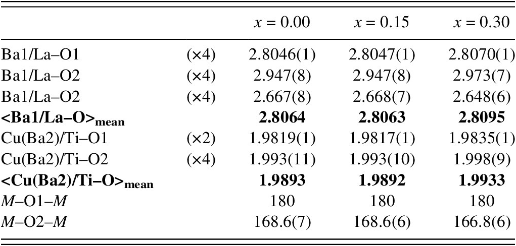
The selected interatomic distances (Å) and angles (degrees) are given in Table III. The average distances <Ba1/La–O> and <Cu(Ba2)/Ti–O> obtained after the Rietveld refinement of the studied compositions are roughly identical to those found in other perovskite compounds containing Ba2+, La3+, Cu2+, and Ti4+ cations, such as BaLa2CuTi2O9 with Ba/La–O = 2.804 Å and Cu/Ti–O = 1.99 Å; BaNd2CuTi2O9 with Ba/Nd–O = 2.794 Å and Cu/Ti–O = 1.99 Å; BaPr2CuTi2O9 with Ba/Pr–O = 2.796 Å and Cu/Ti–O = 1.99 Å (Iturbe-Zabalo et al., Reference Iturbe-Zabalo, Igartua, Aatiq and Pomjakushin2013); La2CuTiO6 with Cu/Ti–O = 2.02 Å (Gomez-Romero et al., Reference Gomez-Romero, lacin, Casan, Fuertes and Martinez1993); and BaLaCrMoO6 with Ba/La–O = 2.8402 Å (Musa-Saad, Reference Musa-Saad2014). They are also very close to those calculated from Shannon’s effective ionic radii: Ba2+ (1.61 Å), La3+ (1.36 Å), Cu2+ (0.73 Å), Ti4+ (0.605 Å), and O2− (1.40 Å) for 12- and 6-fold coordination (Shannon, Reference Shannon1976). The bond M–O2–M angles (M = Cu(Ba2)/Ti) were calculated to be 168.6(7)° for (x = 0.00), 168.6(6)° for (x = 0.15), and 166.8(6)° for (x = 0.30). They are almost identical to the Cu/Ti–O2–Cu/Ti = 167.2(3)° angle observed in BaLa2CuTi2O9 (Iturbe-Zabalo et al., Reference Iturbe-Zabalo, Igartua, Aatiq and Pomjakushin2013). The structural view of the tetragonal compositions BaLa2Cu1−x Ba xTi2O9 (x = 0.00, 0.15, and 0.30) with space group I4/mcm (No. 140) is made up of octahedra with shared corners in three dimensions (3D), as shown in Figure 4a. Through the c-axis, the MO6 octahedra (M = Cu(Ba2)/Ti) are linked via oxygen atoms O(2) of 4a (0, 0, 1/4) positions, and in the ab-plane, they are connected by O(1) atoms of 8h (x, x + 1/2, 0) positions. Due to the special positions occupied by the Cu(Ba2)/Ti atoms at 4c (0, 0, 0) sites, and oxygen atoms O(1) at 4a (0, 0, 1/4) sites, along the c-axis, the bond angle of (M–O1–M) is constrained to be 180°. The volumes of the MO6 octahedra (M = Cu(Ba2)/Ti) were calculated to be 10.4970 Å3 for (x = 0.00), 10.4954 Å3 for (x = 0.15), and 10.5590 Å3 for (x = 0.30). The volumes of the AO12 polyhedra (A = Ba/La) were calculated to be 51.8616 Å3 for (x = 0.00), 51.8585 Å3 for (x = 0.15), and 51.9539 Å3 for (x = 0.30). Each MO6 octahedron consists of six bond lengths (2× M–O1) and (4× M–O2) with an average distance of 1.9893 Å for (x = 0.00), 1.9892 Å for (x = 0.15), and 1.9933 Å for (x = 0.30), whereas each AO12 polyhedron consists of 12 bond lengths (4× A–O1) and (8× A–O2) with an average distance of 2.8064 Å for (x = 0.00), 2.8063 Å for (x = 0.15), and 2.8095 Å for (x = 0.30).
Selected interatomic distances (Å) and bond angles (°) for the tetragonal BaLa2Cu1−x Ba xTi2O9 phases.

A drawing to visualize the anti-phase tilting of the simple perovskite structure Ba1/3La2/3Cu1/3Ti2/3O3 (x = 0.00) is illustrated in Figure 4c; it contains Cu/TiO6 octahedra tilting in out-of-phase along the [001] p pseudo-cubic direction in the basal ab-plane. The tilt system follows the Glazer notation a 0a 0c − (Glazer, Reference Glazer1972), as derived by Howard and Stokes (Reference Howard and Stokes1998) for simple perovskite structures crystallizing in tetragonal symmetry (space group I4/mcm). The title compositions can exhibit interesting features, such as electrical and photocatalytic properties.
IV. CONCLUSIONS
The crystals of the BaLa2Cu1−x Ba xTi2O9 (x = 0.00, 0.15, and 0.30) perovskite were obtained using the conventional solid-state reaction techniques. Their crystal structures were characterized by room-temperature XRPD and analyzed using the Rietveld refinement method. All of these compositions adopt the tetragonal system (space group I4/mcm) with unit-cell volumes 249.43(1) Å3 for (x = 0.00), 249.42(1) Å3 for (x = 0.15), and 250.05(1) Å3 for (x = 0.30). The tilt system is given by the notation a 0a 0c −; this means that the MO6 octahedra (M = Cu(Ba2)/Ti) exhibit out-of-phase tilting along the [001] p pseudo-cubic axis, with no tilting along the two [100] p and [010] p axes. The MO6 octahedra share the corners via oxygen atoms in three dimensions. Along the c-axis, the octahedra are connected by O(1) atoms of (0, 0, 1/4) positions, whereas in the ab-plane, they are associated by O(2) atoms of (x, x + 1/2, 0) positions. The bond angle of M–O2–M is 168.6(7)° for x = 0.00, 168.6(6)° for x = 0.15, and 166.8(6)° for x = 0.30, whereas the bond angle of M–O1–M is constrained to be 180° by space group I4/mcm. The Cu2+(Ba2+) and Ti4+ cations occupy only one crystallographic B-site. Likewise, the two cations Ba2+ and La3+ have only one crystallographic A-site, as in the simple perovskite structure. These materials can have interesting features, such as electrical and photocatalytic properties.
DEPOSITED DATA
Selected powder patterns from this XRD study have been submitted to the ICDD for inclusion in the Powder Diffraction File. The Crystallographic Information Framework (CIF) files containing the results of the Rietveld refinement (including the raw data) were deposited with the ICDD. The data can be requested at pdj@icdd.com.
CONFLICT OF INTERESTS
The authors have no conflicts of interests to declare.


 Open access