


Introduction
Vascular rings represent a collection of aberrant aortic arch and pulmonary artery anatomy that can cause variable amounts of compression on the trachea and/or oesophagus. They can present with respiratory and/or feeding symptoms across the spectrum of ages. Their heterogeneous anatomy, incomplete understanding of the natural history, and difficulties correlating symptoms with diagnostic studies can create a clinical conundrum. A recent increase in diagnosis in asymptomatic patients due to more readily available cross-sectional imaging and fetal echocardiograms has led to additional challenges regarding who may benefit from surgical correction. Reference Evans, Acherman and Ciccolo1 Here, we aim to provide information on the work up, surgical repair strategies, and changing landscape. Further, we hope to aid the general practitioner as they counsel families with an aortic arch anomaly identified in an asymptomatic patient or on fetal echocardiogram.
Anatomy
A vascular ring is defined as a congenital vascular anomaly causing compression of the trachea and/or oesophagus where the compression may be from either an anatomic ring or a sling-like effect of an aberrant vessel. Reference Backer and Mavroudis2 Thus, while classically described as a complete ring around the trachea and the oesophagus, simple compression by aberrant, or even normal, arterial anatomy is often referred to as a vascular ring (Table 1). Some anatomic variants are associated with a Kommerell’s diverticulum, which can exert mass effect on the oesophagus itself. Therefore, vascular rings represent a collection of anatomic variants, each causing variable amounts of compression, which may result in respiratory symptoms or dysphagia that can present at different ages.
The most common variants of vascular rings and their characteristics

* Denotes operations that usually require cardiopulmonary bypass. KD = Kommerell’s diverticulum; Div = division; Lig = ligamentum; R = right; L = Lleft; SCV = subclavian artery; C = carotid; VATS = video-assisted thoracoscopic surgery; LPA = left pulmonary artery.
Symptoms
Patients most commonly present with respiratory or dysphagia symptoms which are often subtle and non-specific commonly leading to misdiagnosis. A vascular ring should be suspected when treatment for routine diagnoses do not result in appropriate clinical response (Table 2).
Features concerning for symptoms associated with compression caused by a vascular ring

*Concerning refers to whether it is more or less concerning for a vascular ring with associated symptoms caused by compression; not in reference to whether the factor is more concerning for the overall health. URI = upper respiratory tract infection; TX = therapies; BMI = body mass index; R = right; EGD = esophagogastroduodenoscopy; EoE = eosinophilic esophagitis; FLIP = functional luminal imaging probe; Echo = echocardiogram; L = left; CT = computed tomography; MRI = magnetic resonance imaging; PFT = pulmonary function test.
Airway symptoms are more common in younger patients as their airways have less developed cartilage and therefore are more easily compressed. Reference Binsalamah, Ibarra and John3 Stridor, recurrent croup, respiratory distress, and chronic cough may occur. Reference Shah, Mora and Bacha4 Symptoms may initially be misinterpreted as laryngomalacia, bronchiolitis, asthma, or reflux. Persistent stridor past the neonatal period should prompt an evaluation for anatomic abnormalities of the trachea or larynx. A frequent concern is recurrent respiratory infections that are severe or with a long duration, often with a history of noisy breathing that worsens with activity. Some may be misdiagnosed with asthma that is unresponsive to inhalers.
A wide variety of gastrointestinal symptoms can manifest, including feeding refusal, aspiration, slow weight gain, dysphagia to solids, and regurgitation of undigested food. Infants rarely demonstrate dysphagia as the passage of liquid is not impeded, but symptoms can develop as they transition toward a solid diet. They may avoid certain foods or textures, especially difficult to chew foods. Reliance on a pureed texture in a child older than 1 year without an additional explanation should prompt additional work up. Older children often demonstrate prolonged feeding time (compared to siblings or friends) due to chewing for a longer time, eating multiple small meals or are always the last to finish. They describe lifelong difficulty swallowing, unaware it’s abnormal.
The symptoms are often subtle and they are consistent with other pathologies (or even normal development), thus leading to a diagnostic and therapeutic conundrum. The increasing prevalence of identification in asymptomatic patients and via fetal echocardiogram has led to additional challenges. With known aberrant anatomy, the practitioner must distinguish between upper respiratory tract infections that are “too frequent” or a picky eater who is “too picky” in order to determine if the patient is truly symptomatic and thus would benefit from repair.
Diagnostic studies
Patients have often undergone a somewhat variable workup depending on their age and presenting symptoms. Reference Porcaro, Ciliberti and Petreschi5 Below, we provide some of the most commonly performed tests when the paediatrician is evaluating a patient with respiratory complaints or dysphagia and what they may demonstrate to alert the provider to the possibility of a vascular ring. Ultimately, if a vascular ring is suspected, a CT scan should be obtained. Many of the listed tests may be unnecessary and should be considered on an individual basis. Once a vascular ring is identified, the patient should be referred to a paediatric cardiologist who can determine if additional testing is warranted. Importantly, a patient’s symptoms and the degree of compression demonstrated on imaging studies do not always correlate. Reference Oakley, Hurn and Suckling6 Thus, the decision to proceed with repair is largely clinical.
Chest Radiograph: Frequent respiratory tract infections may prompt a chest radiograph, and a right aortic arch may be identified (Figure 1A). Additional work up should be carefully considered but is not mandatory, as a right arch without a vascular ring can be a normal anatomic variant. Further, if the patient is asymptomatic, and even if a vascular ring were to be discovered the patient or family would decline any surgical intervention, a CT scan may be deferred. A right arch with recurrent upper respiratory tract infections or any dysphagia symptoms should prompt a CT scan, however.
(a) Chest radiograph demonstrating a right-sided aortic arch with indentation on the trachea. The white arrow demonstrates the area of tracheal indentation by the right-sided aortic arch. (b) Bronchoscopy demonstrates posterior and rightward compression of the distal trachea. A = anterior; P = posterior; L = left; R = right.

Bronchoscopy: Bronchoscopy is essential not only to demonstrate pulsatile compression of the distal trachea and/or right mainstem bronchus (sometimes even near total occlusion) but to rule out other pathologies of the airway such as a laryngeal cleft, abnormally functioning vocal chords or other aetiologies of tracheal stenosis (i.e. complete tracheal rings). Dynamic 3-phase bronchoscopy should be used to rule-out tracheobronchomalacia.
Esophagram: In the setting of a more common vascular ring (an aberrant subclavian artery or double aortic arch), this demonstrates posterior compression (Figure 2). However, anterior oesophageal compression is pathognomonic for a left pulmonary artery sling. It may show slow transit of contrast with proximal dilation up to the point of narrowing. A solid barium tablet or mixed food esophagram may be considered, if liquid passes easily.
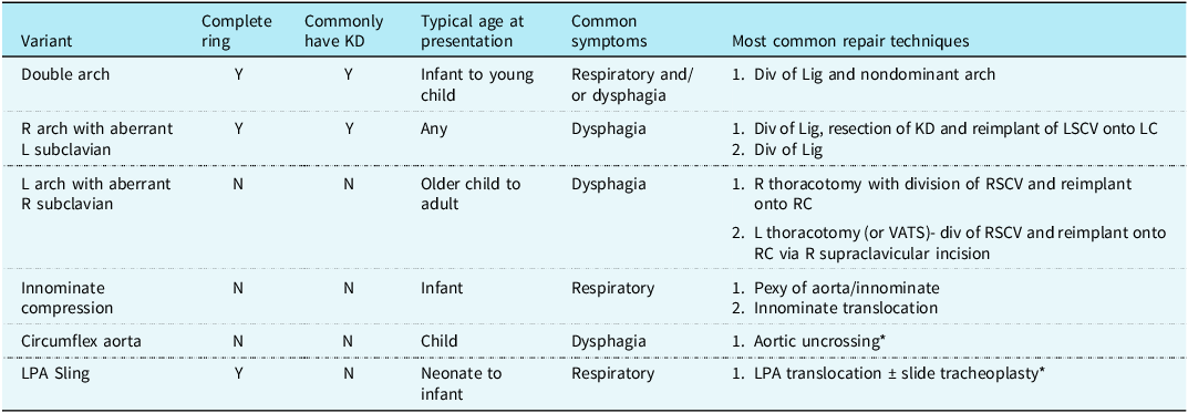
(a) a CT scan with 3D reconstructions oriented posteriorly, demonstrates a right-sided aortic arch with an aberrant left subclavian and a Kommerell’s diverticulum (b) a CT scan 3D reconstructions demonstrating a double aortic arch with an atretic distal left aortic segment with a Kommerell’s diverticulum. (c) the esophagram demonstrates the posterior compression caused by the Kommerell’s diverticulum (marked with a white arrow) of the aberrant subclavian artery. AA = aortic arch; KD = Kommerell’s diverticulum; DA = descending aorta.

Esophagogastroduodenoscopy and Advanced Esophageal Studies: An sophagogastroduodenoscopy may demonstrate pulsatile compression, though this can be subtle and easily missed. Reference Levitt and Richter7,Reference Kemper, Teplitzky, Brown, Mitchell and Shah8 Advanced testing including manometry have been reported, Reference Wellington, Kim, Castell and Xie9 though many demonstrate non-specific abnormalities. Reference Janssen, Baggen and Veen10 Some may consider a functional luminal imaging probe device, which assesses oesophageal dimensions, may demonstrate fixed compression and potentially even pulsatile compression. Reference Becker, Siddique and Nestler11 Importantly, these investigations allow for a before/after comparison since some patients, particularly those with history of prolonged compression, may continue to have symptoms despite surgery that was technically successful.
Echocardiogram: An echocardiogram may be notable for a right-sided aortic arch and the branching pattern may be delineated. Similar to the above, as this would usually be obtained for other reasons, this can lead to the diagnosis of a vascular ring in an otherwise asymptomatic patient. Workup should proceed if any symptoms consistent with a vascular ring are present. However, in the absence of symptoms, no additional workup is necessary.
Cross-sectional imaging: Though the workup for many is somewhat varied, ultimately a CT scan should be performed to confirm and characterise the anatomy. Cross-sectional imaging (CT or MRI) fully delineates the vascular anatomy and can provide clues as to whether there is any compression (e.g. gas within a dilated proximal oesophagus with distal decompression or obvious tracheal narrowing).
While the most common symptom-causing variants involve a right arch (with aberrant left subclavian or double aortic arch), there are less-common variants (i.e. right arch with mirror image branching but retroesophageal ligamentum), or variants that less commonly cause symptoms (e.g. left aortic arch with aberrant right subclavian) that warrant identification. Thus, in the presence of symptoms consistent with compression, CT scan should be considered even other studies suggest absence of a vascular ring.
Genetic Evaluation and Other tests: A right-sided aortic arch, demonstrated by any modality, should prompt consideration for evaluation for DiGeorge syndrome. Numerous other tests may have already been performed, such as a pulmonary function test, pH-impedance probe, or others, which may provide additional information. In most instances, however, these tests are not necessary and would only be indicated for patient-specific reasons.
Surgical indications
Since long-term follow-up studies on children who have undergone or not undergone surgery are lacking, management strategies vary by institution, often depending on institutional experience. Reference Porcaro, Ciliberti and Petreschi5 The clearest indication for surgical repair is symptoms with known anatomy consistent with a vascular ring. However, since these symptoms are otherwise commonly encountered in childhood, verification that they are caused by aberrant anatomy may be challenging. This is especially true in those diagnosed prior to the development of symptoms. Fortunately, as the surgical risks are low and the outcomes good, it may still be reasonable to offer repair with a clear explanation of the procedural risk as well as the anticipated likelihood of symptomatic improvement when a definitive conclusion cannot be reached. A multidisciplinary team approach is valuable in these sometimes challenging cases . Reference Chiu, Zendejas and Baird12–Reference Petreschi, Coretti and Porcaro14
Diagnosis in the asymptomatic—the changing landscape
Recently, there has been a substantial increase in the frequency of diagnosis in asymptomatic patients and on fetal echocardiogram. Reference Evans, Acherman and Ciccolo1,Reference Evans, Acherman, Ciccolo, Berthoty, Mayman and Restrepo15 This diagnosis can prime the parents and practitioners with a possible anatomic basis for any symptoms which may potentially develop. This has led to a younger age at diagnosis and repair. Reference Evans, Acherman and Ciccolo1,Reference Stephens, Eltayeb and Kennedy16,Reference Young, Hornberger and Haberer17 Conversely, knowledge of abnormal anatomy may result in stress to the family or even result in an operation in an otherwise normally developing child due to hypervigilance. To this point, one study noted an increased incidence of right aortic arch with aberrant left subclavian compared to other variants, concluding that this subset may be more common than previously identified, and thus, more likely to be asymptomatic and not require repair. Reference Savla and Weinberg18 Decision making is further complicated by the fact that symptoms do not necessarily correlate with the apparent severity of compression on diagnostic studies. Consultation with appropriate expertise can be helpful when the indication for surgical intervention is less clear.
In the incidentally discovered right-sided aortic arch, the patient and family should be questioned regarding symptoms. If symptoms cannot be elucidated no further diagnostics may be warranted and simply empowering the family with knowledge is sufficient. The same is true of a toddler with a prenatally diagnosed right arch. If symptoms are elucidated, further work up should be pursued to determine if the symptoms correlate with tracheal or oesophageal compression.
For the incidentally diagnosed, reassurance is often the most important thing that can be provided. Even if symptoms are uncovered, or symptoms develop later, the family should be assured that these are not rapidly progressive or emergent; reassurance and elective referral is appropriate and reasonable. This is especially true for families with limited access to medical care.
In the completely asymptomatic, the decision to repair remains unclear. Currently, most clinicians suggest that patients without symptoms should not undergo repair. However, some argue that waiting until symptoms develop allows for continued compression, and therefore, one may not reasonably expect complete symptom resolution following repair. Reference Dodge-Khatami19 Others suggest that diagnosis alone of anatomic variants at a perceived higher risk of compression (e.g. double aortic arch) is sufficient for repair, regardless of symptoms. In addition to surgical repair being less challenging in younger patients due to their size and more pliable vasculature, early evidence seems to suggest that it may result in more effective symptom resolution, Reference Aly, Papneja, Mawad, Seed, Jaeggi and Yoo20–Reference Callahan, Merritt, Canter, Eghtesady, Manning and Abarbanell23 thus the push for earlier repair. Finally, it is important to recognise that repair in adults is more complex and a higher-risk procedure. As incidental and fetal diagnosis become more common, the actual incidence and natural course of these anatomic variants can be better delineated.
In a prenatally diagnosed right-sided aortic arch, a neonatal echocardiogram should be considered. This may help delineate the anatomy, additionally, it can rule out any CHD as there is an association of right aortic arches with CHD. Reference Tawfik, Sobh, Ashamallah and Batouty24 If any respiratory symptoms are present, early airway evaluation should be obtained.
Kommerell’s diverticulum
A Kommerell’s diverticulum is an outpouching of the aorta/proximal aberrant subclavian artery. Though the natural history of a Kommerell’s diverticulum is incompletely understood, it has been shown to increase in size in some patients with evidence of cystic medial necrosis resulting in aneurysm formation or aortic dissection. Reference Luciano, Mitchell, Fraisse, Lepidi, Kreitmann and Ovaert25,Reference Kim, Cambria and Isselbacher26 Rupture seems to be quite rare, though it has been reported. Reference Erben, Brownstein and Velasquez27 Additionally, if a Kommerell’s diverticulum is not resected at the time of vascular ring repair, it can cause a mass effect and result in continued symptoms. In children, resection of the Kommerell’s diverticulum adds little risk; conversely, intervention in an adult may require cardiopulmonary bypass and potentially circulatory arrest. Reference Kouchoukos and Masetti28 For these reasons, many advocate for resection at the time of vascular ring repair. As the natural history becomes better understood, it may have implications for treatment in even asymptomatic patients.
Surgical intervention
The goal of surgery is alleviation of symptoms and prevention of permanent airway or oesophageal dysfunction, achieved through the relief of the compression. Fortunately, the current outcomes are excellent with low morbidity with a high rate of symptom resolution and freedom from reoperation. Reference Binsalamah, Ibarra and John3,Reference Swarnkar, Speggiorin and Austin21,Reference Kim, Cambria and Isselbacher26,Reference Said, Marey and Knutson29–Reference Gikandi, Chiu and Crilley31
Regardless of the anatomy, the principle remains the same, relief of the compression, typically obtained through division of the ring (division of a ligamentum and division of a double aortic arch if present), ideally with resection of a Kommerell’s diverticulum. In children, most are approached via left thoracotomy without cardiopulmonary bypass. In some anatomic variants a right thoracotomy may be preferred. In adults, exposure through a thoracotomy is challenging, therefore, a supraclavicular incision may be necessary as well. Minimally invasive approaches have been performed, involving only the division of nonpatent structures (ligamentum and/or atretic distal aortic arch) when vascular reconstruction is not necessary, but this approach remains less common. Reference Kogon, Forbess, Wulkan, Kirshbom and Kanter32,Reference Riggle, Rice-Townsend and Waldhausen33
Historically, despite a known Kommerell’s diverticulum, many treated vascular rings with division of the ligamentum alone. However this results in a not insignificant risk of recurrent symptoms due to persistent oesophageal compression by the Kommerell’s diverticulum. Reference Backer, Mongé, Russell, Popescu, Rastatter and Costello34 Given the effectiveness and low risk of Kommerell’s diverticulum resection, some advocate for resection at the initial operation, which necessitates reimplanting the aberrant subclavian artery onto the carotid artery. Resection also mitigates the potential risk associated with a Kommerell’s diverticulum in adulthood when repair would be more challenging. Alternatively, some reserve resection for recurrent symptoms after initial repair (division of a ligamentum alone), while others base the decision on the relative size and apparent compression of the Kommerell’s diverticulum.
For those with symptoms caused by a circumflex aorta (right-sided arch with early crossing to a left-sided descending aorta), a more complex operation (aortic uncrossing procedure or descending aortic translocation) may be necessary. Although some advocate for initial division of the ligamentum alone, reserving the more complex procedure only if symptoms persist. Reference Russell, Rastatter and Backer35,Reference Backer, Mongé, Wallen and Eltayeb36
Innominate artery compression syndrome is most often approached via sternotomy or upper mini-sternotomy. Through this approach, a pexy can be performed, which involves securing the vessel to the back of the sternum to elevate it off the trachea relieving the compression Reference Wine, Colman, Mehta, Maguire, Morell and Simons37 or it can be divided and repositioned on the aorta giving it a more gentle path over the trachea. Reference Hawkins, Bailey, Clark and Ivey38 Simultaneous tracheopexy can be incorporated for associated tracheal or bronchial malacia.
Finally, a sling created by a left pulmonary artery originating from the mid-point of the right pulmonary artery (LPA sling) is approached via sternotomy, often necessitating cardiopulmonary bypass. This lesion carries a high incidence of concomitant complete tracheal rings, which must be investigated prior to repair of the sling, as these should be repaired simultaneously, Reference Backer, Russell, Kaushal, Rastatter, Rigsby and Holinger39 though some have advocated for staging, reserving airway intervention for older infants and children.
Outcomes
Outcomes after vascular ring repair are excellent with a low risk of complications or need for reintervention. Reference Yu, Guo and You30 There is a general trend toward symptom improvement in patients requiring surgery. Reference Petreschi, Coretti and Porcaro14,Reference Berry, Padilla and Sorabella40 Symptomatic improvement often begins during the hospitalisation, quieter breathing or being able to more comfortably eat foods that were previously difficult, though full recovery and symptomatic improvement may take weeks. Hospital stay typically lasts from 2 to 5 days and is often dictated by pain control due to the thoracotomy, though use of epidurals can dramatically impact this. Post discharge follow-up is minimal and the patients usually recover well. Paediatrician visits should include an assessment of the incision (thoracotomy incisions can have poor healing) and discussion around the improvement in symptoms and monitoring for any postoperative complications. Importantly, long-term follow-up is essential not only for symptomatic patients who have undergone or not undergone surgery but also for asymptomatic patients, as symptoms may develop over time.
Complications
Vascular ring repairs are performed with low complication rates. As repair may require temporary occlusion of the carotid/subclavian arteries there is a risk of stroke. Fortunately, this is exceedingly rare, however, for that reason some surgeons may prefer a head CT to evaluate the polygon of Willis prior to proceeding with repair. Common complications include chylothorax (5–10%), resulting from an injury to the lymphatics, Reference Binsalamah, Ibarra and John3,Reference Gikandi, Chiu and Crilley31 prolonging the hospitalisation. A chylothorax is more common in older children and usually resolves with restrictions in fat intake and diuretic therapy; rarely, they may require catheter based lymphatic embolisation or a repeat surgical intervention for thoracic duct ligation. Additional complications include vocal cord paresis secondary to recurrent laryngeal nerve injury, diaphragm paresis due to phrenic nerve injury or a Horner’s syndrome due to injury to the stellate ganglion. Reference Naimo, Fricke and Donald41 The majority resolve over time without long-term sequelae.
Risk of recurrent symptoms/need for reintervention
While need for reintervention is low, respiratory symptoms are more likely to be persistent, partially due to the difficulty differentiating respiratory symptoms caused by vascular compression and those inherent to the airway such as tracheomalacia or asthma. Reference Callahan, Merritt, Canter, Eghtesady, Manning and Abarbanell23,Reference Yu, Guo and You30,Reference Ceneri, Desai and Christopher42–Reference Farje, Young, Stein, Eltayeb, Ghadersohi and Hazkani44 Resolution of dysphagia seems to be better, with persistence of dysphagia being less common. Reference Callahan, Merritt, Canter, Eghtesady, Manning and Abarbanell23 However, assessment of symptom resolution and need for reintervention are complicated by many having an “improvement” in symptoms even without “resolution”. Further, care providers may consider the vascular ring “treated” (even in the setting of a less complete operation such as isolated division of the ligamentum) and not consider continued compression as an oetiology for recurrent/persistent symptoms. Thus, patients who may benefit from further intervention may be missed. These considerations are part of the rationale for surgeons preferring a complete repair at the initial operation. Finally, recent data suggest that symptomatic improvement may be increased with earlier repair, which may ultimately result in more pursuing a more complete repair at an earlier age. Reference Swarnkar, Speggiorin and Austin21
Unfortunately, there are instances where the symptoms that resulted in the diagnosis and subsequent treatment of a vascular ring were not actually the result of the aberrant anatomy. Thus, after a complete repair, there is no resolution of symptoms. All attempts should be made to minimise this with any additional preoperative testing and strong consideration should be given for preoperative evaluation by otolaryntology or gastrointestinal subspecialists. If additional concerns are discovered during the preoperative evaluation, this may allow for surgical strategies to address these as well, particularly tracheobronchomalacia. Reference Farje, Young, Stein, Eltayeb, Ghadersohi and Hazkani44
Treatment for persistent/recurrent symptoms
For those who do not experience symptomatic improvement, or have improvement with subsequent recurrent symptoms, repeating the diagnostic workup is necessary. Symptoms may persist due to a residual or untreated Kommerell’s diverticulum, fibrotic bands that were not released at surgery or as a result of post-surgical adhesions, compression by the descending aorta, or trachea/bronchomalacia. Reference Backer, Mongé, Russell, Popescu, Rastatter and Costello34 Some of these may be ameliorated by further surgical intervention. Reference Labuz, Kamran, Jennings and Baird45 Cross-sectional imaging delineates the anatomy, but bronchoscopy, esophagram, and EGD may also be necessary. Operations to help address other oetiologies may include resection of a Kommerell’s diverticulum, lysis of residual fibrotic tissue or newly developed scar tissue, pexy of the ascending or descending aorta, rotational oesophagoplasty, tracheopexy, or the aortic uncrossing or descending aortic translocation procedures. Reference Labuz, Kamran, Jennings and Baird45,Reference Hammond-Jack, Ramakrishnan and Nath46 As these patients have different anatomic variants and have undergone different repairs by different surgeons, they represent a heterogeneous patient population necessitating an individualised approach. Although index repair of a vascular ring is often straightforward, patients with persistent or recurrent symptoms should prompt consideration for referral to a high-volume centre utilising a multidisciplinary approach.
Future directions
Currently, the decision on when to offer surgical repair differs from institution to institution and even practitioner to practitioner. There has been a push towards early referral for repair as the operation is easier and the results likely superior in younger children. Additionally, as the surgical procedure has been demonstrated to be effective and low risk, some advocate for repair any time a vascular ring is identified, even in asymptomatic patients.
The diagnosis and treatment of vascular rings is undergoing a paradigm shift. The increased frequency of diagnosis in asymptomatic patients (particularly fetal) has begun to reshape the landscape. Paediatricians are now primed with the pre-existing knowledge of an anatomic variation that could cause airway or oesophageal symptoms, enabling earlier diagnosis and treatment with improved outcomes. Conversely, parental hypervigilance and anxiety may lead to unnecessary operations in asymptomatic, normally developing children. Importantly, early identification in asymptomatic patients provides an opportunity to study the natural history of these variants and the utility of “expectant management”. Ongoing efforts to follow asymptomatic patients diagnosed incidentally aim to refine management strategies, optimise timing for intervention when indicated, and improve long-term outcomes. Establishing a registry for these patients would enhance our understanding and guide future care.
Financial support
None.
Competing interests
All authors report no relevant competing interests to declare.


 Open access