


Impact Statement
Small interfering RNA (siRNA) therapies hold enormous promise for treating diseases such as cancer. However, delivering siRNA effectively to cells of interest remains a major obstacle. Several nanocarrier systems have been developed to protect siRNA from nuclease degradation and facilitate siRNA transport across the impermeable cellular membrane. However, these delivery platforms predominantly utilize endocytic pathways, limiting cytosolic access, with delivery efficiencies typically below 10%. Furthermore, delivery vehicles encapsulated by siRNA often experience nonspecific interactions in the systemic environment, which contribute to macrophage uptake and reduce the effective delivery of siRNA to the target site. We developed a hyaluronic acid (HA) coated polymeric nanocarrier that packages and protects siRNA while evading immune cells more effectively. The core of the nanocarrier is a guanidinium-functionalized cationic polymer (PONI-Guan) that encapsulates siRNA and facilitates cytosolic access to the cells of interest. The outer HA layer acts as a non-interacting shield that reduces nonspecific binding and reduces macrophage uptake. These HA-coated nanocarriers effectively delivered siRNA to aggressive triple-negative breast cancer cells in vitro with four-fold greater efficiency than to macrophages, demonstrating cellular selectivity. These carriers mediated substantial silencing of STAT-3 gene expression, a key cancer-promoting gene. In vivo studies revealed that HA coating reduced accumulation of the nanocarriers in clearance organs by two-fold, relative to the ‘naked’ PONI-Guan polyplexes, demonstrating improved circulation and reduced immune clearance. The biocompatible nature of HA along with the positively charged polymeric core can be adapted for different gene targets and can be scaled and customized for a range of diseases. With further advancements, HA-shielded nanocarriers could enable safer, more effective gene therapies, supporting global health goals focused on treating cancer and advancing precision medicine.
1. Introduction
Small interfering RNAs (siRNAs) present a promising approach for the treatment of a variety of diseases, including cancers, as siRNAs can target specific mRNA, silencing the corresponding protein expression with high specificity, and minimizing off-target effects [Reference Lee, Kim, Kwon and Roberts1,Reference Kalaimani, Balachandran and Boopathy2,Reference Zhu, Zhu, Wang and Jin3]. This capacity to selectively target the gene of interest has resulted in the development of several siRNA-based therapeutics, including FDA-approved therapeutics such as Patisiran and Givosiran, gaining prominence in clinical applications [Reference Ahn, Kang and Han4,Reference Guo, Li and Yu5].
Although siRNA-based therapies hold significant potential, delivery of cancer-targeting siRNA is challenging due to nuclease degradation, inefficient cellular uptake, and limited circulation time in the systemic environment [Reference Isazadeh, Oruji and Shabani6,Reference Wang, Shigdar and Shamaileh7,Reference Dastgerdi, Dastgerdi and Bayraktutan8]. Several nanocarrier systems have been developed using lipids [Reference El, Garbayo and Amundarain9], polymers [Reference Rehman, Parveen and Sheikh10,Reference Wang, Zhang, Lv and Cheng11], and inorganic materials [Reference Wang, Zhong, Li, Liu and Cheng12,Reference Lin, Revia and Zhang13] to protect siRNA from nuclease degradation and facilitate siRNA transport across the impermeable cellular membrane [Reference Ashique, Almohaywi and Haider14,Reference Yadav, Ali, Thanekar, Pogu and Rengan15,Reference Gao, Cheng and Santos16,Reference Moazzam, Zhang and Hussain17]. However, these delivery platforms predominantly utilize endocytic pathways, limiting cytosolic access, with delivery efficiencies typically below 10% [Reference Chatterjee, Kon, Sharma and Peer18,Reference Guo, Zhang and Huang19]. Further, most nanocarriers exhibit poor pharmacokinetic properties and limited tissue-specific localization. This limitation arises from the clearance of the majority of the administered siRNA from the bloodstream due to opsonization by the reticuloendothelial system (RES) [Reference Cheung and Shoichet20,Reference Xiao, Zoulikha and Qiu21]. To increase therapeutic efficacy despite rapid clearance by the macrophages, there is a need to increase siRNA dosage, increasing the likelihood of off-target effects, thereby limiting siRNA-based therapies [Reference Friedrich and Aigner22,Reference Ranjbar, Zhong, Manautou and Lu23].
Recently, we have demonstrated that poly(oxanorborneneimide) (PONI) polymers bearing cationic guanidinium head groups of varying chain lengths can effectively deliver siRNA to the cytosol of macrophages in vitro and in LPS-induced mice models [Reference Goswami, Nagaraj and Cicek24]. Although siRNA delivery to macrophages holds significant therapeutic promise for several diseases, it also highlights a key challenge: the limited ability of current systems to deliver siRNA to non-phagocytic cells, which is essential for addressing a broader spectrum of disease targets, such as cancer [Reference Zaidi, Fatima and Zaidi25].
To address these challenges, surface modifications with hydrophilic agents, including naturally occurring anionic polysaccharide hyaluronic acid (HA), have been explored [Reference Kotla, Mohd and Larrañaga26,Reference Bokatyi, Dubashynskaya and Skorik27,Reference Van Le and Le Cerf28]. HA has been widely used in biomedical applications, including nanoparticle design, due to its high biocompatibility, biodegradability, and reduced immunogenic response [Reference Masjedi, Ahmadi and Atyabi29,Reference Andretto, Repellin and Pujol30,Reference Zhao, He and Gao31,Reference Dillinger, Guter and Froemel32,Reference Cai, Zhang and Wei33]. HA-coated nanovectors have been utilized to provide a protective shield against protein corona formation in the bloodstream and efficiently evade recognition by the RES, thereby increasing blood circulation time for effective tumor localization [Reference Kotla, Mohd and Larrañaga26,Reference Vetter, Yazdi and Gialdini34]. Despite the considerable potential of HA, the delivery capabilities of HA-coated polyplexes remain largely underexplored [Reference Rohtagi, Garg and Triveni35,Reference Fu, Cai and Chen36].
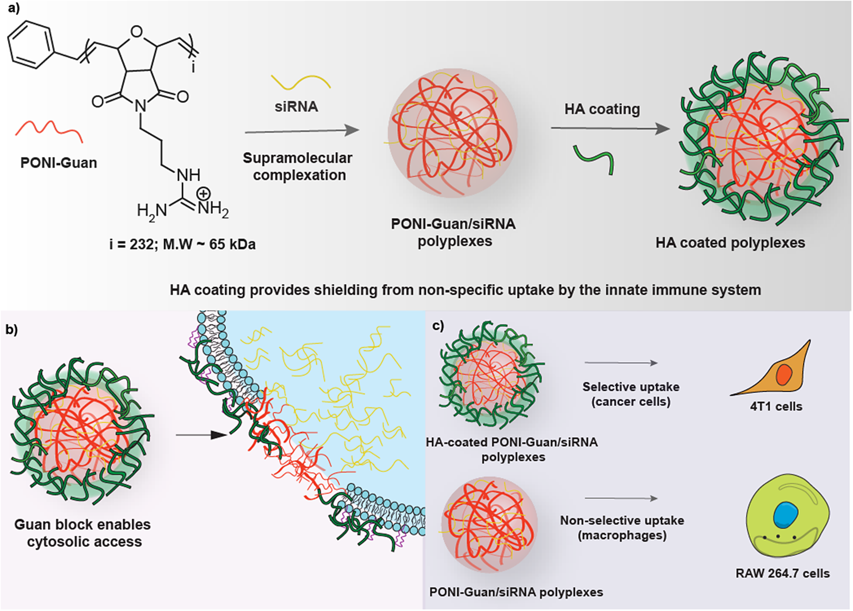
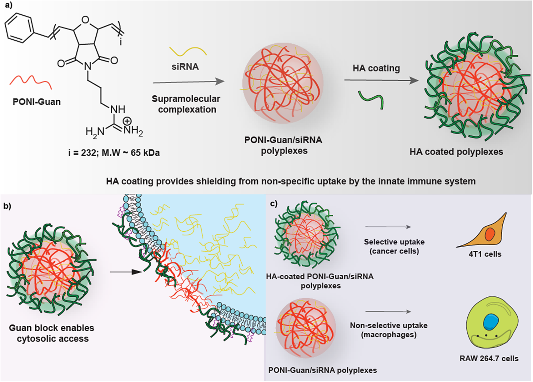
In the present study, we hypothesized that a more efficient delivery to cancer cells could be achieved using HA-coated polyplexes. We utilized PONI-Guan homopolymers to self-assemble with negatively charged siRNA and coated these resulting polyplexes with HA in varying ratios to form HA-coated-PONI-Guan/siRNA (HA-PONI-Guan/siRNA) polyplexes (Fig. 1). These polyplexes demonstrated efficient cytosolic delivery to murine mammary cancer (4T1) cells with a ≈4:1 selectivity relative to RAW 264.7 macrophages. Furthermore, the delivery of STAT3-targeting siRNA to 4T1 cells resulted in efficient gene knockdown (~50%), comparable to that achieved with PONI-Guan homopolymers. This indicates the selective uptake by 4T1 cells while maintaining therapeutic efficacy after the HA coating on the polyplexes. When intravenously administered to 4T1 tumor-bearing mice, HA-PONI-Guan/siRNA polyplexes showed a ~2-fold reduction in accumulation within clearance organs, liver and kidney, compared to the control PONI-Guan polyplexes. These findings collectively demonstrate the potential of HA-coated polyplexes for siRNA delivery to the target cells while avoiding rapid clearance due to macrophage uptake as well as reduced biodistribution to clearance organs, thereby minimizing the potential off-target effects.
Schematic of the study design. PONI-Guan polymers self-assemble with siRNA to form PONI-Guan/siRNA polyplexes. Coating PONI-Guan/siRNA polyplexes with HA leads to highly selective siRNA delivery to cancer cells. HA coating provides (a) a protective shield that prevents RES clearance and (b and c) increased uptake in cancer cells.
2. Methods
2.1. Synthesis of Polymer and Characterization
PONI-Guan homopolymer (~65 kDa) featuring guanidinium groups was synthesized by ring-opening metathesis polymerization (ROMP) as previously described [Reference Lee, Luther and Goswami37,Reference Jeon, Luther and Goswami38]. Briefly, a guanidinium-functionalized PONI-based monomer was synthesized via a four-step synthetic route (1–4) (Scheme S1). The synthesized monomer (4) was dissolved in 1.2 mL dichloromethane (DCM) and transferred to a Schlenk flask. A stir bar was added to a separate Schlenk flask containing Grubbs catalyst (third generation) dissolved in 1 mL of DCM. Afterwards, both the flasks were degassed by multiple freeze–thaw cycles. The monomer was added to the catalyst and stirred for 30 min, under an inert atmosphere. A rapid change in color from greenish to yellowish was observed upon adding the monomer to the catalyst. Ethyl vinyl ether (200 μL) was added at the end to the same flask and stirred for another 15 min to stop the polymerization. The obtained polymer was concentrated, precipitated in a copious amount of hexane, filtered, and washed to afford Guan-Boc (5) polymer with 95% yield. The tert-butyloxycarbonyl (Boc) functionality from Guan–Boc was removed by overnight stirring with a 1:1 mixture of DCM and trifluoroacetic acid (TFA). The resulting solution was evaporated under reduced pressure using DCM to remove excess TFA, affording PONI-Guan (Guan (6)). The obtained polymer was dissolved in Milli-Q water and dialyzed for 2–3 days using Biotech CE dialysis tubing with a 10 kDa molecular weight cut-off (MWCO). Following dialysis, the solution was filtered through a 0.45 μm polyethersulfone (PES) filter and lyophilized to yield a colorless powder. The polymer was characterized via 1H-NMR (nuclear magnetic resonance spectroscopy) for chemical composition and DMF-GPC (gel permeation chromatography), calibrated against poly(methyl methacrylate) (PMMA), for molecular weight distribution.
2.2. Polyplex Preparation and Characterization
PONI-Guan homopolymers (75 μM stock) and siRNA (50 nM, unless otherwise stated) were mixed and incubated for 10 min to form PONI-Guan/siRNA polyplexes. Low molecular weight HA (15–40 kDa) was added to the prepared polyplexes at varying ratios of HA:polymer (1:1, 1:1.25, 1:1.5, 1:1.75, and 1:2), incubated for 5 min to form HA-coated-PONI-Guan/siRNA polyplexes. A concentration of 50 nM siRNA was used for all experiments, unless otherwise stated, and the G/P ratio was kept constant at a ratio of 40 with polymer concentration at 360 nM, based on previous studies [Reference Goswami, Nagaraj and Cicek24]. The polyplex solution was then made up to volume either with Dulbecco’s Phosphate-Buffered Saline (DPBS) for characterization studies or with culture medium containing 10% FBS and 1% antibiotic solution for in vitro experiments.
2.2.1. Size, Zeta Potential, and TEM
Dynamic light scattering (DLS, Malvern Instruments) was used to measure the size and zeta potential of the prepared polyplexes. Transmission electron microscopy (TEM) was used to assess the morphology and size of the polyplexes. Briefly, polyplexes prepared in DPBS were drop-cast onto the TEM grid (300-mesh copper), allowed to air dry for 24 h, and imaged using a Tecnai-T12 instrument.
2.2.2. siRNA Encapsulation Studies Using Agarose Gel Electrophoresis and Ribogreen Assay
Agarose gel electrophoresis was used to evaluate the complexation of siRNA with the polymer. Briefly, HA-coated polyplexes at varying ratios were prepared, resuspended in DPBS, and mixed with gel loading dye (Thermo Scientific, R0611). The solution was loaded into the wells of 0.8% agarose gel containing ethidium bromide (Invitrogen, 15585011), allowed to run for about 30 min at 100 V, 80 mA settings, and imaged using a gel imager.
Heparin displacement assays were performed using Quant-iT Ribogreen RNA Assay Kit (Invitrogen, R11490) to evaluate the siRNA encapsulation efficiency. Measurements were taken every hour after adding the Ribogreen reagent, following the manufacturer’s protocol. This assay measures free siRNA content in each of the wells, which can be used to calculate encapsulation efficiency using the following formula:
2.3. Evaluation of Cellular Uptake by Flow Cytometry
RAW 264.7 and 4T1 cells were seeded in 24-well plates at a seeding density of 30 × 103 cells per well. Twenty-four hours after seeding, cells were treated with HA-coated-PONI-Guan/AF488-siRNA polyplexes (25 nM) for 6 h, after which the culture media were replaced, and the cells were incubated for an additional 24 h. To evaluate the mechanism of uptake, cells were pretreated with small-molecule inhibitors: chlorpromazine (1.5 μg mL−1), imipramine (3 μg mL−1), nystatin (50 μg mL−1), or methyl-β-cyclodextrin (7.5 μg mL−1) for 4 h at 37°C, after which cells were treated with the polyplexes and incubated for an additional 24 h. Cells were washed, trypsinized, spun down to pellets, and resuspended in DPBS for flow cytometry analysis on the LSRFortessa 5 Laser instrument. At least 10,000 events were acquired for each sample according to the instrumentation protocol. Data were collected for three biological and three technical replicates per biological replicate.
2.4. Evaluation of Fluorescence Knockdown in Reporter Cell Lines
GFP-expressing HEK 293T (deGFP HEK 293T) cells were seeded in 24-well plates at a seeding density of 20 × 103 cells per well, 24 h before the experiment. Cells were treated with HA-coated-PONI-Guan/si_GFP (50 nM) polyplexes, media replaced after 6 h, and allowed to incubate for about 36 h. Cells were harvested at the end of the experiment for flow analysis as previously described.
2.5. Cellular Growth Inhibition Studies
4T1 and RAW 264.7 cells were seeded in 96-well plates at 5 × 10³ cells per well. On the day of the experiment, cells were treated with HA-coated-PONI-Guan/si_STAT3 (50 nM) polyplexes, media replaced after 6 h, and allowed to incubate for about 36 h. At the end of the experiment, Alamar blue assay was performed to measure cell viability.
2.6. STAT3 Knockdown and Analysis Using Quantitative Real-Time Reverse Transcription PCR
4T1 and RAW 264.7 cells were seeded in 6-well plates at a seeding density of 150 × 103 cells per well. On the day of the experiment, cells were treated with HA-coated-PONI-Guan/si_STAT3 (50 nM) polyplexes, media replaced after 6 h, and further incubated for 36 h. Cells were harvested using Trizol reagent and the extraction of RNA was performed using Pure Link RNA Kit. Following RNA conversion to cDNA, qRT-PCR was performed using iTag Universal SYBR Green Supermix on a CFX Connect Real-Time System. β-actin expression levels were used to normalize STAT3 mRNA expression.
2.7. In Vivo Studies
2.7.1. Animal Care
Animal studies were performed under Protocol Number 5965 following the guidelines of the Institutional Animal Care and Use Committee (IACUC) at the University of Massachusetts, Amherst. Five-week-old female BALB/c mice were purchased from the Jackson Laboratory and used for the biodistribution studies. Prior to performing any procedure, all mice were allowed to acclimate for 1 week in the animal facility. Tumor transplantation was performed by injecting 4T1 cells of density 1 × 10⁵ cells into the second mammary fat pad of the BALB/c mice. The mice were monitored, and bodyweight measurements were taken every day. Vernier calipers were used to measure tumor volume (mm3) using the formula: (long diameter × short diameter2)/2.
2.7.2. Biodistribution of HA-Coated Polyplexes
HA-coated-PONI-Guan/Cy5-siRNA polyplexes were intravenously injected in the 4T1 tumor-bearing mice, once the tumor volume reached about 100 mm3. Mice were euthanized 6 h from the time of injection, tumors and major organs were collected, and imaged using the IVIS imaging system. Living image software was used to subtract the background fluorescence and calculate average radiant efficiency.
2.7.3. Analysis of Hepatic Function
Blood was collected to assess the liver health of the mice. Serum was separated from the blood samples in serum separator tubes (BD Microtainer Capillary Blood Collector, BD 365967) as per the manufacturer’s protocol. Serum alanine aminotransferase (ALT) levels were measured using a Teco Diagnostics kit (A524150).
3. Results and Discussion
3.1. Characterization of HA-PONI-Guan/siRNA Polyplexes
PONI homopolymer (~65 kDa) featuring guanidinium groups was synthesized (Scheme S1) by ring-opening metathesis polymerization (ROMP) and characterized (Supplementary Figs. S1–S3) as previously described [Reference Lee, Luther and Goswami37,Reference Jeon, Luther and Goswami38]. PONI-Guan polymers and siRNA self-assembled via a simple co-incubation process to yield PONI-Guan/siRNA polyplexes, mediated by electrostatic interactions (details mentioned in the methods section). The guanidinium to phosphate (G/P) ratios for the polyplexes were kept constant at G/P 40, as previously reported [Reference Friedrich and Aigner22]. These polyplexes were then coated with low molecular weight HA at varying ratios of HA to polymers (1:1, 1:1.25, 1:1.5, 1:1.75, and 1:2) to generate HA-coated PONI-Guan/siRNA polyplexes, details of which are described in the experimental methods section. The complexation of siRNA with PONI-Guan polymers in HA-coated polyplexes was evaluated using gel mobility shift assays, demonstrating complexation with siRNA (Supplementary Fig. S4).
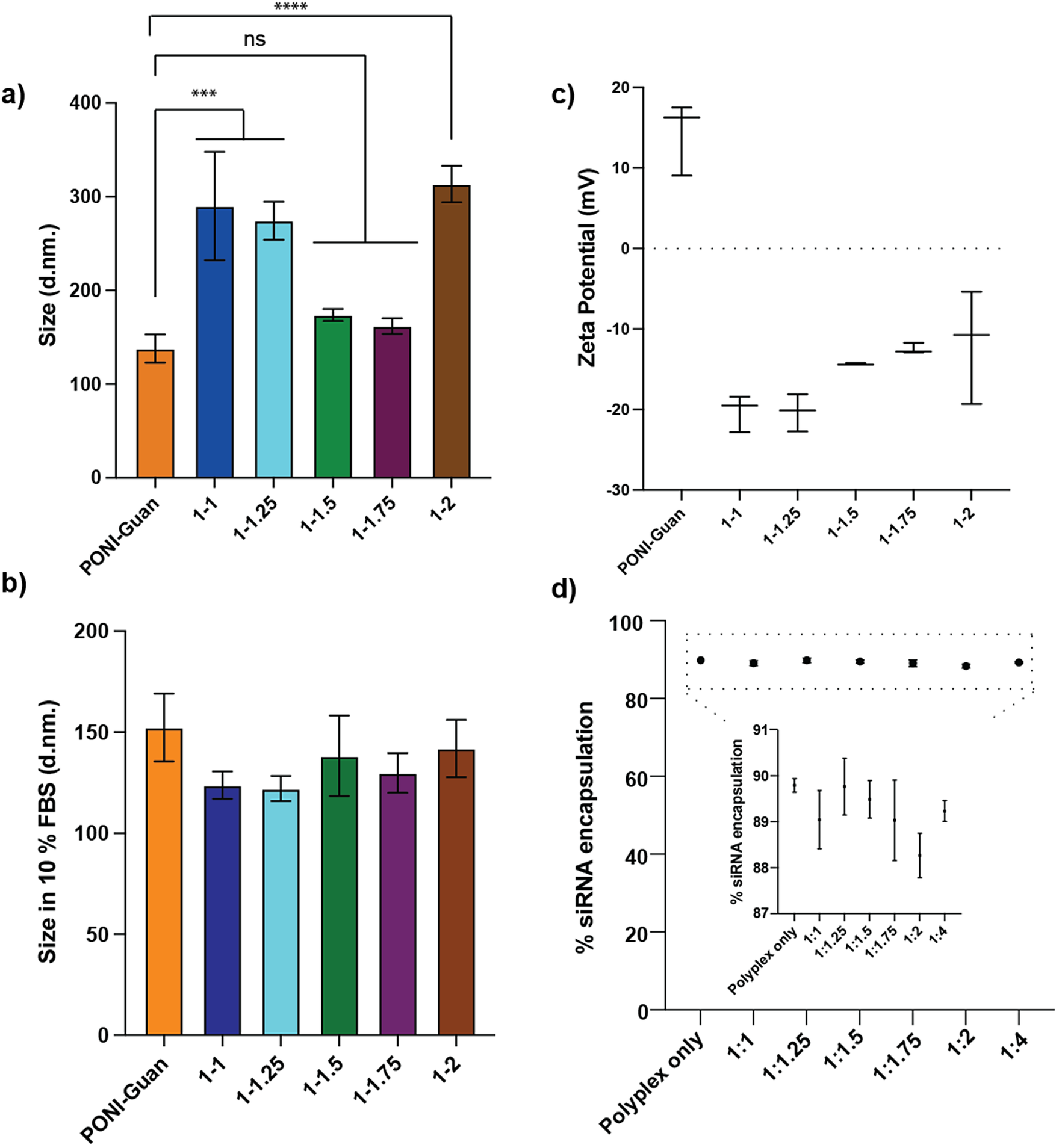
PONI-Guan/siRNA polyplexes exhibited ~130 nm sizes, while coating polyplexes with HA showed an increase in size (~300 nm) at ratios 1:1 and 1:1.25, similar sizes at 1:1.5 and 1:1.75, and an increase in size (~300 nm) at 1:2 (Fig. 2a). The observed increase in sizes can be attributed to the partial binding of HA around the polyplexes, where native HA largely remains in the coiled confirmation, leaving other binding sites accessible for interactions with neighboring polyplexes, resulting in aggregation [Reference Martens, Remaut and Deschout39]. Size measurements in the presence of fetal bovine serum (10% FBS) yielded similar sizes for all the polyplexes with and without HA (Fig. 2b). Transmission electron microscopy (TEM) analysis of HA-coated polyplexes further confirmed sizes as well as core–shell-like morphology, consistent with successful HA encapsulation around the polyplexes (Supplementary Fig. S5). PONI-Guan/siRNA polyplexes show a surface charge of ~+14 mV. Zeta potential analysis demonstrated that HA-coated polyplexes retained a negative surface charge (from −20 to −12 mV) across decreasing HA concentrations relative to PONI-Guan/siRNA polyplexes (1:1, 1:1.25, 1:1.5, 1:1.75, 1:2) (Fig. 2c), supporting the continued presence of HA on the polyplex surface.
Characterization of HA-PONI-Guan/siRNA polyplexes. (a) Sizes of HA-coated PONI-Guan/siRNA polyplexes (50 nM siRNA) at varying HA to polymer ratios in (a) DPBS. (b) 10% FBS. (c) Zeta potential of HA-PONI-Guan/siRNA polyplexes (50 nM siRNA) at varying HA to polymer ratios in 10 mM NaCl. (d) siRNA encapsulation efficiency in polyplexes at varied HA concentrations with PONI-Guan polyplex as a control. Standard deviation plotted from three experimental replicates. ***p < 0.0001, corresponding to a 95% confidence interval (CI).
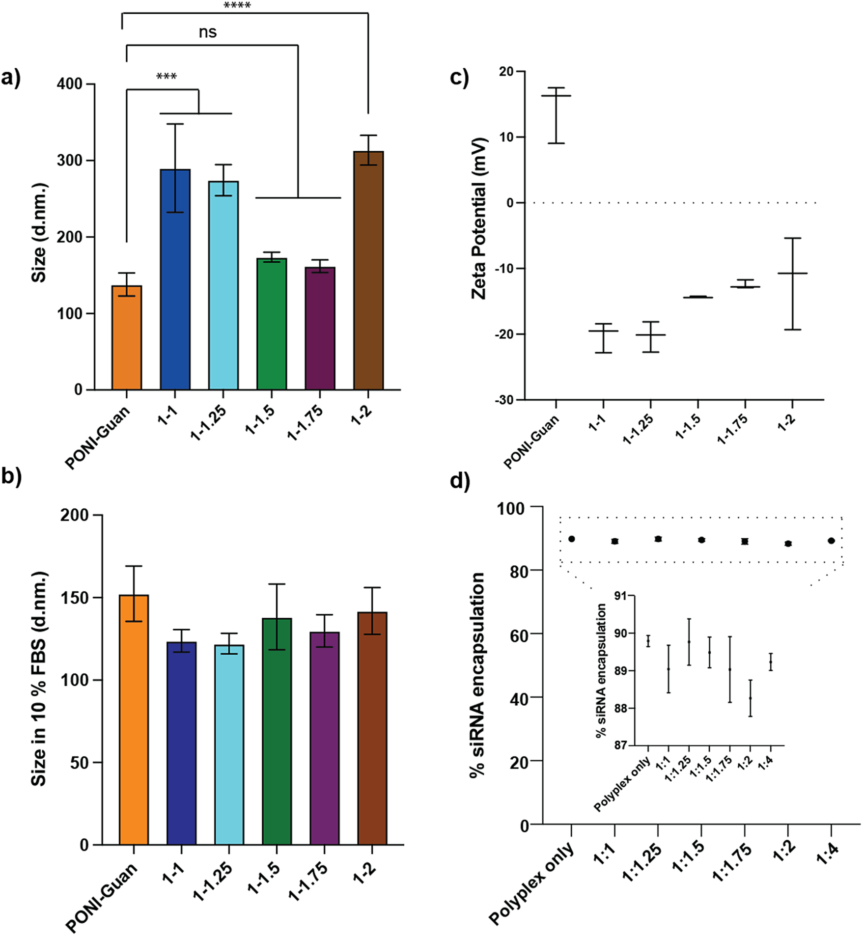
Encapsulation efficiency of siRNA was evaluated using Ribogreen assay, with all polyplexes demonstrating >85% encapsulation of siRNA (Fig. 2d).
3.2. Selective siRNA Delivery to Tumor Cells by HA-Coated Polyplexes and Evaluation of Uptake Mechanism
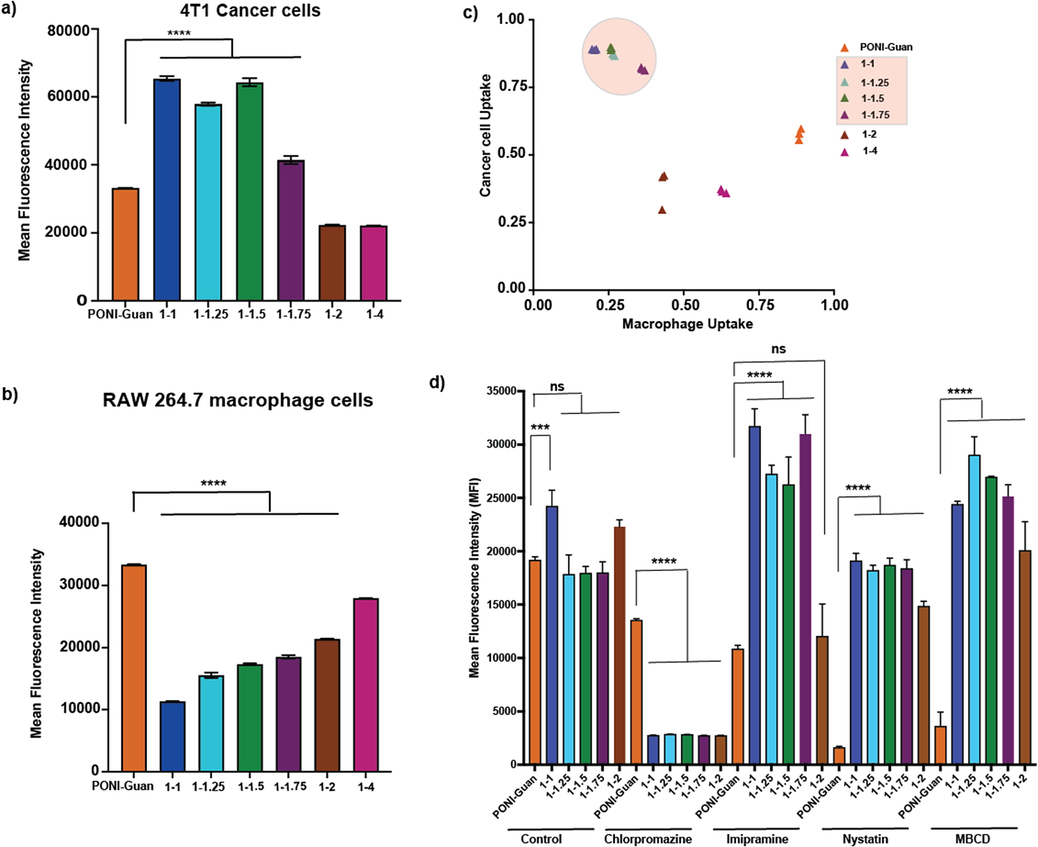
Small interfering RNA (siRNA) delivery to the cytosol is necessary for activity [Reference Hu, Zhong and Weng40,Reference Sajid, Moazzam, Kato, Yeseom Cho and Tiwari41]. Murine mammary carcinoma (4T1) cells and macrophages (RAW 264.7) were utilized to evaluate the delivery efficiency and selectivity. HA-coated polyplexes at varying HA to polymer ratios (1:1, 1:1.25, 1:1.5, 1:1.75, 1:2, and 1:4) were generated with AF488-siRNA for uptake studies. Both cell lines were incubated with HA-coated polyplexes for 24 h, after which cells were harvested to quantify siRNA uptake efficiency using flow cytometry. A series of gates was applied for analysis as shown in Supplementary Fig. S6 and mean fluorescence intensity was derived from FITC-A histograms (Fig. 3a and b) along with percentage cellular uptake (Supplementary Fig. S7). PONI-Guan polyplexes showed efficient cellular uptake in both 4T1 and RAW 264.7 cell lines, while HA-coated polyplexes at ratios 1:1, 1:1.25, 1:1.5, and 1:1.75 showed significantly higher uptake in 4T1 cells than in macrophages. Ratios 1:2 and 1:4 showed similar uptake to PONI-Guan polyplexes in 4T1 cells owing to the reduced concentrations of HA. HA-coated polyplex uptake efficiency was further evaluated across varying ratios by plotting percentage AF488-siRNA uptake in 4T1 cancer cells versus RAW 264.7 macrophages (Fig. 3c). This scatter plot demonstrated that HA-coated polyplexes at ratios 1:1 to 1:1.75 showed the most efficient cancer cell uptake and reduced macrophage uptake, with HA:polymer ratio 1:1 showing a ≈ 4:1 selectivity against macrophages.
Evaluation of cellular uptake by flow cytometry. (a) Quantification of HA-coated-PONI-Guan/AF-488 siRNA (25 nM siRNA) uptake in 4T1 cancer cells. (b) Quantification of HA-coated-PONI-Guan/AF-488 siRNA (25 nM siRNA) uptake in RAW 264.7 macrophages. (c) Scatter plot demonstrating maximal AF-488 siRNA uptake between 4T1 and RAW 264.7 cells using varying ratios of HA-PONI-Guan/siRNA polyplexes. (d) Evaluation of siRNA uptake after incubation with small-molecule inhibitors targeting endocytosis or cholesterol dependence using flow cytometry. Error bars represent the standard deviation of three experimental replicates; ***p < 0.0001, corresponding to a 95% confidence interval.
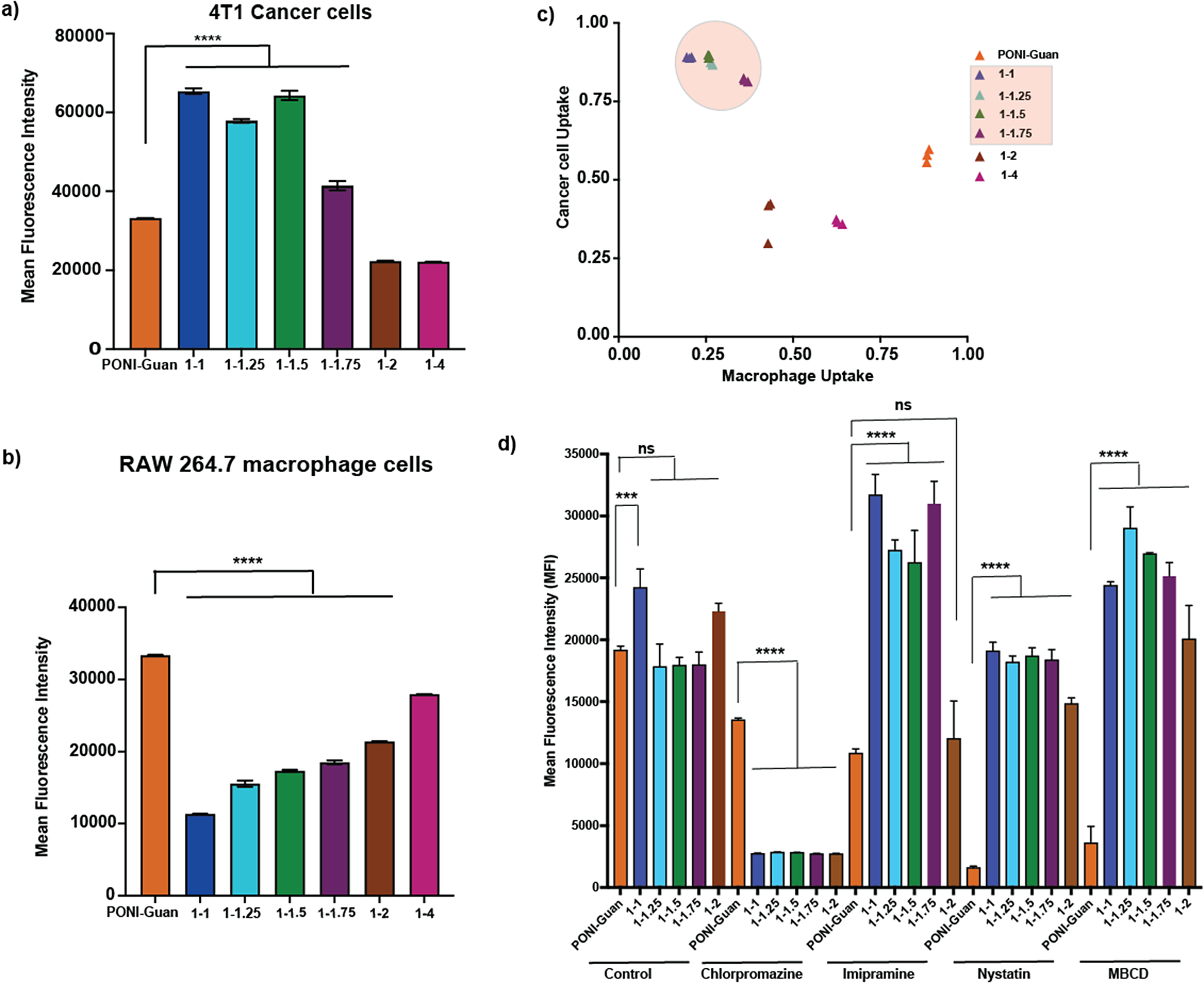
To investigate the mechanism of siRNA uptake, small-molecule inhibitors were used: nystatin and methyl-β-cyclodextrin (MBCD) to inhibit cholesterol-dependent pathways, and imipramine and chlorpromazine to inhibit micropinocytosis/clathrin-mediated endocytosis, respectively, in 4T1 cells and studied using flow cytometry (Fig. 3d, Supplementary Fig. S8). PONI-Guan/siRNA polyplexes were most affected in the presence of MBCD/nystatin, demonstrating cholesterol dependence, consistent with our previous studies [Reference Friedrich and Aigner22]. On the contrary, HA-PONI-Guan/siRNA polyplexes showed reduced uptake in the presence of chlorpromazine and nystatin, suggesting a complex mechanism of uptake involving multiple pathways.
3.3. Evaluation of Fluorescence Reporter Gene Silencing by HA-Coated Polyplexes
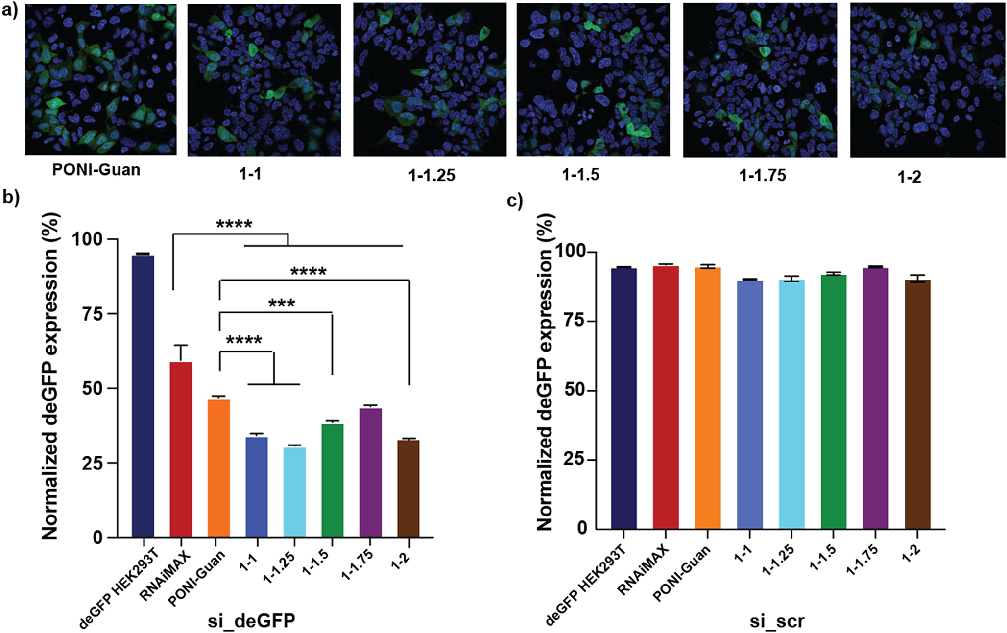
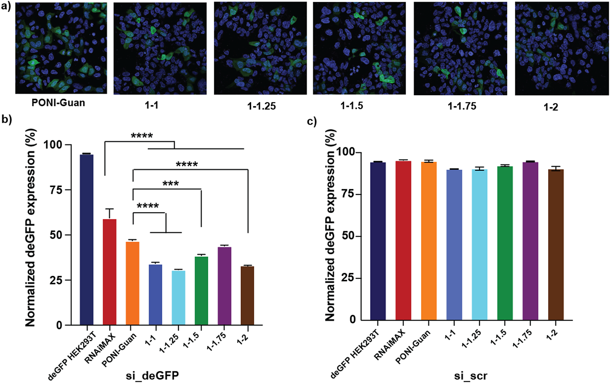
The knockdown efficacy of HA-PONI-Guan/siRNA polyplexes was further assessed in deGFP HEK 293T, a GFP-expressing reporter cell line. Confocal laser scanning microscopy showed that deGFP HEK 293T cells treated with HA-coated-PONI-Guan/si_deGFP polyplexes at varying ratios demonstrated efficient knockdown in GFP expression (Fig. 4a). Quantification of fluorescence expression using flow cytometry revealed that ratios 1:1 and 1:1.25 showed maximum knockdown efficacy (~70%), while PONI-Guan/si_deGFP polyplexes demonstrated ~55% knockdown of GFP expression (Fig. 4b). Further, HA-PONI-Guan/siRNA polyplexes at the optimized ratios demonstrated significantly greater knockdown efficacy compared to the commercial transfection reagent (RNAiMAX). As the concentration of HA decreased, gene knockdown levels were comparable to those observed with PONI-Guan/si_deGFP control. Treatment with scrambled siRNA had no effect on GFP expression (Fig. 4c).
Fluorescence reporter gene silencing with HA-coated polyplexes in deGFP HEK 293T cells. (a) Confocal microscopy images depicting knockdown of GFP expression in deGFP HEK 293T cells using HA-coated-PONI-Guan/si_deGFP and PONI-Guan/si_deGFP (50 nM siRNA) polyplexes. (b) Quantification of GFP knockdown by HA-coated-PONI-Guan/si_deGFP polyplexes using flow cytometry. (c) Quantification of GFP knockdown by HA-coated-PONI-Guan/si_scr polyplexes using flow cytometry. Standard deviation plotted from three experimental replicates. ***p < 0.0001, corresponding to a 95% confidence interval.
3.4. Silencing of STAT3 Gene Expression In Vitro
Following the efficient delivery of HA-PONI-Guan/siRNA polyplexes, we further evaluated the therapeutic efficacy of the system. STAT3 was chosen as the therapeutic target due to overexpression in breast cancers [Reference Manore, Doheny, Wong and Lo42,Reference To, Dmello, Richards, Ernst and Chand43,Reference Hu, li, Fu, Zhao and Wang44]. It plays a pivotal role in orchestrating crosstalk between tumor cells and tumor-associated macrophages, thereby playing a crucial role in driving malignant progression [Reference Dinakar, Kumar and Mudavath45,Reference Wang, Man and Huo46]. Suppressing STAT3 gene expression impairs tumor growth by promoting apoptosis [Reference Long, Fei, Chen, Yao and Lei47].
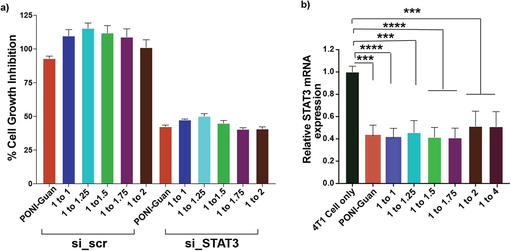
4T1 cancer cells and RAW 264.7 macrophages were treated with either HA-coated-PONI-Guan/si_STAT3 polyplexes or PONI-Guan/si_STAT3 (50 nM) polyplexes. A maximum inhibitory activity of ~50% was achieved by HA-coated polyplexes at all ratios in 4T1 cells (Fig. 5a). An analysis of STAT3 mRNA levels revealed ~60% gene knockdown with PONI-Guan/si_STAT3 and varying ratios of HA-coated-PONI-Guan/si_STAT3 polyplexes, indicating that the HA coating does not compromise the therapeutic efficacy (Fig. 5b).
HA-coated polyplex mediated STAT3 siRNA delivery in 4T1 cancer cells. (a) Graph depicting % cell growth inhibition incubated with HA-coated-PONI-Guan/si_STAT3 (50 nM) polyplexes at varying ratios for 48 h, quantified using Alamar blue assay. (b) Relative STAT3 mRNA expression after incubation with HA-coated-PONI-Guan/si_STAT3 (50 nM) polyplexes, evaluated using qRT-PCR with β-actin as internal control. (c) Quantification of relative invaded cell number from cells treated with either HA-coated-PONI-Guan/si_STAT3 (50 nM) or HA-coated-PONI-Guan/si_scr (50 nM) polyplexes at varying ratios using the cell invasion assay. Standard deviation plotted from three experimental replicates. ***p < 0.0001, corresponding to a 95% confidence interval.
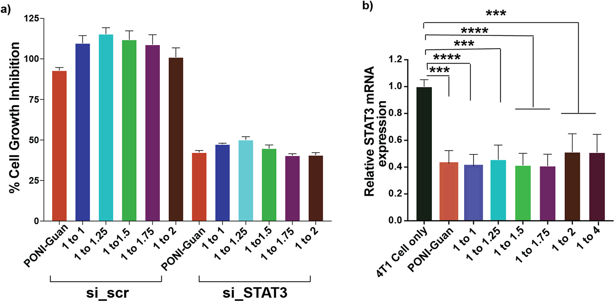
In contrast, RAW 264.7 macrophages demonstrated only ~20% cell growth inhibition with HA-coated-PONI-Guan/si_STAT3 polyplexes at a ratio of 1:1. Ratios 1:1.25, 1:1.5, and 1:1.75 demonstrated about 27% growth inhibition, while ratio 1:2 showed 40% inhibition. PONI-Guan/si_STAT3 polyplexes demonstrated ~56% inhibition, closer to the ratio of 1:2 due to the low concentrations of HA (Supplementary Fig. S9).
3.5. Biodistribution of HA-Coated Polyplexes in 4T1 Tumor Model
Encouraged by the in vitro results, we assessed the biodistribution of HA-PONI-Guan/siRNA polyplexes in vivo using a 4T1-induced orthotopic breast cancer model. Orthotopic implantation of cancer cells directly into the mammary fat pad replicates key aspects of human primary tumor growth and metastatic progression [Reference Pillar, Polsky, Weissglas-Volkov and Shomron48].
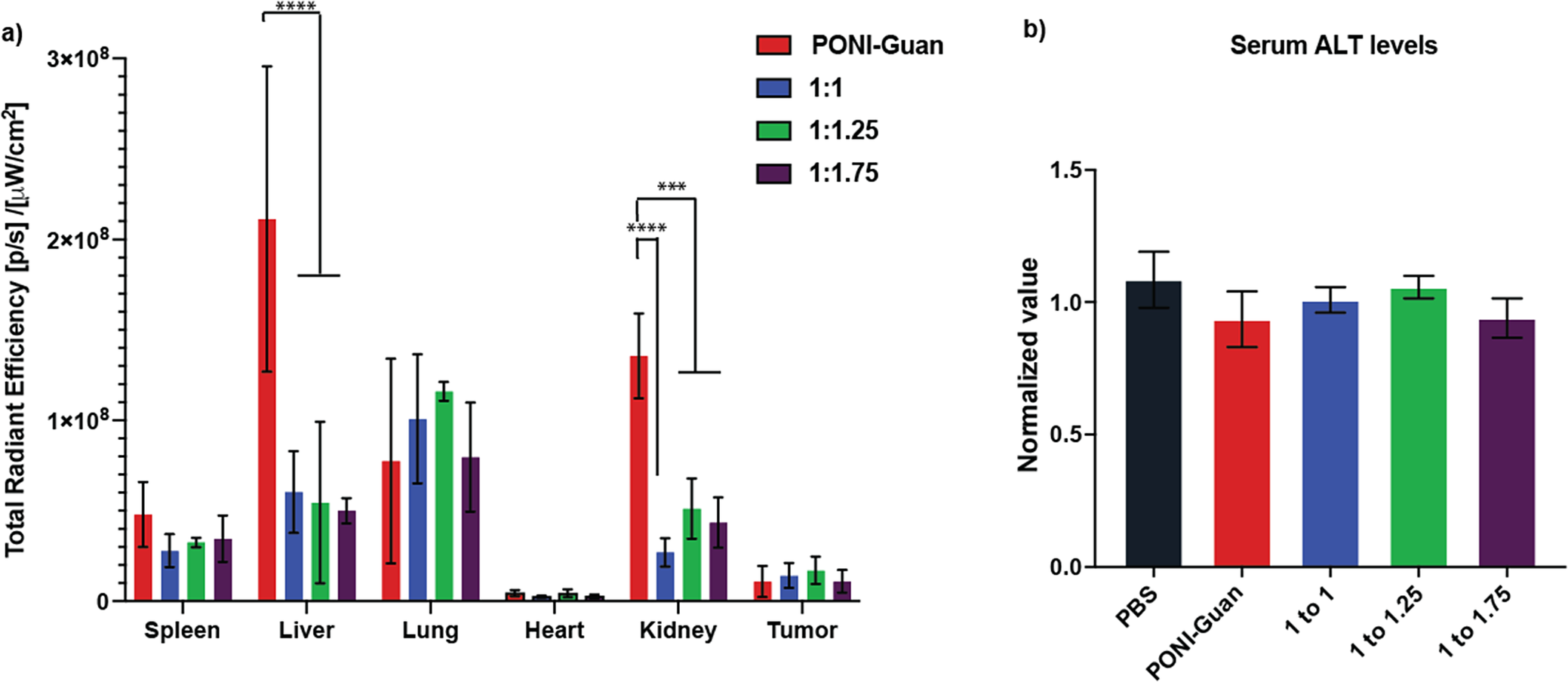
The in vivo biodistribution of HA-PONI-Guan/Cy5-siRNA (0.14 mg kg−1) polyplexes in 4T1 tumor-bearing mice was imaged using an in vivo imaging system (IVIS). The polyplexes were intravenously injected via the tail vein, following which mice were sacrificed after 6 h, major organs collected, and imaged ex vivo using IVIS (Supplementary Fig. S10). The PBS group was used as a control to account for autofluorescence. Biodistribution results revealed that HA-coated-PONI-Guan/Cy5-siRNA polyplexes accumulated significantly less in clearance organs – liver and kidney (Fig. 6a). Quantitative ROI (region of interest) analysis indicated that HA-PONI-Guan/Cy5-siRNA polyplexes accumulated in the liver and the kidney at levels approximately two-fold lower than PONI-Guan/Cy5-siRNA polyplexes, supporting our hypothesis of reduced RES clearance. It should be noted that tumor uptake showed insignificant differences across conditions, and the outcome that is currently under study. Further, analysis of alanine aminotransferase (ALT) levels from the separated serum samples indicated no significant liver toxicity in the experimental groups, highlighting the safety of systemic delivery (Fig. 6b).
In vivo biodistribution of HA-coated-PONI-Guan/Cy5-siRNA (0.14 mg kg−1 siRNA) polyplexes in 4T1-tumor-bearing BALB/c mice. (a) Tissue-specific biodistribution of HA-coated-PONI-Guan/Cy5-siRNA polyplexes (ratios 1:1, 1:1.25, and 1:1.75) at 6 h time point, following systemic administration (n = 3). Fluorescence intensity in organs was quantified using regions of interest (ROIs) generated by Live Image 4.2 software. (b) Liver function assessment using ALT analysis from blood plasma samples. Standard deviation plotted from three experimental replicates. ***p < 0.0001, corresponding to a 95% confidence interval.

4. Conclusion
These studies highlight the potential of HA-coated polyplexes for effective siRNA delivery, while avoiding RES clearance. The HA coating forms a protective layer around the PONI-Guan/siRNA polyplexes, preventing phagocytic clearance. By varying the ratios of HA, we identified the optimal HA concentrations required for efficient siRNA delivery to cancer cells while evading macrophage uptake. Our in vitro results revealed that HA-coated polyplexes facilitated selective siRNA uptake in 4T1 cancer cells relative to RAW 264.7 macrophages, achieving efficient STAT3 gene knockdown (~50%) in 4T1 cells. Furthermore, in vivo biodistribution studies showed reduced accumulation of HA-coated polyplexes in clearance organs, confirming our hypothesis that the negatively charged HA coating shields PONI-Guan/siRNA polyplexes from RES clearance relative to cationic polyplexes. While further investigation is necessary to improve the tumor-targeting capabilities, these studies in the orthotopic breast cancer model demonstrate the potential of HA-coated polyplexes as delivery systems for cancer therapy.
Impact Assessment
Small interfering RNA (siRNA) therapies hold enormous promise for treating diseases such as cancer. However, delivering siRNA effectively to cells of interest remains a major obstacle. Several nanocarrier systems have been developed to protect siRNA from nuclease degradation and facilitate siRNA transport across the impermeable cellular membrane. However, these delivery platforms predominantly utilize endocytic pathways, limiting cytosolic access, with delivery efficiencies typically below 10%. Furthermore, delivery vehicles encapsulated by siRNA often experience non-specific interactions in the systemic environment, which contribute to macrophage uptake and reduce the effective delivery of siRNA to the target site.
We developed a hyaluronic acid (HA)-coated polymeric nanocarrier that packages and protects siRNA while evading immune cells more effectively. The core of the nanocarrier is a guanidinium-functionalized cationic polymer (PONI-Guan) that encapsulates siRNA and facilitates cytosolic access to the cells of interest. The outer HA layer acts as a noninteracting shield that reduces non-specific binding and reduces macrophage uptake. These HA-coated nanocarriers effectively delivered siRNA to aggressive triple-negative breast cancer cells in vitro with four-fold greater efficiency than macrophages, demonstrating cellular selectivity. These carriers mediated the substantial silencing of STAT3 gene expression, a key cancer-promoting gene. In vivo studies revealed that the HA coating reduced the accumulation of nanocarriers in clearance organs by two-fold, relative to PONI-Guan polyplexes, demonstrating improved circulation and reduced immune clearance.
The biocompatible nature of HA, along with the positively charged polymeric core, can be adapted for different gene targets and can be scaled and customized for a range of diseases. With further advancements, HA-shielded nanocarriers could enable safer, more effective gene therapies, supporting global health goals focused on treating cancer and advancing precision medicine.
Author contributions
Conceptualization: H.N., A.G., V.M.R.; Funding acquisition: V.M.R.; Investigation: H.N., A.G.; Methodology: H.N., A.G., Y.A.C., N.N., R.G., M.A.H., E.L., D.R., C.-M.H.; Project administration: V.M.R.; Validation: H.N.; Visualization: H.N., V.M.R.; Writing - original draft: H.N., V.M.R.; Writing - review & editing: H.N., A.G., N.N., V.M.R.
Acknowledgments
The authors express their gratitude to Dr. Amy S. Burnside and the Flow Cytometry Core Facility, and Dr. James Chambers and the Light Microscopy Core Facility at UMass, Amherst.
Financial support
This work was supported by the National Institute of Health (NIH) (Project Number: R01 EB022641) (V.M.R), D.R. was supported by Chemistry-Biology Interface Predoctoral Training Grant (Project Number: T32GM139789), Y. A. C. was partially supported by the IALS Translational Research Fellowship at the University of Massachusetts Amherst, C.-M. H. was partially supported by a fellowship from the University of Massachusetts as part of the Chemistry–Biology Interface Training Program (National Research Service Award T32 GM139789) and a fellowship from PPG.
Competing interests
The authors declare that Vincent Rotello is Editor-in-Chief of Cambridge Materials: Health. He was not involved in the editorial handling of this manuscript, or in the decision to publish. The peer review process was managed independently by the Associate Editor Gang Zheng.
Data availability statement
The authors confirm that the data supporting the findings of this study are available within the article [and/or its supplementary materials].
Open peer review
To view the open peer review materials for this article, please visit http://doi.org/10.1017/S2978170126000002.
Supplementary Material
To view supplementary material for this article, please visit http://doi.org/10.1017/S2978170126000002.


 Open access


Comments
Dear Editor,
Attached is a manuscript entitled “Hyaluronic acid-coated polymer-siRNAnanovectors for evading macrophage uptake and STAT3 gene silencing in breastcancer cells.”
This research describes an approach to highly efficient cytosolic siRNA deliveryusing hyaluronic acid (HA)-coated nanovectors generated from guanidiniumfunctionalized PONI-Guan polymers. These nanovectors employ the guanidiniumblock to complex siRNA and form PONI-Guan/siRNA polyplexes. HA serves as anon-interacting protective shield on the polyplexes that prevents macrophageuptake.
Our work demonstrates the potential of these HA-coated polyplexes for efficientsiRNA delivery to 4T1 triple-negative breast cancer cells in vitro, with a 4:1selectivity relative to macrophages. Further, HA-coated polyplexes demonstratedefficient STAT3 gene knockdown in 4T1 cells. Intravenous administration ofHA-coated polyplexes in 4T1 tumor-bearing mice showed significant 2-folddecreased accumulation in clearance organs, in comparison to the PONI-Guanpolyplexes. Collectively, HA-coated polyplexes provide an effective strategy forselective siRNA delivery to tumor cells while avoiding macrophage uptake. We areconfident that Cambridge Materials: Health is an ideal venue for the publicationof this article.
If any other information is required, please let me know.
Best wishes,
Vincent Rotello
University Distinguished Professor
Charles A. Goessmann Professor of Chemistry