


Introduction
The bacterium Escherichia coli, more often known as E. coli, is common in the human and animal digestive systems as well as in the environment, including plants, water, and soil. Reference Kashef, Djavid and Shahbazi1 Among human infections, wounds, otitis media, and bloodstream infections, it ranks first among these pathogens and is the most prevalent cause of UTIs. Reference Gebre-Sellassie2 In underdeveloped nations, E. coli is the leading etiology of water & food-borne diarrhea in humans, and it kills a lot of kids younger than five. Reference Turner, Scott-Tucker, Cooper and Henderson3
E. coli has been shown to be resistant to several antibiotics, and this is becoming an increasingly serious problem in both industrialized and poor nations. Bacterial resistance to antibiotics is on the rise, which makes illness treatment more difficult. Reference Bell, Turnidge, Gales, Pfaller and Jones4 It is common practice to treat people with severe symptoms without doing bacteriological investigations in as many as 95% of instances. Reference Dromigny, Nabeth, Juergens-Behr and Perrier-Gros-Claude5
The frequency and susceptibility profiles of Escherichia coli can vary greatly depending on factors such as geography, demographics, and the environment.
This study intended to ascertain the frequency and antibiotic susceptibility of E. coli from various clinical samples in the Jazan area.
Materials and methods
The antibiotic susceptibility and culture findings of isolates from sputum, wound swabs, and urine were examined in this retrospective cross-sectional study. It was conducted through a multi-center approach involving various healthcare facilities in the Jazan Region, ensuring representative sampling from January 2023 to December 31, 2023, across all public and private hospitals in the area.
Inclusion criteria: All clinical samples positive for E. coli obtained from patients in the sharing centers in Jazan Region. Clinical sources include urine, blood, wound swabs, sputum, and other relevant specimens.
Methods
All participants were subjected to the following
Laboratory procedures
As part of standard operating procedure, samples were collected in sterile containers to ensure aseptic collection. The clean-catch midstream urine sample was collected in the morning using sterile wide-mouth glass containers. Calibrated wire loops were used to inoculate urine samples onto cystine lactose electrolyte-deficient medium, MacConkey agar, and blood agar (HiMedia Laboratories Pvt. Limited, India). After that, the samples were aerobically incubated at 37 ºC for 24 h. Positive cultures were analyzed using standard microbiological methods to identify uropathogens. When a urine culture yielded more than 105 colony-forming units/ml of urine, it was considered significant bacteriuria. Reference Magliano, Grazioli, Deflorio, Leuci, Mattina and Romano12 Using sterile cotton swabs, pus was recovered from the wound. Sterile, wide-mouthed containers were used to collect sputum samples. Plates of MacConkey agar, blood agar, and chocolate agar were infected with specimens (HiMedia, India). After 24 and 48 hours of incubation at 37ºC in an aerobic environment, the plates were evaluated.
Microscopic examination
Gramme staining and careful examination were used to identify colonies that had been obtained from blood agar or Mac Conkey’s agar plates.
Antimicrobial susceptibility tests
Following standard operating procedure, Mueller-Hinton agar was subjected to antimicrobial susceptibility testing using the Kirby-Bauer disc diffusion technique. Automated procedures with BD Phoenix and Vitck equipment. The National Committee for Clinical Laboratory Standards’ standards were followed for analyzing the resistance data. As a quality control measure, antimicrobial susceptibility testing made use of E. coli ATCC 25922 and S. aureus ATCC 25923 reference strains. Traceability and confirmation of Escherichia coli by use of time-tested microbiological techniques. The Kirby-Bauer disc diffusion technique or automated systems performed antimicrobial susceptibility testing according to Clinical and Laboratory Standards Institute (CLSI) guidelines. Ampicillin, Ciprofloxacin, Trimethoprim/sulfamethoxazole, Ceftriaxone, and Meropenem are among the often prescribed antibiotics that should be included in the testing. The inhibitory zone width was measured to the nearest millimeter after a 24-hour incubation at 37°C. Isolates were classified as susceptible, intermediate, or resistant according to the criteria set by the Clinical and Laboratory Standards Institute. Standards offered by CLSI for interpretation. 6
Data collection: Demographic data (age, gender) & clinical information (source of specimen, hospital ward) recorded for each patient. Antimicrobial susceptibility results are documented systematically, including zone diameters and interpretation.
Data analysis
A descriptive study was conducted to ascertain the distribution and frequency of E. coli isolates from various clinical sources. Calculation of antimicrobial susceptibility rates for each antibiotic tested. Statistical analysis to explore associations between clinical variables and antimicrobial resistance patterns (eg, chi-square test, logistic regression).
Ethical considerations
Protection of patient confidentiality and privacy throughout the study. Informed consent was acquired from patients or parents before sample collection. Adherence to ethical standards in research involving human subjects. The reasearch approved by Jazan Armed Force Hospital Ethical committee.
Quality control
Standardization of laboratory procedures and adherence to quality control measures to ensure accuracy and reliability of results. Regular calibration and quality assurance of laboratory equipment and antimicrobial disks.
Results
Escherichia coli is among the most common bacteria that can cause illness. Reference Zhou, Zhou, Zheng, Gong, Li and Jin7 Severe health complications, including extended hospital stays and treatment failures, are caused by E. coli antibiotic resistance patterns, which continue to be a major public health concern globally. Reference Salam, Al-Amin, Salam, Pawar, Akhter and Rabaan8 E. coli prevalence and antibiotic susceptibility from clinical samples in Jazan were the focus of this study.
Shows distribution of positive culture isolates.

Shows distribution of specimen in studied group.

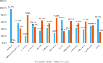
Shows distribution of antimicrobial susceptibility pattern of E. coli isolates.

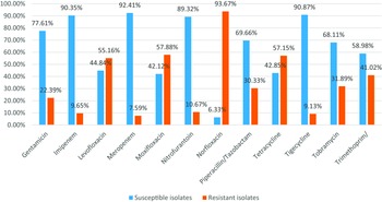
Shows distribution of antimicrobial susceptibility pattern of E. coli isolates.

This study reported that 41 patients (20.56%) their age was less than 20, 67 patients (33.57%) their age ranged from 20 to 40, 92 patients (45.88%) their age ranged from 40 to 60, 53 patients (26.51%) were males, and 147 patients (73.49%) were females.
In addition, Naqid et al. Reference Naqid, Balatay, Hussein, Saeed, Ahmed and Yousif9 identified the sensitivity pattern of Escherichia coli bacteria obtained from several clinical sources in Duhok city, Iraq. A total of 454 samples were collected, with 321 women and 133 males included. The age range of the participants was ten to sixty. Urine samples from females were found to have higher clinical isolates of Escherichia coli compared to male samples.
Consistent with prior studies, this finding shows that females are more likely to get UTIs caused by E. coli. Reference Assafi, Ibrahim, Hussein, Taha and Balatay10,Reference Yismaw, Abay, Asrat, Yifru and Kassu11
The reason this microbe is more common in girls than males is because germs can’t travel as far via their shorter urethras to reach the bladder and infect the bladder more easily during sexual activities. Reference Magliano, Grazioli, Deflorio, Leuci, Mattina and Romano12 Low socioeconomic position and inadequate personal hygiene exacerbate these risk factors for UTI. Reference Akram, Shahid and Khan13
Our results showed that 25.64% of patients had positive culture E. coli, the majority of isolates were obtained from the urine samples in 171 patients (85.49%), followed by pus in 18 patients (9.06%), and sputum in 11 patients (5.45%).
The findings of this study were in line with those of Naqid et al. Reference Naqid, Balatay, Hussein, Saeed, Ahmed and Yousif9 who reported that the majority of the E. coli isolates were obtained from urine samples (418; 92.2%). This was followed by wound samples (18; 3.9%), cervical samples (7; 1.5%), blood samples (4; 0.9%), semen samples (3; 0.7%), ascitic samples (2; 0.4%), and cerebral spinal fluid samples (2; 0.4%).
As well, the present study agreed with Eltabey et al., Reference Eltabey, Ibrahim, Zaky and Saleh14 who reported that the common isolates were obtained from the urine samples.
Our findings demonstrated that there were high sensitivity rates to Amikacin, Tigecycline and Imipenem by (97.49%), (90.87%) and (90.35%) respectively, while there were high resistance rates to Norfloxacin, Ampicillin and Cefotaxime by (93.67%), (79.60%) and (71.65%) respectively.
The results were in agreement with those of Naqid et al., who had previously shown that 88.3% of the E. coli strains they had identified were resistant to ampicillin, 63.9% to ceftriaxone, and 63.9% to cefepime. However, it was shown that E. coli was quite sensitive. Reference Naqid, Balatay, Hussein, Saeed, Ahmed and Yousif9
Consistent with other studies, we found that imipenem had a profound effect on E. coli isolates obtained from urine. Reference Adinortey, Amewowor, Otwe, Galyuon and Asante16
Odongo et al. also determined that Escherichia coli isolates were highly susceptible to cefotaxime/clavulanic acid (100%) and nitrofurantoin (70%), but very resistant to cefuroxime (100%), ceftazidime (100%), nalidixic acid (90%), and ciprofloxacin (90%). The results of this study were in agreement with those of Odongo et al. Reference Odongo, Ssemambo and Kungu17
Furthermore, according to Kasanga et al. the majority of the isolates tested positive for resistance to ampicillin (81.4%), sulfamethoxazole/trimethoprim (70.7%), ciprofloxacin (67.9%), levofloxacin (64.6%), ceftriaxone (62.3%), and cefuroxime (62%), Yet, amikacin (100%), imipenem (99.5%), nitrofurantoin (89.3%), ceftolozane/tazobactam (82%), and gentamicin (72.1%) were all extremely effective against E. coli isolates. Reference Kasanga, Shempela, Daka, Mwikisa, Sikalima and Chanda18
Eltabey et al. also discovered that almost all of the tested isolates were responsive to Amikacin (90%) and Meropenem (92%); however, all of the isolates were resistant to Amoxicillin/clavulanic acid (100%) and Cefotaxime (44%). Reference Eltabey, Ibrahim, Zaky and Saleh14
Distribution of positive culture isolates in all studied patients

This table shows that 25.64% of patients had positive culture E. coli, 22.39% of patients had positive culture Klebsiella pneumoniae, 19.58% of patients had positive culture Staphylococcus aureus, 10.82% of cases had +ve culture Staphylococcus epidermidis, 5.09% of patients had positive culture Staphylococcus haemolyticus, 4.96% of patients had positive culture Enterobacter cloacae, 3.12% of patients had positive culture Enterococcus faecalis and 1.34% of patients had positive culture Proteus mirabilis (Table 1).
Distribution of demographic data in patients with positive culture E. coli

This table shows that 279 patients (20.56%) their age <20, 456 patients (33.57%) their age ranged from 20 to 40, 623 patients (45.88%) their age ranged from 40 to 60, 360 patients (26.51%) were males, and 998 patients (73.49 %) were females (Table 2).
Distribution of specimens in patients with positive culture of E. coli

The majority of isolates were obtained from the urine samples in 1161 patients (85.49%), followed by pus in 123 patients (9.06%) and sputum in 74 patients (5.45%) (Table 3).
E. coli isolates’ responsiveness to various antibiotics

There were high sensitivity rates to Amikacin, Tigecycline, and Imipenem by (97.49%), (90.87%), and (90.35%), respectively, while there were high resistance rates to Norfloxacin, Ampicillin & Cefotaxime by (93.67%), (79.60%), and (71.65%) respectively (Table 4).
Conclusion
Escherichia coli bacteria isolated from different types of human tissues showed distinct antibiotic sensitivity patterns. A considerable degree of resistance to commonly used antibiotics was demonstrated by these microorganisms. Of the medicines tested, amikacin, tigecycline, and imipenem were the most effective against the different strains of E. coli. Instead, the clinical isolates of E. coli exhibited increased levels of resistance to the antibiotics norfloxacin, ampicillin, and cefotaxime. As a result, it is suggested that antibiotic sensitivity testing be carried out by medical professionals in order to choose the antibiotics that are the most effective.
Financial support
There was no funding for this submission.
Competing interests
All authors have no conflict of interest.




 Open access

