


Introduction
More than a billion people worldwide are infected with soil-transmitted gastrointestinal parasites, such as Trichuris trichiura, which cause significant morbidity, particularly in tropical and subtropical regions (Chen et al. Reference Chen, Gong, Chen, Li and Zhou2024). Experimental infections with Trichuris muris in laboratory mice are therefore frequently used as a model system to advance our understanding of host-parasite interactions and the immune response to infection (Klementowicz et al. Reference Klementowicz, Travis and Grencis2012). These laboratory studies show that infection outcome is strongly influenced by the host genetic background and infection dose: in resistant strains, high doses induce robust Th2 responses leading to parasite expulsion (Bancroft et al. Reference Bancroft, Else, Humphreys and Grencis2001; Cliffe and Grencis Reference Cliffe and Grencis2004), whereas low doses elicit Th1 responses, often resulting in chronic infections (Bancroft et al. Reference Bancroft, Else and Grencis1994).
While these laboratory studies have provided fundamental insights, recent research shows that immune responses in naïve laboratory animals can differ substantially from those of their wild counterparts, limiting the generalisability of these findings (Mair et al. Reference Mair, McNeilly, Corripio-Miyar, Forman and Else2021). For example, laboratory mice exposed to natural outdoor environments show increased susceptibility to infection (Leung et al. Reference Leung, Budischak, Chung The, Hansen, Bowcutt, Neill, Shellman, Loke and Graham2018), and comparative studies indicate that wild Mus musculus domesticus often exhibit weaker immune responses than laboratory mice (Mair et al. Reference Mair, Fenn, Wolfenden, Lowe, Bennett, Muir, Thompson, Dieumerci, Logunova, Shultz, Bradley and Else2024). Together, these findings underscore the need to assess whether laboratory insights extend to natural host-parasite systems.
In wild rodent populations, infections with Trichuris spp. are common (Brouat et al. Reference Brouat, Kane, Diouf, Bâ, Sall-Dramé and Duplantier2007; Msoffe et al. Reference Msoffe, Thomas, Rija, Nzalawahe, Katakweba, Misinzo and Mnyone2025), yet controlled experimental infections in these natural host-parasite combinations remain scarce (Behnke and Wakelin Reference Behnke and Wakelin1973). One relevant host is the multimammate mouse, Mastomys natalensis, a widely distributed African rodent and a major agricultural pest (Leirs et al. Reference Leirs, Kirkpatrick, Sluydts, Sabuni, Borremans, Katakweba, Massawe, Makundi, Mulungu, Machang’u and Mariën2023; Thomas et al. Reference Thomas, Msoffe, Van Houtte, Mhamphi, Mariën, Sabuni, Makundi, Nzalawahe, Machang’u and Leirs2023). Field studies in Tanzania report prevalences of T. mastomysi up to 60%, typically at low parasite loads (Vanden Broecke et al. Reference Vanden Broecke, Bernaerts, Ribas, Sluydts, Mnyone, Matthysen and Leirs2021; van de Ven et al. Reference van de Ven, Broecke, Ribas, Leirs, Sabuni and Mariën2025), making M. natalensis a well-suited natural system to experimentally test host-parasite interactions.
To investigate how infection dose influences early establishment of T. mastomysi and worm burden in M. natalensis, wild-caught individuals were experimentally infected with embryonated eggs harvested from naturally infected conspecifics. Two controlled infection regimes were applied via oral gavage: (1) a single high-dose administration and (2) repeated low-dose administration. By using wild hosts under controlled infection conditions, we provide a functional measure of host susceptibility in a natural host–parasite system and improve our understanding of how Trichuris spp. persist in their natural wild hosts.
Materials and methods
Experimental design
The study was conducted from late June to late August 2024 in Morogoro, Tanzania. Fifty-four wild Mastomys natalensis were captured using Sherman LFA live traps (Sherman Live Trap Co., Tallahassee, FL) placed around the campus of Sokoine University of Agriculture (SUA). The animals were transported to the Institute of Pest Management (PMC) at SUA, where their sex and body weight were recorded. Fresh faecal samples were collected from each individual and screened for Trichuris spp. eggs using a salt-sugar flotation technique. All animals were housed individually in standard laboratory cages (28 × 11.5 × 12cm) with wood shavings as bedding and supplemented with cotton cylinders and cardboard tunnels. Food and water were provided ad libitum.
To obtain Trichuris eggs, 24 Trichuris-positive individuals were euthanised, and adult worms were recovered from the caecum, from which eggs were harvested as described by Antignano et al. (2011). The eggs were stored in foil-covered flasks at room temperature (~22–26°C) for six weeks to allow embryonation.
The remaining M. natalensis were kept in quarantine for six weeks to reduce the likelihood of acute infections and allow acclimatisation to laboratory conditions. During quarantine, two rounds of oral anthelmintic treatments were administered at two-week intervals (weeks 2 and 4) using a combination of ivermectin (2 mg/kg) and pyrantel pamoate (100 mg/kg). This treatment regimen was based on previous work showing that both compounds reduce gastrointestinal nematode burdens in M. natalensis, including Trichuris (van de Ven et al. Reference van de Ven, Broecke, Ribas, Leirs, Sabuni and Mariën2025). However, other helminths, such as cestodes, may persist. Ectoparasites were removed by brushing and cages were cleaned at each treatment.
After six weeks, all eggs in the flasks were fully embryonated (containing first-stage larvae; see Supplementary Figure S1), as confirmed by light microscopy. A small subset was examined to assess embryonation and egg viability. Quarantined mice were randomly assigned to three groups of ten individuals each, with equal numbers of males and females per group: (1) single oral gavage of 100–200 embryonated eggs, (2) weekly oral gavage of 20 embryonated eggs (‘trickle’ infection; Bancroft et al. Reference Bancroft, Else, Humphreys and Grencis2001; Glover et al. Reference Glover, Colombo, Thornton and Grencis2019), and (3) an untreated control group. Oral egg doses were based on the estimated viability per batch; the actual number of infective eggs may have varied. Faecal samples were collected weekly throughout the experiment to monitor gastrointestinal helminth infections that may have persisted despite anthelmintic treatment.
At 21 days post-infection, mice were euthanised with an overdose of isoflurane, followed by cervical dislocation to confirm death. The entire gastrointestinal tract was preserved in absolute ethanol and subsequently examined for helminths under a stereomicroscope (Ribas et al. Reference Ribas, Chaisiri, Morand, Hugot, Haukisalmi and Henttonen2011). All helminths were isolated and stored in 70% ethanol, and Trichuris was identified morphologically and molecularly.
Molecular analysis
Trichuris larvae obtained from one experimentally infected individual were used for molecular analysis to confirm the species identity of the eggs used in the experimental infections. Genomic DNA was extracted using the NucleoSpin tissue kit (Macherey-Nagel GmbH & Co., Duren, Germany) and eluted in 60 μL of DNase/RNase-free water. PCR amplification was performed in 15 μL reactions containing 0.2 μM of each primer, 0.2 mM dNTPs, 2 mM MgCl₂, 1X Green GoTaq® Flexi buffer, 1 U GoTaq® G2 Flexi DNA Polymerase, and 1.5 μL DNA template. The thermal cycling conditions were as follows: initial denaturation at 94°C for 3 min; 40 cycles of 94°C for 30 s, 52.5°C for 30 s, and 72°C for 1 min, followed by a final extension at 72°C for 5 min (Ribas et al. Reference Ribas, López, Makundi, Leirs and de Bellocq2013). The ITS-1, 5.8S, and ITS-2 regions (~1,100 bp) were amplified using NC5 (5′-GTAGGTGAACCTGCGGAAGGATCATT-3′) and NC2 (5′-TTAGTTTCTTTTCCTCCGCT-3′) primers. The PCR product was visualised on a 1.4% agarose gel and sequenced using the forward primer at the VIB Genetic Service Facility (University of Antwerp, Belgium). BLASTn analysis confirmed the highest similarity to Trichuris mastomysi, consistent with previous sequences from Mastomys in Tanzania (e.g. JX683517.1, JX683518.1, JX683520.1; Ribas et al. Reference Ribas, López, Makundi, Leirs and de Bellocq2013).
Statistical analyses
Infection success, defined as the presence or absence of Trichuris larvae at necropsy, was analysed as a binary response using Firth’s bias-reduced logistic regression, with infection regime (high dose, low dose, control) as the main explanatory variable. This approach was chosen due to the relatively small sample sizes and complete separation in the control group, in which no infections were observed (see Supplementary Table S1). Host sex, body weight, and the presence of other helminths were included as covariates.
Infection intensity, defined as larval counts, was analysed among infected individuals only using zero-truncated negative binomial regression, with infection regime included as the main explanatory variable. The control group was excluded from this analysis because no control animals were infected. Host sex, body weight, and the presence of other helminths were included as covariates. Effect sizes are reported as incidence rate ratios (IRR) with 95% confidence intervals.
All statistical analyses were performed in R version 4.5.1 (R Core Team 2025) using the ‘logistf’ package for Firth’s penalised logistic regression (Heinze and Schemper, Reference Heinze and Schemper2002) and ‘VGAM’ package for zero-truncated negative binomial regression (Yee Reference Yee2010).
Results
Infection success under different infection regimes
At necropsy (21 dpi), only larval stages of Trichuris mastomysi were recovered from the caecum, and no adult worms were detected in any individual. This is consistent with the expected pre-patent period of Trichuris spp., in which adult worms typically appear from ~32 days post-infection onwards (Klementowicz et al. Reference Klementowicz, Travis and Grencis2012). In two individuals in the high-dose group, low-intensity Trichuris eggs were detected during faecal screening. However, necropsy revealed no evidence of patent infection in either case, as one individual harboured no detectable larvae and the other carried only non-egg-producing larval stages. This may reflect transient egg passage. To account for this, infection success was therefore assessed both including and excluding these individuals, yielding very similar qualitative patterns (Table 1). Other helminths were present in all treatment groups, with gastrointestinal cestodes detected in 43.33% of individuals and Syphacia spp. in 10%, and were included as covariates in the statistical analyses.
Infection success and mean intensity of Trichuris in experimental Mastomys natalensis. Infection success is shown both for all individuals and after excluding individuals with low-intensity egg shedding detected in faeces prior to necropsy (‘excl. egg+’). Exact 95% binomial confidence intervals (95% CI) are shown for infection success. Mean intensity ± standard deviation (SD) and range are reported for infected individuals only.

Table 1. Long description
From left to right, the Group column lists High, Low, and Control. For High, infection success is 8 out of 10 (80 percent), infection success excluding egg plus is 7 out of 8 (87.5 percent), 95 percent confidence interval is 44 to 97 percent, mean intensity plus or minus standard deviation is 75.6 plus or minus 25.1, and range is 42 to 118. For Low, infection success is 9 out of 10 (90 percent), infection success excluding egg plus is 9 out of 10 (90 percent), 95 percent confidence interval is 55 to 100 percent, mean intensity plus or minus standard deviation is 15.1 plus or minus 7.6, and range is 5 to 27. For Control, infection success is 0 out of 10 (0 percent), infection success excluding egg plus is 0 out of 9 (0 percent), 95 percent confidence interval is 0 to 31 percent, mean intensity plus or minus standard deviation and range are both dash, indicating no data.
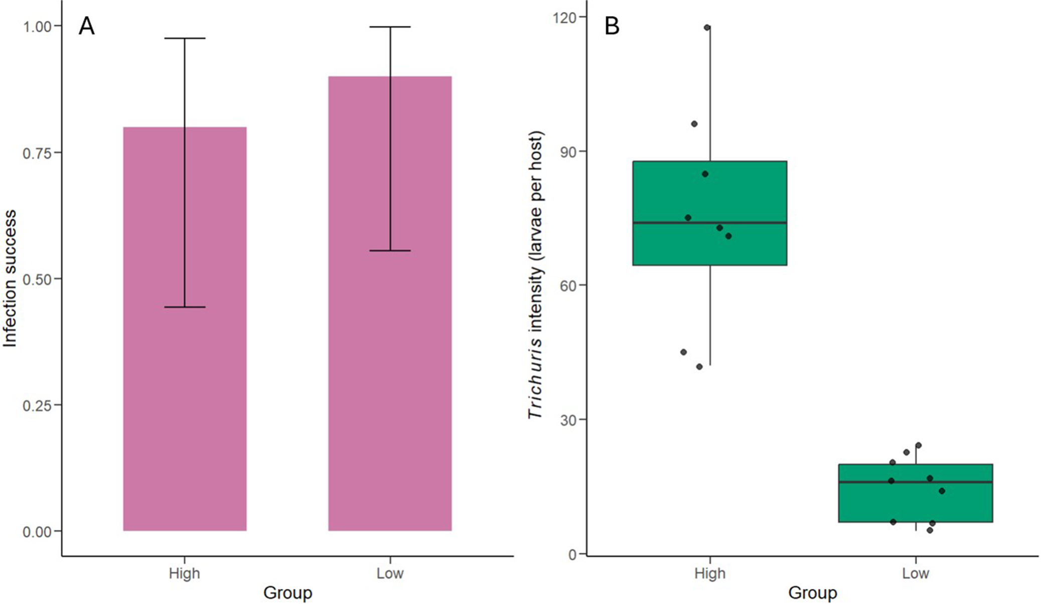
Infection success was high in both treatment groups, with 90% in the low-dose group and 78–87.5% in the high-dose group, while no infections were observed in the control group (Figure 1A). Using a Firth bias-reduced logistic regression model, both treatment groups were significantly associated with infection success relative to controls (low dose: OR = 73.2, 95% CI [6.30–10,398], p <0.001; high dose: OR = 42.2, 95% CI [4.05–5,621], p <0.001; see Supplementary Table S2). Neither sex, body weight, nor co-infection status significantly predicted infection success (all p >0.6).
(A) Infection success (with 95% binomial confidence intervals) and (B) boxplots of Trichuris mastomysi larval burdens in wild-caught Mastomys natalensis following experimental infection under different infection regimes. Boxes represent the interquartile range, the centre line indicates the median, whiskers show the spread of the data, and points represent individual animals.

Figure 1. Long description
Panel A on the left is a bar graph with y-axis labeled Infection success from 0.00 to 1.00 and x-axis labeled Group with categories High and Low. Both bars are shaded pink and reach similar heights, around 0.75, with overlapping error bars extending from about 0.50 to 1.00. Panel B on the right is a boxplot with y-axis labeled Trichuris intensity (larvae per host) from 0 to 120 and x-axis labeled Group with categories High and Low. The High group shows a boxplot centered near 80 larvae per host, with a wider spread and several individual data points scattered above and below. The Low group shows a boxplot centered near 20 larvae per host, with a narrower spread and individual points clustered closely. Both panels compare the same two groups, showing similar infection success but much higher Trichuris intensity in the High group.
Infection intensity among infected hosts
Among infected individuals, mean intensity was substantially higher in the high-dose group (mean ± SD: 75.6 ± 25.1 larvae; Figure 1B) than in the low-dose group (15.1 ± 7.6 larvae), consistent with a dose-response relationship. The difference was supported by the zero-truncated negative binomial regression, which showed that larval intensity was significantly lower in the low-dose group than in the high-dose group (IRR = 0.20, 95% CI [0.14–0.28], p <0.001; see Supplementary Table S3). Sex and body weight were not associated with larval intensity, while co-infection with other helminths showed a non-significant tendency toward lower Trichuris intensity (p = 0.096).
Discussion
Experimental infections with Trichuris muris in inbred laboratory mice have shaped current concepts of resistance and susceptibility to helminth infection. Applying a comparable experimental approach in a natural host-parasite system, we infected wild-caught Mastomys natalensis with T. mastomysi and quantified early parasite establishment under different exposure regimes.
This study demonstrates that T. mastomysi can successfully establish in wild-caught M. natalensis under controlled experimental conditions and that infection dose strongly influences larval burden during early infection. Infection success was high in both exposure groups and absent in controls, indicating that the recovered larvae resulted from the experimental inoculations rather than background infection. Among infected hosts, larval intensity was higher in the high-dose group than in the repeated low-dose group, suggesting a dose-dependent effect on early parasite establishment.
These findings are broadly consistent with previous work on Trichuris systems, where infection dose and host context can both influence parasite establishment (Bancroft et al. Reference Bancroft, Else and Grencis1994, Reference Bancroft, Else, Humphreys and Grencis2001; Leung et al. Reference Leung, Budischak, Chung The, Hansen, Bowcutt, Neill, Shellman, Loke and Graham2018; Mair et al. Reference Mair, Fenn, Wolfenden, Lowe, Bennett, Muir, Thompson, Dieumerci, Logunova, Shultz, Bradley and Else2024). In this wild host, susceptibility may therefore reflect not only intrinsic host factors, but also prior exposure history and environmental challenges.
Dose primarily influenced the number of larvae that successfully established rather than the likelihood of infection, a distinction that is biologically relevant. Observational studies in wild hosts often report relatively low helminth burdens (Vanden Broecke et al. Reference Vanden Broecke, Bernaerts, Ribas, Sluydts, Mnyone, Matthysen and Leirs2021; van de Ven et al. Reference van de Ven, Broecke, Ribas, Leirs, Sabuni and Mariën2025), suggesting that low parasite burdens in natural populations may, in part, reflect relatively low exposure to infectious stages. However, the present study focused on a single early time point, preventing assessment of later parasite development and persistence (Behnke and Wakelin Reference Behnke and Wakelin1973; Glover et al. Reference Glover, Colombo, Thornton and Grencis2019). Extending the infection duration in future studies will be important to determine whether larvae successfully mature to adult stages, and thus whether initial exposure dose ultimately determines final parasite burden.
Host sex and body weight were not associated with either infection success or infection intensity, although the relatively small sample size could limit the detection of covariate effects. Co-infection showed a non-significant tendency toward lower Trichuris intensity, which may warrant further investigation in larger studies. Co-infections are very common in natural populations, and interactions between parasite species may arise through competition for resources or immune-mediated effects (Fenn et al. Reference Fenn, Wolfenden, Young, Goertz, Lowe, MacColl, Taylor and Bradley2020; Graham Reference Graham2008).
By integrating controlled experimental infection with a wild-caught host, this study provides an experimentally grounded perspective on Trichuris infection dynamics in a natural rodent system. Our results show that wild M. natalensis are highly permissive to early T. mastomysi establishment under controlled exposure and that infection dose strongly influences early parasite burden. These findings highlight how biological and ecological drivers can shape infection dynamics in natural populations. Semi-controlled experimental systems therefore provide an important complement to mechanistic laboratory experiments and observational field studies.
Supplementary material
The supplementary material for this article can be found at http://doi.org/10.1017/S0022149X26101618.
Acknowledgements
We thank the staff at the Institute of Pest Management (Sokoine University of Agriculture, Morogoro, Tanzania), particularly Geoffrey Sabuni, Shabani Lutea, Omary Kibwana, Ramadhani Iddy, and Baraka Edson, for their invaluable assistance during the fieldwork and experiment. During the preparation of this work the authors used ChatGPT to improve readability. All content generated by the tool was subsequently reviewed and edited by the authors, who take full responsibility for the final content of the publication.
Financial support
This work was supported by the Research Foundation – Flanders FWO through a senior research project (grant number G065720N).
Competing interests
The authors declare there are no conflicts of interest.
Ethical standard
All experimental procedures involving animals were approved by the SUA Committee responsible for overseeing research involving animals and conducted in accordance with the SUA Code of Conduct for Research and Ethics (2023) and Animal Welfare Act (2008). The study protocol was also reviewed by the University of Antwerp Ethical Committee for Animal Experimentation (2024–2029) in accordance with EEC Council Directive 2010/63/EU.



 Open access

