

Introduction
The primary role of the neurologist in the emergency department (ED) when confronted with a potential exacerbation of a neuroimmunologic condition is first, to rule out potential “mimics,” and second, to treat acute episodes of true neurologic dysfunction.
True neuroimmunologic emergencies are relatively rare ED presentations.
Most patients with a known neuroimmunologic condition such as multiple sclerosis (MS) present to the ED with a non-neurologic issue or a “pseudorelapse.”Reference Abboud, Mente and Seay1
MS Relapses
A relapse (also referred to as an exacerbation, flare, or attack) is defined as “new or acutely worsening neurological symptoms lasting 24–48 hours and separated by at least 30 days from the onset of the last relapse.”Reference Abboud, Mente and Seay1, Reference Buckley, Kennard and Swash2 These symptoms must be constant (not off and on) and accompanied by an objective change in neurologic exam, with no other explanation (such as fever, infection exhaustion, or dehydration).
Relapse vs. Pseudorelapse
Relapse
New or worsening constant symptoms lasting at least 24–48 hours.
Usually associated with inflammation and demyelination in the central nervous system (CNS) on magnetic resonance imaging (MRI; enhancing the lesion in a post-gadolinium T1-weighted image).
Post-contrast imaging must be acquired 5 minutes after gadolinium is administered.
Separated from the last exacerbation by at least 30 days.
Pseudorelapse
Temporary flare-up of old symptom(s) lasting less than 24 hours, typically described as “off-and-on” symptoms.
Symptoms are not associated with CNS inflammation (no enhancement on MRI).
Possible triggers: increased core body temperature due to infection, exposure to heat and humidity, overexertion, bladder or bowel fullness, and/or stress.
Symptoms usually return to baseline when the trigger is eliminated, though it may take days in the case of infection or physical exhaustion.
Assessment of a Relapse
Determining whether a person is having a true relapse can be challenging, but is important before initiating steroid treatment. Evaluation for underlying causes is imperative:
Screen for underlining infections with a urinary albumin to urinary creatinine ratio (UAUC), complete blood count (CBC), and possibly chest x-ray. Frequently, pseudorelapses are related to an underlying infection, most commonly a urinary tract infection (UTI) or upper respiratory infection (URI). It is important to note that patients with MS often do not have the normal signs and symptoms of a UTI, and a urine test must be performed despite a lack of presenting classic UTI symptoms.
If presenting symptoms are acute versus subacute, consider evaluation for a vascular cause.
Symptoms that rarely represent an MS relapse include low back pain, chest pain, altered mental status, aphasia, and seizures; thus, other causes should be considered in the differential diagnosis and further evaluation is necessary.
Differential Diagnosis
The differential diagnosis is discussed in more detail in Chapter 5.
There are multiple manifestations of autoimmune CNS conditions among patients who present to the ED.
An important step in determining the correct etiology is establishing the onset and chronology of symptom evolution.
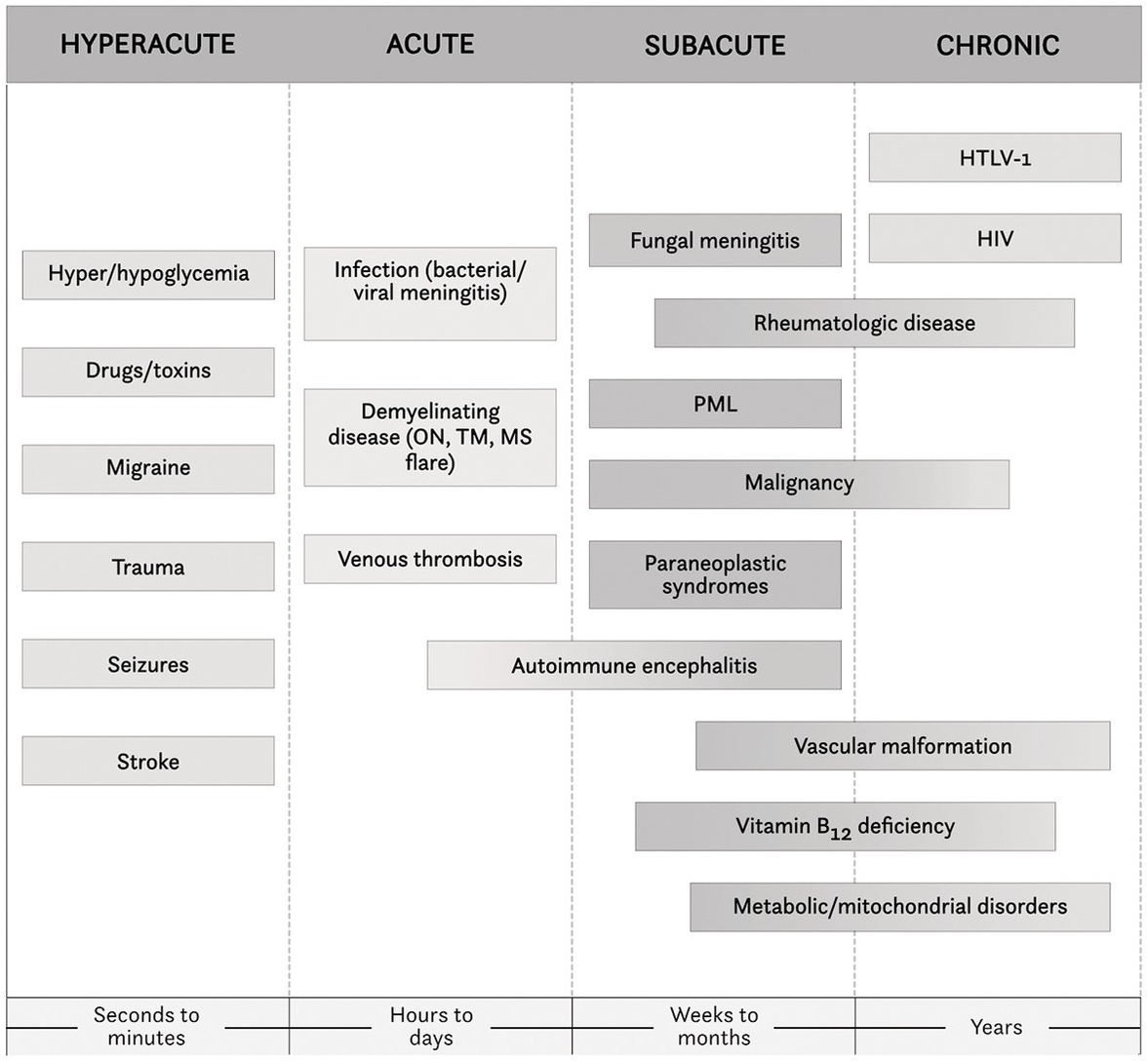
A characteristic feature of acute demyelinating disease is gradual worsening of symptoms over the course of hours to days. A timeline of common alternative diagnoses based on chronological progression of symptom severity is shown in Figure 1.1.
If the patient’s symptoms are of maximal severity at onset (within seconds to minutes), it is critical to rule out a vascular cause (i.e., stroke).
Patients who experience a slow and gradual onset of symptoms are less likely to present to the ED. However, it is important to keep in mind that an acute event, such as a fall, may increase patients’ awareness of their slowly progressing symptoms, thereby prompting an ED visit for an unrelated reason.
Clinicians should also consider whether the patient has any associated non-neurologic symptoms.
One of the most common reasons for patients to present to the ED is recrudescence of previous symptoms (Uhthoff’s phenomenon) in the setting of a febrile illness.
In these cases, patients will typically return to their neurologic baseline after recovering from the illness.
Always consider ruling out a UTI, even if the patient does not complain of dysuria, hematuria, or frequency.
Always consider meningitis or encephalitis as possible diagnoses in the right clinical context.
Chronic immunomodulation/suppression in patients with neuroimmunologic conditions, such as MS, can increase their risk of serious infections.
Patients may not always present with the classic symptoms for these conditions, such as fever, headache, encephalopathy, or neck stiffness.
Uncommon and opportunistic infections should always be ruled out.
Cerebrospinal fluid (CSF) studies should always be performed in the right clinical context.
In individuals with space-occupying lesions, the possibility of an abscess should be considered.
Other symptoms that should significantly broaden the differential diagnosis include unintentional weight loss, rashes, swollen joints, and symptoms suggestive of multi-organ system involvement, which would warrant a more aggressive diagnostic workup when present.
Once possible alternative diagnoses have been ruled out, the next step is to initiate treatment for the confirmed acute relapse.
Common diagnoses according to the time course of symptom onset.
Treatment of an Acute Relapse
Not every acute demyelinating relapse requires acute treatment with corticosteroids.
Typically, steroids are reserved for patients with symptoms that are severe enough to affect their ability to work or function at home.
In MS patients with severe relapses and significant acute functional disability, clinical trials have demonstrated a benefit of treatment with high-dose intravenous (IV) corticosteroids.Reference Buckley, Kennard and Swash2, Reference Dowling, Bosch and Cook3
“High” doses of steroids range from 1,000 to 2,000 mg over 3–5 days.Reference Abbruzzese, Gandolfo and Loeb4–Reference Sellebjerg, Frederiksen, Nielsen and Olesen9
The most common regimen is 1,000 mg of IV methylprednisolone (IVMP) for 3 days for an average mild relapse (or for 5–7 days with moderate to severe presentations).
An alternative dosing regimen consists of a 3- to 5-day course of 1,250 mg of oral prednisone.Reference Sellebjerg, Frederiksen, Nielsen and Olesen9
However, the highest widely available dosing formulation of prednisone is 50 mg tablets (25 pills per day).
The first high-dose steroid treatment must be under medical supervision, since steroids can cause a wide range of mostly mild, but potentially severe, physical and psychiatric side effects (Table 1.1).
If a patient tolerates the first dose of IVMP without complications and inpatient physical therapy is not indicated, oral steroids may be used to complete the course.
With or without steroid treatment, patients generally begin to recover days to weeks after reaching peak disability, though steroids typically improve the rate of recovery.
If the patient fails to respond to the initial steroid treatment and continues to worsen, a longer course of steroids may be considered.
If the patient’s relapse is severe and not responding to IVMP after 3–5 days, plasma exchange should be considered (typically divided into five separate treatments).Reference La Mantia, Eoli and Milanese10, Reference Beck, Cleary and Anderson11
There is little evidence to support treatment with adrenocorticotropic hormone (ACTH) or intravenous immunoglobulin (IVIG) when patients do not respond to steroids,Reference Bindoff, Lyons, Newman and Saunders12, Reference Alam, Kyriakides, Lawden and Newman13 unless there is an allergy to them.
Because some patients may report a subjective worsening of symptoms following high-dose steroid treatment, any suspected lack of response should be confirmed with objective findings on neurologic exam.
Aside from treating an MS relapse with corticosteroids, providers may have to treat commonly associated symptoms when present.
A review of all possible symptoms is beyond the scope of this chapter (see Chapter 2), but some examples include:
Bothersome paresthesias, which can be treated with gabapentin or tricyclic antidepressants (TCAs).
Trigeminal neuralgia, which is commonly treated with carbamazepine, gabapentin, or duloxetine.
Diplopia and ophthalmoplegias, which may be conservatively treated with the use of an eye patch.
Weakness and gait imbalance, which may require intensive physical therapy depending on severity.
Common side effects
Euphoria
Insomnia
Dysphoria
Anxiety
Depression
Increased appetite
Metallic taste
Lower-extremity edema
Headache
Myalgia
Easy bruising
Acne
Gastrointestinal distress/heartburn
Flushing
Palpitations
Uncommon side effects
Anaphylaxis
Cataract formation
Mania
Osteoporosis
Osteonecrosis/aseptic necrosis
Psychosis
Worsening comorbidities
Diabetes mellitus
Hypertension
Mood disorders
Peptic ulcer disease
Common ED Presentations of Neuroimmunologic Conditions
Optic Neuritis
Optic neuritis is a common acute presentation in the ED. It may occur as a manifestation of MS or as an isolated event.
Symptoms usually progress over the course of hours to days.Reference Martinelli, Rocca and Annovazzi15
The cardinal symptom of optic neuritis is decreased visual acuity and pain with eye movement.
Complete vision loss is uncommon, occurring in fewer than 3% of cases.
Classic signs of optic neuritis include:
Afferent pupillary defect.
Decreased visual acuity.
Decreased red color saturation (in approximately 85% of patients).
Papillitis (in about one-third of patients).
At some point in the course of their disease, about half of MS patients will develop optic neuritis.
Optic neuritis (typically unilateral) may be the initial presenting symptom in about 15–20% of patients with MS.
Bilateral optic neuritis is more common in neuromyelitis optica spectrum disorder (NMOSD) and in myelin oligodendrocyte glycoprotein (MOG) antibody disease compared to multiple sclerosis.
In the acute setting, the same practice guidelines for treatment of acute relapses (see “Treatment of an Acute Relapse” section) are followed.
Generally, patients notice improvement in their vision within 3 weeks from onset, when untreated.
After an initial attack of optic neuritis, about 38% of patients will develop MS at 10 years.
If the patient does not present with the typical features of optic neuritis, further workup should be performed.
Particular consideration should be given to vascular processes such as amaurosis fugax or temporal arteritis if the symptoms develop suddenly and the pain is not of typical character.
The differential diagnoses for optic neuritis are broad. Table 1.2 lists the most common causes, but this list is not an all-inclusive.
| Disease | Characteristics |
|---|---|
| Multiple sclerosisReference Le Page, Veillard and Laplaud16 |
|
| Neuromyelitis optica spectrum disorder (NMOSD)Reference Weinshenker, O’Brien and Petterson17 |
|
| Acute disseminated encephalomyelitis (ADEM) |
|
| Non-arteritic ischemic optic neuropathy (NAION)Reference Cortese, Chaudhry and So18, Reference Dudesek and Zettl19 |
|
| Arteritic ischemic optic neuropathy (AION)Reference Gettig, Cummings and Matuszewski20 |
|
| NeurosarcoidosisReference Cortese, Chaudhry and So18 |
|
| Granulomatosis with polyangiitis (Wegener’s granulomatosis)Reference Thompson, Banwell and Barkhof23 |
|
| Behçet syndromeReference Akaishi, Nakashima and Sato24 |
|
| Inflammatory bowel diseaseReference Joseph and Scolding25 |
|
| Susac’s syndromeReference Gonzalez-Gay, Barros and Lopez-Diaz26 |
|
| Systemic lupus erythematosus (SLE)Reference Petzold and Plant27 |
|
| Infectious/parainfectiousReference Tang and Wei28, Reference Greco, Marinelli and Fusconi29 |
|
| Toxic exposuresReference Dalvi, Yildirim and Yazici30 |
|
| Nutritional deficienciesReference Sedwick, Klingele, Burde and Behrens31, Reference Garcia-Carrasco, Mendoza-Pinto and Cervera32 |
|
| Leber hereditary optic neuropathy (LHON)Reference McGlasson, Wiseman and Wardlaw33 |
|
Acute Transverse Myelitis
The incidence of acute transverse myelitis is estimated to be as high as 3.1 per 100,000 person-years.Reference Purvin, Sundaram and Kawasaki34
Transverse myelitis affects women more commonly than men.
The symptoms largely depend on the location of the inflammatory lesion.
Patients may present with a combination of sensory, pyramidal, and autonomic symptoms below the level of the lesion.
Symptoms will typically evolve over a few days.
The transverse myelitis can be complete but is more commonly partial.
Partial transverse myelitis causes asymmetric symptoms:
Patients commonly present with a hemi-sensory level.
Table 1.3 summarizes common causes of transverse myelitis.
| Disease | Characteristics |
|---|---|
| Multiple sclerosisReference Le Page, Veillard and Laplaud16 |
|
| Neuromyelitis optica spectrum disorder (NMOSD)Reference Weinshenker, O’Brien and Petterson17 |
|
| Acute disseminated encephalomyelitis (ADEM) |
|
| NeurosarcoidosisReference Cortese, Chaudhry and So18 |
|
| Behçet syndromeReference Akaishi, Nakashima and Sato24 |
|
| Systemic lupus erythematosus (SLE)Reference Petzold and Plant27 |
|
| Rheumatoid arthritisReference Kahloun, Abroug and Ksiaa35 |
|
| SclerodermaReference Sharma and Sharma36 |
|
| Nutritional deficiencies |
|
| Lyme disease |
|
Intranuclear Ophthalmoplegia and Other Brainstem Syndromes
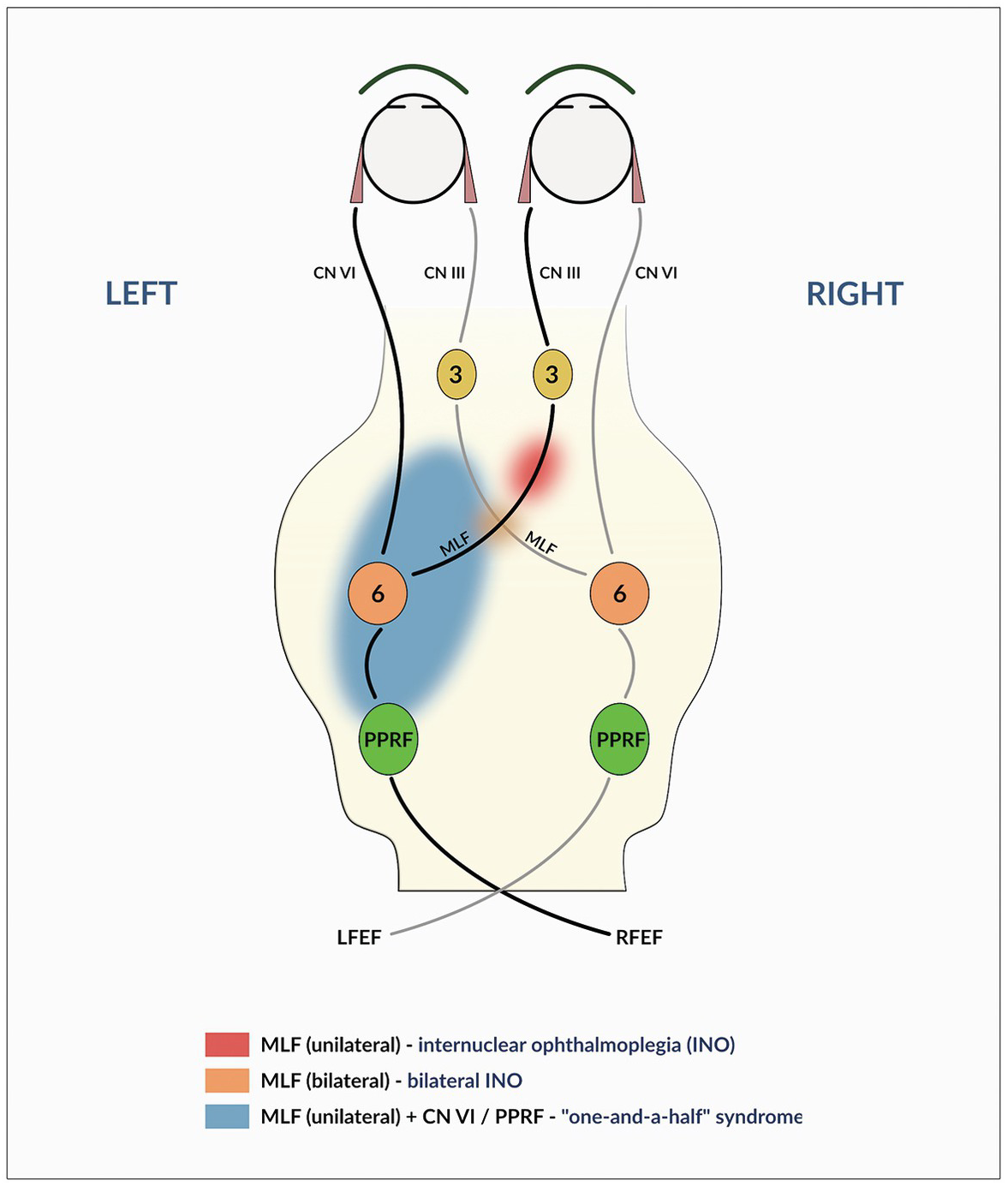
Intranuclear ophthalmoplegia (INO) and other ophthalmoplegias are common presentations in MS and NMOSD (Figure 1.2).
INO is caused by damage to the medial longitudinal fasciculus (MLF).
Patients with INO are unable to fully adduct the affected eye when looking to the opposite side. The abducting eye usually has nystagmus on end gaze.
A common initial complaint is double vision (diplopia).
In addition to affecting individual nuclei, lesions can involve multiple structures simultaneously.
Bilateral INO, where neither eye can adduct, can be seen if both MLFs are affected (Figure 1.2).
If the sixth cranial nerve and bilateral MLFs are affected, abduction of unaffected eye will be the only movement seen on examination. This is known as the “one-and-a-half syndrome” (Figure 1.2).
MS can affect any of cranial nerves that control individual eye movements.
A Maddox rod can be extremely useful in determining the pattern of ophthalmic weakness.
Infratentorial lesions can affect the coordination of eye movements, leading to nystagmus, vertigo, and dizziness in about 20% of MS patients.Reference Chavala, Kosmorsky, Lee and Lee37
Lesions in the vestibular nuclei, cranial nerve 8 at the site of entrance into the brainstem, brachium conjunctiva, and cerebellum can cause a presentation that may mimic vestibular neuritis or benign paroxysmal positional vertigo (BPPV).Reference Chavala, Kosmorsky, Lee and Lee37, Reference Sawicka-Pierko, Obuchowska and Mariak38
Vertigo in the MS patient can also be related to BPPV rather than to a new lesion,Reference Sawicka-Pierko, Obuchowska and Mariak38 and therefore should always be tested by way of the Dix–Hallpike maneuver.
Trigeminal neuralgia or tic douloureux.
Described as a sudden, severe, shooting, electrical pain that radiates down the path of the fifth cranial nerve.
This pain is typically very brief, on the order of seconds, but can occur multiple times in close succession.
The pain can be provoked by minimal stimuli such as gentle touch, chewing, or wind blowing over the affected area.Reference Meyerson, Van Stavern and McClelland39
While the disease commonly results from compression of the fifth cranial nerve by a vascular structure, it can often occur secondary to MS lesions in the pons.
If trigeminal neuralgia develops in a younger adult or if it presents bilaterally, diagnostic workup for demyelinating disease should be pursued.
Tumefactive Demyelinating Lesions
The terms “tumefactive demyelination” and “tumefactive MS” are oftentimes used interchangeably. However, they are not synonymous, and not all patients with tumefactive lesions meet the diagnostic criteria for MS (see Chapter 4).
Tumefactive demyelination is a relatively rare occurrence.
The average annual incidence is 3 cases per 1 million people.
Although tumefactive demyelinating lesions may pose a neurologic emergency, most patients typically do not present emergently to the hospital.
Potential complications from tumefactive lesions include:
Significant edema surrounding the lesion leading to mass effect, which carries the risk for possible herniation.Reference Klein, Ray and Carpenter40
Acute stroke due to vascular occlusion.
Seizures due to cortical involvement.Reference Sofat, Malik and Higgens41, Reference Torabi, Patel and Wolfe42
The management of emergent tumefactive demyelinating lesions is similar to emergent management of a CNS tumor with mass effect; it may require neurosurgical intervention and intensive care unit monitoring.
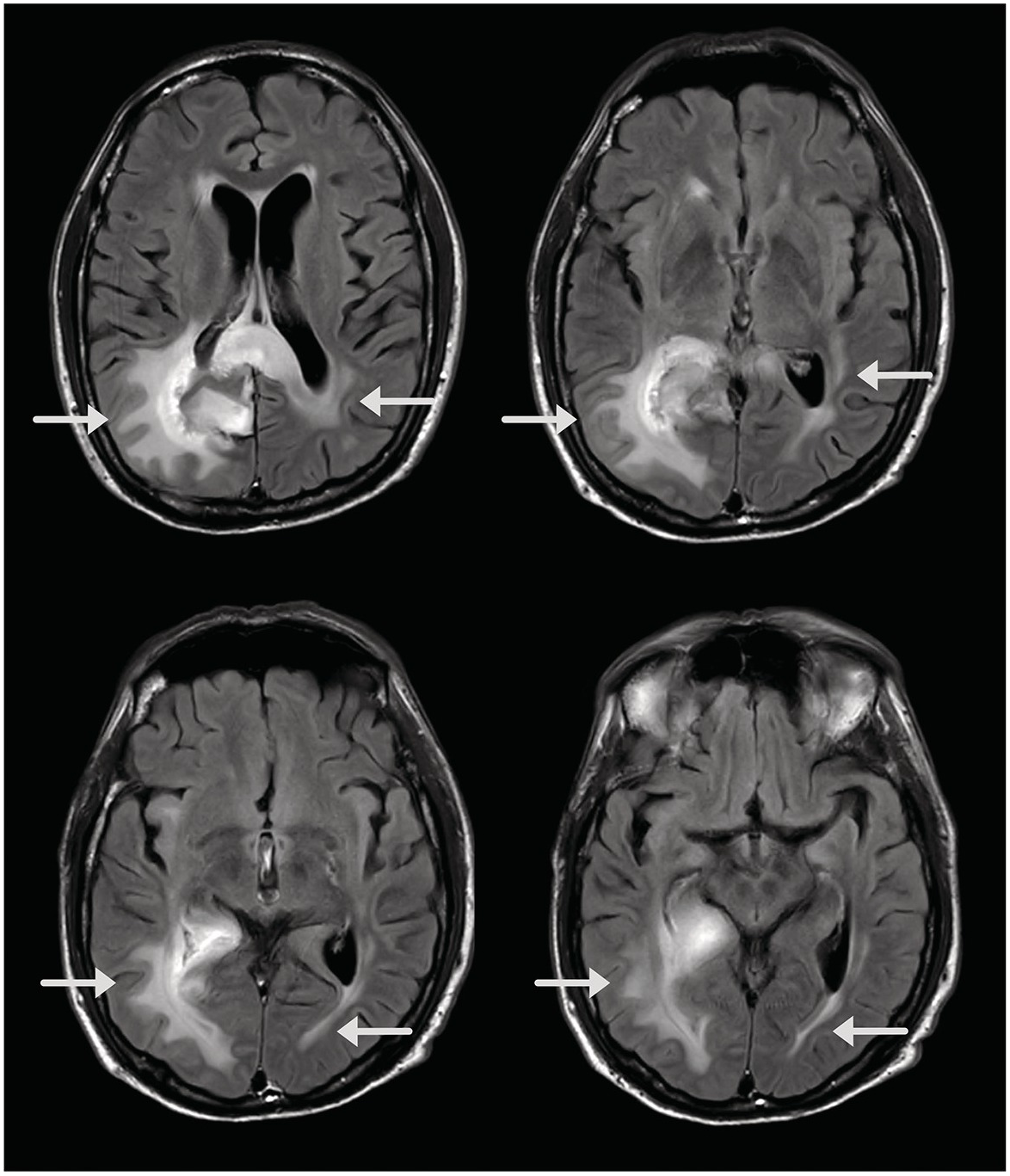
MRI typically shows (Figure 1.3):
Large (greater than 2 cm) lesions with little mass effect and surrounding edema.
Open-ring gadolinium enhancement of lesions.
The lesions are typically solitary, but multiple tumefactive lesions may occur in some instances.
Differentiating between a primary CNS tumor and a tumefactive lesion is challenging,Reference Frohman, Kramer and Dewey43 especially in the absence of brain lesions typical for MS. In such cases, a diagnostic brain biopsy may be considered.
Histopathologic specimens of tumefactive demyelinating lesions typically show perivascular infiltrates of mononuclear inflammatory cells with intermingled macrophages.
The risk of tumefactive MS is increased in patients who are weaning off highly potent disease-modifying therapies, most specifically natalizumab and fingolimod.Reference Anagnostou, Mandellos and Limbitaki44
Close clinical monitoring is warranted when discontinuing these medications.
Indications for Hospitalization
Most acute demyelinating episodes do not require hospitalization.
Hospital admission should be considered if:
The patient is unable to care for himself/herself at home due to the severity of the neurologic symptoms.
The patient requires a medication that can be administered only in a hospital setting (e.g., IV).
The patient has a life-threatening infection.
The cause of the patient’s presentation is unclear and a potentially life-threatening or permanently disabling cause is being considered in the differential diagnosis.
IV steroids are being administered for the first time and could cause an anaphylactic reaction.
Patient has uncontrolled diabetes mellitus and glucose monitoring/insulin administration may be required.
Conclusion
Most confirmed MS exacerbations can be managed in the outpatient setting. Pseudo-exacerbations should be ruled out by excluding infections. Patients with new-onset demyelinating syndromes require admission unless they can be seen by a neurologist within 1–2 weeks and the symptoms are mild and progressing slowly (i.e., paresthesias).
An MS relapse is defined as a clinical worsening or change in neurologic symptoms that persists for more than 24 hours.
Pseudo-exacerbations (including Uhthoff's phenomenon) should be considered before diagnosing a true relapse.
Not every acute demyelinating relapse requires treatment with steroids. These treatments are reserved for patients with symptoms that are severe enough to affect their ability to work or function at home.
Optic neuritis and transverse myelitis are common ED presentations of demyelinating disease.
Patients should be hospitalized when unable to care for themselves due to the severity of their symptoms, when they require IV therapy, or when their clinical presentation is unclear and a potentially life-threatening condition may be possible.


