


INTRODUCTION
Intensity-modulated radiotherapy (IMRT), allowing for highly conformal dose distribution to the tumour, whereas sparing the adjacent normal structures, is becoming standard treatment for prostate and head-and-neck (H&N) cancer since the late 1990s and early 2000.Reference Zelefsky, Fuks and Happersett1–Reference Butler, Teh and Grant3 However, rapid dose falloff of IMRT plans generally requires accurate treatment delivery therefore accurate margins and reproducible setup are paramount.Reference Manning, Wu and Cardinale4, Reference Hong, Tome and Chappell5
Image-guided radiotherapy (IGRT) has been developed and used to ensure accurate interfraction patient setup and dose delivery.Reference Houghton, Benson and Tudor6–Reference Kupelian, Langen, Willoughby, Zeidan and Meeks11 Commonly used in-room X-ray imaging technologies for IGRT includes: kilovoltage, megavoltage cone beam computed tomography (KVCBCT and MVCBCT) and fan beam computed tomography (such as KVFBCT and MVFBCT).Reference Kupelian, Lee and Langen12–Reference Meeks, Harmon and Langen18
The Elekta Oncology Systems Synergy medical linear accelerator is among the first commercially available treatment units with integrated KVCBCT system implementing X-ray-based volumetric imaging system (XVI, Elekta Oncology Systems, Norcross, GA, USA) capable of acquiring patient images in the treatment position. Interfraction displacements can be quantified and adjusted using KVCBCT so that daily treatment dose can be accurately delivered. On the other hand, helical tomotherapy (HT, Accuray Inc., Sunnyvale, CA, USA), employing on-board megavoltage computed tomography of energy of 3·5 MV photon beam via a fan beam (MVFBCT), also enables pre-treatment imaging and adjustments for image-guided IMRT.
In this study, we report our IGRT experience using the Elekta Synergy KVCBCT and HT MVFBCT and to address interfraction variations in patient setup during the course of prostate and H&N cancer IMRT treatment. The daily shifts obtained from these two image guidance (IG) in the mediolateral (ML), craniocaudal (CC) and anteroposterior (AP) dimensions were analysed and compared. The purposes of the work are (1) to explore if/how different image modalities affect patient repositioning and the clinical target volume to planning target volume (CTV-to-PTV) margins for two commonly IGRT-treated sites such as prostate and H&N cancers; (2) to investigate if there is a significant difference in patient interfraction setup during early, middle and late treatment courses; and (3) to explore whether it is possible to reduce daily IG to a less frequent pattern.
MATERIALS AND METHODS
Patient characteristics and setup procedures
All H&N and prostate cancer cases in this study were treated with image guided IMRT. Two CT-based IG modalities, KVCBCT and MVFBCT, were investigated for 132 H&N and prostate patients. A total of 1,097 and 957 pre-treatment scans for prostate, 974 and 632 scans for H&N on a TomoTherapy unit or an Elekta Synergy accelerator were investigated. For prostate cases, most patients received a prescription dose of 70–75·6 Gy with a daily fraction dose of 1·8–2·5 Gy; for the H&N patients, the prescription dose was in the range of 45–66 Gy with daily fraction dose of 1·8–2·2 Gy.
Our routine image-guided radiotherapy procedure included setting up patients to the lasers based on skin tattoos (for prostate) and the marks on the mask (for H&N), acquisition of pre-treatment CT images, image registration and making appropriate shifts if necessary. The patients were ensured in proper position with immobilisation device: thermoplastic mask for H&N and custom alpha cradle for prostate treatments. Pre-treatment scans, always covering PTV and a few slices up and down in CC direction, were registered to the reference image set (the planning image set) to verify patient positioning using either bony structure and/or soft tissue in three dimensions. We started with automatic registration using bony structure for all cases, except for prostate cases treated on Elekta Synergy using KVCBCT, which started with soft tissue using automatic registration.Reference Borgefors19, Reference Hristov and Fallone20 All auto-registration were followed by visual inspection and manual adjustment to align PTV with planning CT. Therefore, all cases under investigation used anatomy-based manual registration, although different methods for automatic registration were used as a starting point. For H&N cases, automatic registration using bony structure was usually adequate for both modalities. For prostate, however, manual adjustments were common in both IG modalities because of internal organ movement. The goal of the manual adjustment was to align PTV with the reference. All daily registration and corresponding shifts were reviewed by the physicians.
Data/statistical analysis
Analysis of variance (ANOVA) was employed to explore how different image modalities affect patient repositioning in ML, CC and AP directions. Statistics analyses were done using SAS (version 9.2, SAS Institute Inc., Cary, NC, USA). p-Value of 0·05 or smaller was considered as statistically significant.
CTV-to-PTV margin
The setup errors in ML, CC and AP directions were used to calculate the population-based CTV-to-PTV margin, when IGRT is not used, using the following equation (assuming minimum dose to 95% CTV for 90% of patients): margin 2·5∑ + 0·7σ,Reference Van Herk21 where the systematic variation ∑ is the standard deviation (SD) of the average setup shifts per patient in the group of patients and the random variation σ is the root mean square of the SD of the day-to-day setup positions of all patients. A uniform margin is defined as the calculated maximal margin among three translational directions for each image modality.
Time dependence on patient repositioning in the course of treatment
We broke the entire treatment course into three equal intervals in chronic order: early-, middle- and late-treatment stage to investigate if patient setup errors (and margins) varied with treatment progressing. We calculated the interfraction shifts and the CTV-to-PTV margins over the three defined stages for patients imaged with both modalities. ANOVA was used to test whether there is a significant difference in three defined treatment stages.
Frequency of IG
Daily IG corrects setup displacements and ensures dose delivery accuracy; however, it inevitably introduces an extra imaging dose and prolongs the patient's treatment time on table. To explore the possibility of reducing daily IG to a less frequent pattern, such as every other day or every few days, we investigated the occurrence and distribution of large displacements. To identify large displacements, a three-dimensional shift vector (R), calculated as the square root of the sum of square of the shifts in ML, CC and SI directions, was constructed for each fraction. For both sites and both IG modalities, criteria of 5 and 10 mm were used as cutoffs. Days with shift vector greater than cutoffs were identified as the days that IG should not be omitted.
In addition, daily shift data were re-sampled assuming pre-treatment CT scans were performed less frequently, e.g., every other fraction, every three, four or five fractions. Considering average daily shifts as the baseline, we subtracted the baseline shifts from the averaged shifts based on the re-sampled frequencies. A residual difference (Δ), defined as the difference in averaged displacements between daily pre-treatment CT scan and assumed pre-treatment CT scan given at other frequencies, was calculated. A t-test was performed to test whether less frequent pre-treatment IGRT scans would result in significant difference compared with setup guided by daily IGRT scans. The CTV-to-PTV margins were calculated if IGRT given at different frequencies.
RESULTS
Figure 1 shows the average shifts and SDs in ML, CC and AP direction using KVCBCT and MVFBCT for prostate patients. Patient #1–36 are prostate cases scanned with KVCBCT, whereas #37–72 are the cases scanned with MVFBCT. In general, both MVFBCT and KVCBCT yielded similar average shifts, whereas MVFBCT shows larger variability compared with KVCBCT in three translational dimensions.
Average shifts and standard deviations in mediolateral, craniocaudal and anteroposterior direction for prostate cancer pateints treated with KVCBCT and MVFBCT. Thirty-six patients from each modality are analysed. Abbreviations: KVCBCT, kilovoltage cone beam computed tomography; MVFBCT, megavoltage fan beam computed tomography.

Table 1 shows the average shifts and SDs in ML, CC and AP directions using KVCBCT and MVFBCT image scan. The SDs for KVCBCT were generally smaller than MVFBCT for both prostate and H&N. At a significance level of 0·05, no significant differences were observed between MVFBCT and KVCBCT scans in all three translational directions (Figure 2).
The average daily shifts and SDs for 72 prostate and 60 H&N cases imaged using KVCBCT and MVFBCT in this analysis

Abbreviations: SD, standard deviation; H&N, head-and-neck; KVCBCT, kilovoltage cone beam computed tomography; MVFBCT, megavoltage fan beam computed tomography.
Registered pre-treatment images with the planning CT images for (a) KVCBCT on an Elekta Sygnery-S Linac and (b) MVFBCT on a TomoTherapy unit. Daily computed tomographies (shaded) and the planning images (non-shaded) are shown. Abbreviations: KVCBCT, kilovoltage cone beam computed tomography; MVFBCT, megavoltage fan beam computed tomography.
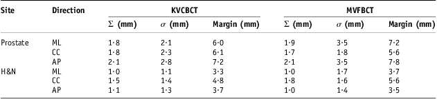
Table 2 shows systematic positioning error (Σ), random positioning error (σ) and calculated CTV-to-PTV margin. For the same treatment site, MVFBCT generally yielded comparable systematic error but larger random positioning error compared with KVCBCT. The same image modality resulted in larger Σ and σ for prostate than H&N cases in all translational directions. The systematic errors are 1·9, 1·7, 2·1 mm and 1·0, 1·8, 1·0 mm for prostate and H&N cases using MVFBCT guidance, which were comparable with results by Den et al.Reference Den, Doemer and Kubicek22 for H&N IMRT using KVCBCT.
Systematic (Σ) and random (σ) setup errors and the calculated CTV-to-PTV margins using KVCBCT and MVFBCT for prostate and H&N cases
Abbreviations: KVCBCT, kilovoltage cone beam computed tomography; MVFBCT, megavoltage fan beam computed tomography; H&N, head-and-neck; ML, mediolateral; CC, craniocaudal; AP, anteroposterior.
Averaged patient shifts in the course of treatment
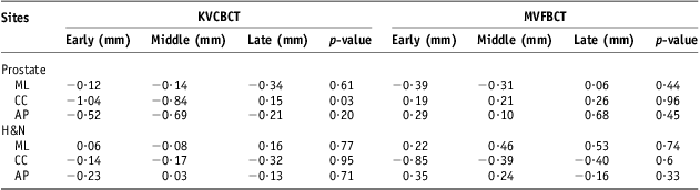
Table 3 shows the average shifts in three equally distributed stages in the treatment course for the prostate and H&N patients. For both sites with either MVFBCT or KVCBCT as daily setup guidance, no statistically significant differences were found in ML and AP directions. A value of p = 0·03 was found in CC direction for prostate patients with KVCBCT, indicating progressive smaller shifts towards the end of treatment course. It might be understood by gradual improvement of positioning accuracy by therapists along the treatment course.
Average shifts for prostate and H&N patients during the early, middle and late treatment course using KVCBCT and MVFBCT
Note: p-values among the early, middle and late treatment stage were calculated using ANOVA model.
Abbreviations: H&N, head-and-neck; KVCBCT, kilovoltage cone beam computed tomography; MVFBCT, megavoltage fan beam computed tomography; ANOVA, analysis of variance; ML, mediolateral; CC, craniocaudal; AP, anteroposterior.
The calculated CTV-to-PTV margins in ML, CC and AP directions for prostate are 5·6, 6·0, 7·2 (early), 6·0, 6·2, 7·2 (middle) and 6·4, 6·3, 7·4 (late) for KVCBCT, compared with 7·3, 5·9, 8·2 (early), 7·4, 5·6, 8·0 (middle) and 7·2, 5·8, 7·6 (late) for MVFBCT. For H&N, the calculated CTV-to-PTV margins in ML, CC and AP directions are 3·1, 5·1 and 3·2, 3·5, 5·0 and 4·1, 3·3, 4·5 and 3·8 for KVCBCT, 3·6, 5·9 and 3·4, 4·1, 6·0 and 3·7, 3·8, 5·2 and 3·6 for MVFBCT at early, middle and late stages.
IG frequency
Table 4 shows the percentage of the days with large displacement for pooled prostate and H&N fractions. In general, cases with MVFBCT as IG had larger shifts than those with KVCBCT as IG. If 5 mm was used as the cutoff, 35·1% of prostate treatments imaged with KVCBCT and 52·2% on MVFBCT would have shift vector >5 mm and IG on those days should not be omitted. If 10 mm was used as the criterion, 3% of prostate treatments for KVCBCT and 10·8% of the prostate treatment for MVFBCT should use IG. Displacements in H&N treatments were generally smaller because of the rigid anatomic structure of H&N. However, 8·6% of the treatment with KVCBCT as IG and 17·4% with MVFBCT as IG would need IG if 5 mm criterion was used.
Percent of fractions with shift vector (R) are >5 and 10 mm

Note:  $$--><$>R = \sqrt {{{x}^2} + {{y}^2} + {{z}^2} } $$$
, where x, y and z are shift in ML, CC and SI direction, respectively.
$$--><$>R = \sqrt {{{x}^2} + {{y}^2} + {{z}^2} } $$$
, where x, y and z are shift in ML, CC and SI direction, respectively.
Abbreviations: KVCBCT, kilovoltage cone beam computed tomography; MVFBCT, megavoltage fan beam computed tomography; H&N, head-and-neck.
We further investigated the pattern of the days with large setup displacements. It appeared that the occurrence of the large displacements was random throughout the treatment course for each patient. Given the large percentage, the days with significant displacements and stochastic nature of their occurrence, we conclude that daily IG should still be optimal.
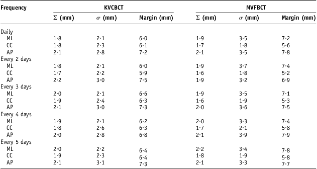
Figure 3 shows residual differences in mean shifts in ML, CC and AP direction for prostate cases assuming pre-treatment CT scans were given daily, every two, three, four and five fractions. For each direction, from left to right (a–e), the symbols show the IGRT were given in different frequencies. The first symbol (a) in each direction represents the baseline assuming daily IGRT was given. Using the daily IGRT average shifts as baseline, the maximum absolute residual shifts were 0·3, 0·1 and 0·4 mm for MVFBCT, and 0·1, 0·4 and 0·2 mm for KVCBCT, respectively. Among different frequencies, significant differences were found if IGRT was given in every 2 days for prostates, and significant difference in lateral and longitudinal directions were also seen for H&N cases (data not shown). Table 5 shows the calculated CTV-to-PTV margins for prostate cases assuming IGRT given at different frequencies, such as daily, every 2, 3, 4 and 5 days.
Residual differences (Δ) in average shifts in ML, CC and AP directions for prostate cases assuming pre-treatment IGRT scans were given daily (a), and every 2 (b), 3 (c), 4 (d) and 5 (e) fractions. For each direction, from left to right, the symbols (a–e) show the IGRT was given in different frequencies. Abbreviations: ML, mediolateral; CC, craniocaudal; AP, anteroposterior; IGRT, image-guided radiotherapy; MVFBCT, megavoltage fan beam computed tomography; KVCBCT, kilovoltage cone beam computed tomography.
The calculated CTV-to-PTV margins for prostate cases assuming IGRT given at different frequencies, such as every 2, 3, 4 and 5 days
Abbreviations: CTV, clinical target volume; PTV, planning target volume; IGRT, image-guided radiotherapy; KVCBCT, kilovoltage cone beam computed tomography; MVFBCT, megavoltage fan beam computed tomography; ML, mediolateral; CC, craniocaudal; AP, anteroposterior.
DISCUSSION
We retrospectively analysed patient interfraction setup variations for prostate and H&N IMRT treatment using KVCBCT and MVFBCT as IG. IGRT data were acquired on two accelerators including Elekta Syngery and TomoTherapy. Elekta Synergy integrates a kV X-ray source and an amorphous silicon flat-panel detector into the gantry to provide photon energy of 100–120 kVp for pre-treatment cone beam imaging. Helical tomotherapy MVFBCT, however, employing the actual treatment beam (with reduced energy of 3·5 MV photons) from the linear accelerator as the X-ray source for image acquisition, enables pre-treatment fan beam imaging and adjustments for IGRT. The KVCBCT system is superior to MV-based imaging modalities in general.Reference Stutzel, Oelfke and Nill23 In this study, we found no statistically significant difference in terms of average daily shifts between KVCBCT and MVFBCT image scans for H&N and prostate patients. However, superior image quality of KVCBCT scans resulted in smaller random setup errors in translational directions for both H&N and prostate (Table 2) as compared with MVFBCT. As a result, larger CTV-to-PTV margin in MVFBCT may be needed than that of KVCBCT for both prostate and H&N IMRT if determined from IGRT data. Our analysis (Table 2) also suggested anisotropic margins in three translational directions for KVCBCT and MVFBCT. Uniform margins (maximal margins among three translational directions) of 7·8 mm for MVFBCT and 7·2 mm for KVCBCT in prostate IMRT, and 5·6 mm for MVFBCT and 4·8 mm for KVCBCT in H&N IMRT may be appropriate. One of the limitations of this work is no rotational deviations were considered in margin calculation because of the unavailability of such data. Although the rotational correction may slightly offset the deviations in translational shifts, we expected a negligible impact according to Li et al.Reference Li, Qi and Pitterle24
Although IGRT increases the precision and reproducibility of radiotherapy treatments, concerns have been raised for its additional doses to patient, which might lead to unnecessary toxicity for sensitive structures. Ding GX et al.Reference Ding and Coffey25 concluded that extra dose to radiosensitive organs can be amounted to 300 cGy over an entire treatment course if KVCBCT scans are acquired daily, the average MVFBCT imaging dose on tomotherapy will be even higher (roughly 2–5 cGy per scan) to achieve acceptable image quality for patient alignment. In accordance with the as low as reasonably achievable principle in radiation protection, we explored the option of acquiring the image in less frequent fashion, while maintaining the accuracy of dose delivery. This analysis indicates that daily IG scan still remains as the optimal choice to eliminate possible significant patient positioning error not only for prostate but also for H&N, consistent with Kuplian et al.Reference Kupelian, Langen, Willoughby, Zeidan and Meeks11
CONCLUSION
KVCBCT and MVFBCT image may provide different image quality in IGRT for H&N and prostate. However, no statistically significant difference in average interfraction patient setup was seen between KVCBCT and MVFBCT image guidance. In addition, we observed no statistical difference in early, middle and late stage of the treatment course. Daily IGRT is optimal to ensure accurate dose delivery.
Our data suggests that the CTV-to-PTV margin, when determined from IGRT data, may be varied for different imaging modalities in H&N and prostate irradiation. In the absence of IGRT, the calculated uniform CTV-to-PTV margins (based on the maximal deviations in three translational directions) of 5·6 mm for H&N and 7·8 mm for prostate, respectively, may be appropriate using MVFBCT, while for KVCBCT, a smaller margin of 4·8 mm for H&N and 7·2 mm for prostate, respectively, may be considered. Image modality with better image quality is always encouraged in clinical practice if possible.
Acknowledgement
The authors wish to thank the physicians in Medical college of Wisconsin and University of Colorado, Denver.
Conflict of interest
There is no conflict of interest for all authors.



