
1.1 Beginnings
There is a widely held view that the discipline of palaeopathology began in 1774 with the publication of Esper’s account of fossil bones found in caves in Bavaria, although there is very little hard evidence to support this view, as I will discuss further below. What is almost certain is that an interest in, and fascination with, bones has a very much longer history, perhaps because in them resides the last tangible evidence for individual existence. The wish to have one’s bones buried either close to those of a loved one, or (if not that, then) in one’s own country, for example, can be traced back at least as far as the Iliad and the Trojan War; ‘… inter my bones not far from thine …’ urges the ghost of Patroclus to Achilles,Footnote 1 while Nector suggests to Atreides that ‘… the friends of the dead … bring their bones home to their children ….’Footnote 2 For none was the desire for the return of one’s bones to home soil expressed more fervently than by those who died during the Crusades, although the practice predated them, having earlier been followed on behalf of others who had died far from home, especially by the Germans who died in foreign lands, and later by the French and the English.Footnote 3 Following their death in the Holy Land, the bodies of the rich and the nobility might be boiled and the flesh removed so that their bones could be taken back for burial by those who survived them. This practice was condemned by the Church, and it was eventually forbidden by Pope Boniface VIII in the bull Detestande feritatis issued on 27 September 1299. In its place Boniface decreed that all those who died in a foreign catholic country should be immediately buried at the site chosen by the deceased, or, failing that, temporarily buried at or near the place of death and transferred to the final place selected for burial only after the body had turned to ashes, a process that might be long delayed.Footnote 4
During the medieval period, a rather more macabre interest in the skeleton was displayed by artists depicting memento mori such as the Three Quick and the Three Dead, the danse macabre and skeletons with a caption taking a form such as: ‘As you were, so was I, and as I am, so shall you be’.Footnote 5 In depictions of the Three Quick and the Three Dead, three richly dressed princes or noblemen are seen riding to the hunt, only to be greeted by themselves as skeletons; as we are, so shall you be. The lesson of all the various forms of memento mori was presumably to focus the medieval mind on the vainglories of earthly life and, in its place, prepare it for the rewards that will hopefully come in the life hereafter. Many illustrations of this kind are to be found in churches and cathedrals throughout Europe, and it is often said that this art form was particularly stimulated by the coming of the Black Death, which was certainly enough to remind one that life was indeed transitory and uncertain.Footnote 6 Another popular image, the danse macabre or dance of death, first appeared in the Cimetriére des Innocents in Paris in 1424 and quickly spread throughout the rest of Europe. In these pictures, death is shown as a skeleton, sometimes wearing a crown on his head, inviting people of all kinds to a dance that leads inevitably to the grave; again the intention seems to be to act as a reminder of the closeness of death and the necessity to be prepared for it at all times.Footnote 7
1.2 Relics
Nor should we overlook the importance to the medieval mind of relics, the physical remains, usually, but not exclusively, bones of saints and other holy men and women, which were imbued with special powers and with special access to the divine. The cult of the relic can be traced back to the persecution of the Christians during the second and third centuries, and by the eighth century relics were required for the consecration of altars; by the twelfth and thirteenth centuries body parts were required for the procedure of canonisation to take place.Footnote 8 Churches were built or altered to contain relics,Footnote 9 and a huge trade in relics grew up throughout the medieval world and so, naturally, did fraud. Many bones were certainly not from those to whom they were attributed, and in many cases, they were not even human. Nevertheless, great collections were made, most notably perhaps by Charlemagne, whose collection came to reside in Aachen Cathedral,Footnote 10 and by Frederick Barbarossa, who took the bones of the Magi from Milan and presented them to Cologne Cathedral in 1164. Relics attracted visitors to the churches and cathedrals in which they lay, and pilgrims might travel many miles to see part of a saint or the reliquary in which it was housed. The churches profited greatly from the relic business and so did others. Chaucer’s Pardoner, for example, made more money in a day by letting some poor person see the relics he carried with him than that man made in two months, even though the relics on display were from no saint but were nothing more than pig’s bones.Footnote 11
1.3 Curiosities
The artists who created the various forms of memento mori referred to above clearly had some knowledge of the anatomy of the human skeleton, even if they are rarely accurate as to detail.Footnote 12 Although the dance of death persisted in some parts of Europe into the nineteenth century, most of these art forms had disappeared by the early modern period, but for a short time in the seventeenth and eighteenth centuries, human bones were incorporated on gravestones in place of the otherwise overly comfortable or sentimental images that were the norm.Footnote 13 The skull was now the element most frequently depicted, sometimes with both full face and side view being shown, giving it a quasi-three-dimensional appearance. It is usually reasonably accurately depicted, but when other bones are shown as well, they are often hard to recognise and sometimes are more like animal than human bones, which supposes that the stonemasons may not have been so well informed anatomically as their medieval forebears and, instead, tended to take their models from the butcher rather than the ossuary.
Among the bones that were returned for burial from distant lands, or were removed by the medieval gravediggers to be placed in ossuaries, there must have been some with pathological abnormalities, but there seem to be no early published accounts of such material. What are to be found, however, are published accounts of extraordinary skeletons, appearing many years before Esper’s supposed foundation stone was laid in the palaeopathological edifice, and almost always the authors were medical men. One might cite, for instance, Bernard Connor’s account in 1695 of a skeleton ‘all the Bones whereof were so united as to make but one continued Bone without Articulation ….’Footnote 14 Connor encountered this unusual skeleton during his travels on the continent, and he supposed that it had come from a graveyard or charnel house. What is almost certain, however, is that this was a case of ankylosing spondylitis, probably the first to have been described, and that Connor recognised the difficulty in respiration that would have resulted to the individual during life consequent upon the fusion of the costo-transverse and costo-vertebral joints.Footnote 15 Some ten years previously, Thomas Molyneux, on his own journeys through Europe, had come across a ‘prodigious large’ Os Frontis in the medical school at Leyden that was more than twice the size of several normal bones with which he compared it. Extrapolating from this to the general size of the skeleton (although by what means he neglects to tell us), Molyneux supposed that his subject must have been at least eleven or twelve feet tall and ‘the greatest Monster the World ever saw’.Footnote 16 The gigantic theme was further explored some years later by William Cheselden, better known for his operation for the bladder stone than for his anthropology. In a very short report (of fourteen lines) Cheselden commented on some, presumably Romano-British, bones that had been unearthed at St Albans. The left femur was reported to be twenty-four inches long, the right, twenty-three inches, and each tibia was twenty-one inches long. According to Cheselden’s calculations the bones must have belonged to a man who was ‘eight foot high’.Footnote 17
These specimens and others like them were regarded as mere curiosities by those who described them, and in this respect they were like the other anatomical curiosities collected, mostly by medical men, during the seventeenth and eighteenth centuries, a number of which formed the basis of a collection belonging to the Royal Society, found to be sadly neglected by a Committee for Inspecting the State of the Repository in the first quarter of the eighteenth century.Footnote 18
1.4 And So, Back to Esper
It seems likely that Esper’s position as the father of palaeopathology has its origins in a remark made by Moodie in his Paleopathology.Footnote 19 At page 62, he writes,
The earliest reference, in paleontological literature to the pathological nature of fossil bones was by E. J. C. Esper (1742–1810), Professor at Erlangen, in 1774 as cited by Goldfuss. Esper described on the lower half of the femur of a cave bear (Ursus spelaeus), what he regarded as an osteosarcoma.
Moodie made two errors in this passage. First, it was not Eugen Johann Christoph Esper who was the author of the work referred to, but his older brother, Johann Friedrich.Footnote 20 Second, the lesion referred to was most certainly never referred to by Esper as an osteosarcoma. What Esper actually wrote, referring to an illustration of the bone in question, was
Dass es der Rest von einem osse Femoris, und zwar ab der untere Theil desselbigen ist, zeigt sich von selbst. Nur muss ich sagen, dass die grosse Dicke von c. gegen d, ein Callus ist, mit welchem die Natur einem Bruch dieser Röhre wieder geheilt.Footnote 21
Roughly translated, the second sentence reads: I must say that the large thickening from c to d is a callus, with which Nature has again healed the break.
Goldfuss,Footnote 22 to whom Moodie also refers, did indeed mention Esper and gave the correct reference to his book (on page 144 of his own work) and says (on page 276):
Esper giebt (tab 14 f2 p74) die Abbildung eines Hüftknochens, welcher Spuren eines Bruches zeigt, den ein Callus wieder verband.Footnote 23
Or: Esper provides the illustration of a hip-bone, which shows traces of a break, which a callus has connected again.
Various authors who have followed Moodie have perpetuated at least one of the errors in the passage from his book quoted above. There is little doubt that Esper described what he thought was a healed fracture, although the illustration of it is not terribly convincing There also seems to be some doubt as to whether all those who wrote after Moodie had read the original; Moodie himself certainly had not, as he himself confessed to Henry Wellcome in a letter written in 1931.Footnote 24
In 1774 one E. J. C. Esper, a professor in one of the schools, possibly a medical school at Erlangen, published a pamphlet [sic] in which he described what he took to be an osteosarcoma on the lower half of a cave bear femur. This was probably a healed fracture. I have never seen the pamphlet but it should be possible to secure a copy.
Moodie, who was working for Wellcome at the time, no doubt hoped that Wellcome would be able to obtain the pamphlet, which is, in fact, a quarto volume of some 148 pages, but there is no evidence that he did so. It seems likely that Moodie took his information from Mayer, whose paper he had read. In this paper, Mayer wrote that Esper had described a femur of a cave bear ‘und zwar an dessen oberem abgebrochenen Ende ein Osteosarcoma des Knochens’.Footnote 25 Mayer felt that the lesion was, in fact, a healed fracture with some necrosis, and it was this opinion that Moodie duly recorded (on page 62 of Paleopathology) and that he relayed to Wellcome. Quite how Mayer arrived at the notion that Esper had described an osteosarcoma is by no means clear; I can find no evidence that Esper referred to anything like it anywhere in his original publication,Footnote 26 and it seems unlikely that an explanation for this misunderstanding will now be forthcoming.
1.5 An Obsession with Skulls
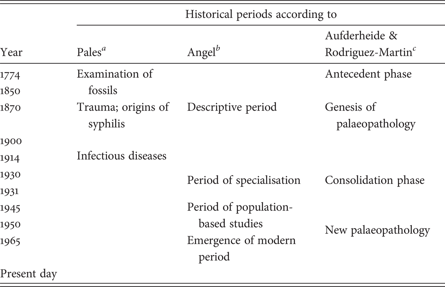
A number of authors have tended to divide the early period of palaeopathology into various phases. For example, Pales described three phases, the first between 1774 and 1870, which was devoted to the study of fossil faunas; the second from 1870 to 1900, when trauma and the origin of syphilis were pre-eminent among the study of human remains; and the third from 1900 given over to the study of infectious diseases.Footnote 27 Angel, by contrast, proposed a different scheme.Footnote 28 Activities prior to the First World War he considered to form a descriptive phase, the between-war period he termed the period of specialisation, while the 1950s ushered in the beginning of population-based studies, and the 1960s heralded the modern period. Angel considered that the modern period was announced by three events, the publication of Jarcho’s symposium on palaeopathology, held in Washington in 1965; the publication of Diseases in Antiquity (edited by Brothwell and Sandison); and the founding of the Paleopathology Association by the Cockburns and others in 1973.Footnote 29 A different scheme again has been proposed by Aufderheide and Rodríguez-Martin in which the beginnings of palaeopathology are dated from 1870 to the start of the First World War; a consolidation phase occupied the inter-war years, with the ‘new’ palaeopathology emerging at the end of the Second World War and continuing to the present.Footnote 30
Whatever scheme may be preferred by the student of the history of palaeopathology, and none of the divisions within them coincides or is consistent between them (see Table 1.1), the theme that was actually pre-eminent in the study of human remains in the latter part of the nineteenth century and for almost the first half of the twentieth was an obsession with skulls and race; pathology was very much second fiddle to the greater part of the work that went on then. The theoretical underpinning for this work on race seems to have stemmed from Blumenbach’sFootnote 31 publications, in which he claimed to be able to characterise humankind into five distinct races on the basis of his analysis of their morphology, especially that of the skull,Footnote 32 and also from Gall’s invention of ‘cranioscopy’, by which means various moral and mental faculties of an individual could be determined from the shape of the skull. Cranioscopy later became phrenologyFootnote 33 and, according to Luyendijk-Elshout, was the stimulus for the development of craniology during the first half of the nineteenth century,Footnote 34 although I think that the use of skulls in determining racial characteristics was probably of more importance. Whatever the impetus, the discovery of the Native American burial mounds during the push west in the United States provided an enormous collection of material on which to work, and none was more energetic in the cause than Samuel George Morton.Footnote 35 Morton was a physician in Philadelphia who, although he did not venture into the field himself, nevertheless amassed an enormous number of skulls from other collectors – apparently at the cost of between ten and fifteen thousand dollarsFootnote 36 – and which was to become widely acclaimed as one of the wonders of the scientific world. Morton used his collection to categorise the various races on the basis of their mean cranial capacity (as a supposed indication of their intellect), which he measured originally by filling the skull with sieved white mustard seed, having first occluded the various orifices to prevent the seed escaping, and then measuring the volume of the seed used. The cranial capacity was equivalent to the volume of seed needed to fill the skull to the level of the foramen magnum. Later Morton substituted the mustard seed with lead shot, one-eighth of an inch in diameter, to try to obtain the volume with greater accuracy. The results of his labours, obtained from the measurement of many hundreds of skulls, were published in an immensely influential book and arrived at the very satisfactory ranking (to him) of the races from Caucasian at the top, then descending to Mongolian to Malay to American (native American, that is) and, finally, Ethiopian in fifth place.Footnote 37 Morton’s data have been much discredited in recent years, having attracted especial opprobrium from Stephen Jay Gould, who noted a number of methodological errors, most notably Morton’s failure to separate male from female measurements. While not going so far as to accuse Morton of deliberate fraud, Gould nevertheless thought, on the basis of his reanalysis of the data, that some subconscious ‘finagling’ had gone on to ensure that the results concurred with ‘a truth passionately held …’ before the study was undertaken. From his reworking of the data, Gould found that Native Americans actually held first rank, while modern Caucasians were relegated to a miserable third (out of six).Footnote 38
a n 27.
b n 28.
c n 30.
Following Morton, the mania for collecting skulls seemed to know no bounds. After the Civil War in America had ended, the army turned its attentions to the Native Americans in the west. The Surgeons General urged their medical officers to collect skulls typical of the Indian tribes or those with rare pathological conditions for the Army Medical Museum. It has been estimated that thousands, perhaps hundreds of thousands, of skulls were amassed and disposed of to various institutions by this means, often simply to sit on shelves and collect dust for many years to come.Footnote 39 Nor was Europe exempt from the mania, and large collections were brought together, probably none greater in extent than that due to Joseph Barnard Davis.
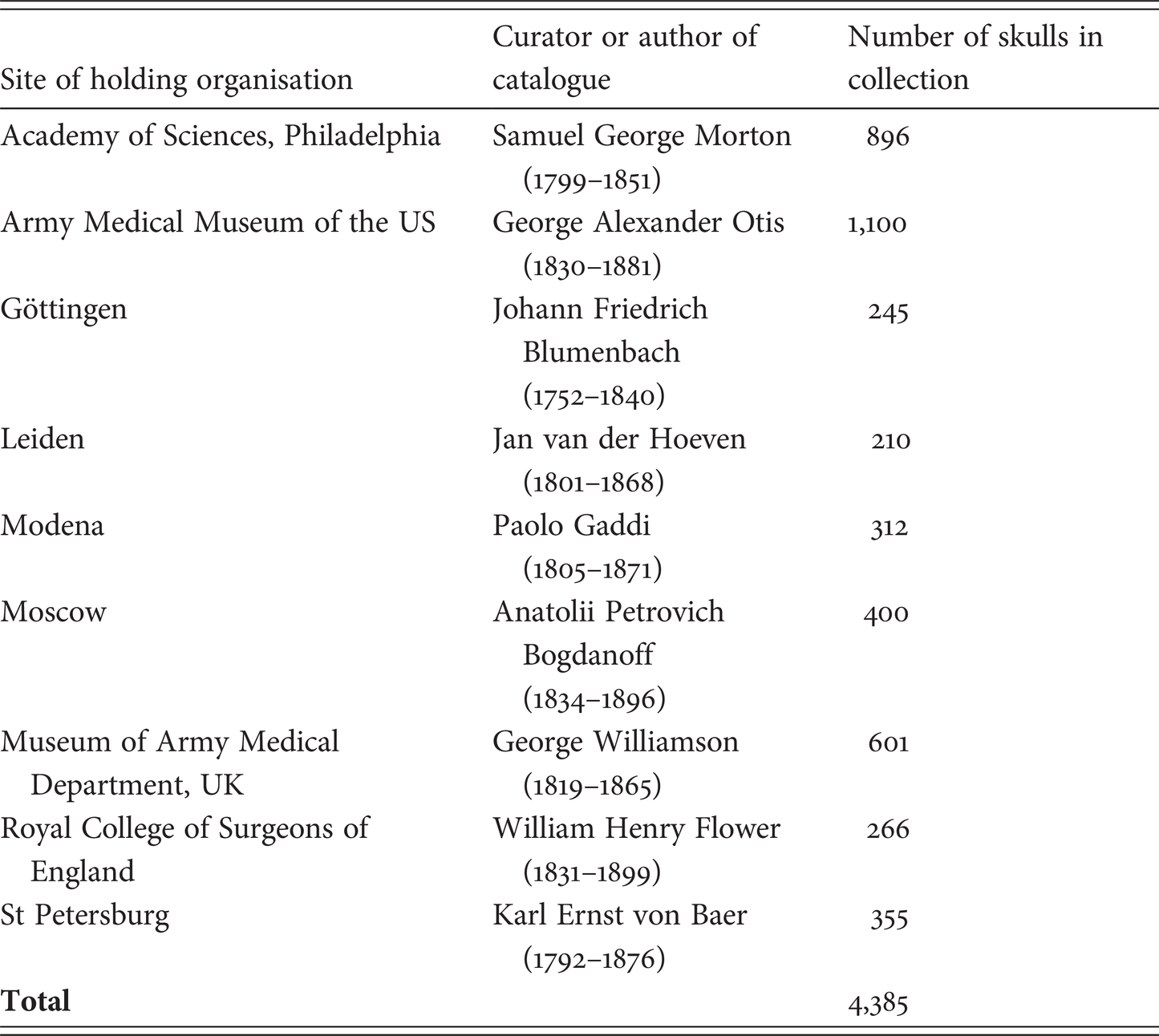
Davis was a physician in Stoke-on-Trent in Staffordshire, a place more famous for its potteries than its anthropology. Like Morton, he acquired his collection second-hand; exhorting men he knew in foreign countries (especially those of the British Empire) to find and send him skulls, otherwise buying collections from other craniologists, including that of Professor van Lidth of Utrecht, which included the skulls of Napoleonic soldiers. There was, indeed, a considerable traffic in human skulls, what Luyendijk-Elshout refers to as the ‘commercium craniorum’.Footnote 40 Davis, like Morton, was concerned with measuring cranial capacity, this time using clean Calais sand of specific gravity 1.425, weighing the sand and then calculating the volume. He published a catalogue of his collection, including all the measurements he made, some 25,000 in all, in 1867 and a supplement to it in 1875.Footnote 41 In the introduction to the catalogue he lists some of the other collections he knew of and held in various museums in Europe and in America (Table 1.2), accounting for more than 4,000 skulls in total. When he died in 1881 his obituarist damned his efforts with faint praise: ‘… all his works showed that his strong points were untiring energy in collecting and recording of specimens, rather than any deep power of observation, judgement, or induction’.Footnote 42 The work that the craniologists were undertaking was indeed not universally admired, as Davis himself remarked, in the preface to the Supplement,
… some of great repute have declared that the distinctions between skulls can scarcely be regarded as of any scientific value, others … urge me to introduce general summaries, which shall mark out the features of families of crania, and make each race to be distinguished readily. Were such a such a course feasible, it would be one most agreeable to take; but, whilst almost all skulls are distinguishable, the immense difficulty of drawing out such summaries as could be of any value in a large number of instances, stands in the way.Footnote 43

* Data from Davis, Thesaurus craniorum and Supplement (n 41).
Davis, like Morton, thought that he was able to differentiate between some races on the basis of their estimated brain weight. From his work he arrived at what was presumably a highly acceptable result, that is, that French brains weighed on average sixty-six grams less than English; not so happily, German brains were heavier by some seventy-nine grams, but this Davis attributed to the fact that this followed from the unusual size of the German skulls!Footnote 44 No finagling here, then. In 1880, shortly before his death, he arranged for his collection to be transferred to the Museum of the Royal College of Surgeons of England, in Lincoln’s Inn Fields, into the care of the conservator, William Flower.Footnote 45
Probably the most highly statistical approach to craniometry was contributed by Karl Pearson working in the Galton Laboratory at University College London in the first quarter of the twentieth century.Footnote 46 Pearson and his group published a great many papers mostly in Biometrika, which Pearson and others founded in 1901; they became convinced that neither skull volume, nor the cranial index correlated with mental ability, and while this may not have been the direct cause, nevertheless after their work deriding the traditional craniological observations was published, the obsession with skulls began to fade away.Footnote 47
1.6 The First Palaeopathologist
The obsession with skulls and notions of race did not entirely eclipse other palaeopathological work, including, in the latter part of the nineteenth century, a contribution by probably the most important pathologist ever to do so, Rudolf Virchow (1821–1902). Virchow was more interested in anthropology than in palaeopathology and published over a hundred papers on the former, many dealing – not surprisingly – with the morphology of the skull. His first mention of palaeopathology occurs in a footnote to a paper of 1870 in which, as a throwaway remark, he states that ‘while in Balve, I saw a dorsal vertebra of a cave bear which had been greatly deformed by … spondylitis deformans’, by which he meant that the vertebra showed the presence of marginal osteophytes. He did not return to the pathology of the cave bear until many years later in a publication that dealt with lesions, which he thought were analogous to human osteoarthritis and which he referred to as cave-gout (Höhlengicht). He also famously wrote about the Neanderthal skeleton, which he considered was afflicted by osteoarthritis and by rickets, and of the exostoses and hyperostosis in Pithecanthropus.Footnote 48 It is doubtful, however, that he would ever have described himself as a palaeopathologist or expressed any particular interest in the subject. On the other hand, it is possible, even likely, that Marc Armand Ruffer (1859–1917) would have done so, had he ever been asked. After qualifying in medicine at University College London, Ruffer worked at L’Institute Pasteur with Pasteur and Metchnikoff before becoming the first director of the British Institute of Preventive Medicine in 1891 and then accepting the chair of bacteriology in the medical school in Cairo in 1896. There he came into contact with Grafton Elliot Smith (1871–1837), who was the professor of anatomy and who examined the bodies excavated by George Reissner (1867–1942) in advance of the flooding of the Nubian cemeteries by the raising of the Aswan dam in 1907. Ruffer specialised in the histological examination of mummified tissues and published a number of papers between 1910 and 1919, some appearing after his death at sea in 1917.Footnote 49
The excavations at Nubia, which provided Ruffer with some of his earliest material, uncovered many hundreds of skeletons, which were examined by Elliot Smith, although he had been determined not to be drawn into Egyptology when he arrived in Cairo in 1900 to take up the chair of anatomy. After returning from a holiday in his native Australia in 1902, however, he found sixty-four crates of human remains waiting for him, his reluctance to become involved apparently having been overcome by the possibility of determining the race of the Nubians from their skeletons. To help him with the work, he enlisted the support of Frederic Wood Jones (1879–1954), via Wood Jones’s old teacher Arthur Keith (1866–1955). Once Wood Jones arrived, he found himself often working alone in the desert while Elliot Smith was in Cairo running his department. Wood Jones himself returned to London in 1907 and was replaced by Douglas Derry (1874–1961), who was a member of Elliot Smith’s department. None of those involved with the Nubian material was particularly interested in palaeopathology, although Smith later claimed to have started Ruffer off on his studies.Footnote 50 The results of the examination of the Nubian material were published in a series of Bulletins published between 1907 and 1910 and in a large report, almost certainly written largely by Wood Jones.Footnote 51 None of these publications was meant to be other than interim reports, and the intention was to study selected items more fully, either in Cairo or in London, but then the principals moved on to other things, Elliot Smith to the chair of anatomy in Manchester, Wood Jones to be demonstrator in anatomy at St Thomas’s Hospital in London, and Derry to University College Hospital.Footnote 52 The Report of 1911 is a profound disappointment for the palaeopathologist; it is often difficult to know to which diseases the authors refer, and it is impossible to gain any impression of the frequency with which they occurred. The authors tended to concentrate on those diseases that they considered to be of particular importance, such as tuberculosis or gout, or that occurred so often that they could not be ignored, and of these, osteoarthritis and fractures are the prime examples. Nor can these deficiencies be made good because the Report contains too little information on the pathology and virtually no numerical data, and the original field notes have apparently been lost.Footnote 53
The man who seems first to have been given the actual title of palaeopathologist was Roy Moodie, who has made an appearance here several times already. He was born in Bowling Green, Kentucky, in 1880 and educated at the universities of Kansas and Chicago before taking a post as associate professor of anatomy in the University of Illinois. In 1923 he removed to Los Angeles, where he was sometime research professor in the Dental College of the University of Southern California; he died in 1934.Footnote 54
Moodie was a prolific publisher and a great synthesiser; his Palaeopathology is a vast compendium on what was then known about the subject.Footnote 55 The book was not without its critics, however. One of the contemporary reviewers of the book said that the author tended to be ‘digressive’ and that some of his diagnoses were uncertain.Footnote 56 Percy Stocks, who worked in Pearson’s laboratory, was much more scathing. ‘[T]he book is spoilt by much repetition and much fanciful deduction from insufficient evidence’, while Moodie’s suggestion that certain fossils showed that the animals died from poisoning on account of backward curves to their necks, ‘rather tempted us to laughter’.Footnote 57 Moodie was appointed palaeopathologist by Henry Wellcome, whom he seems to have met and begun to correspond with early in 1928, probably when Wellcome was in America giving evidence to the Senate Committee on Foreign Affairs.Footnote 58 On 2 March 1928, Moodie was writing to Wellcome to ask if he would give him a thousand dollars a year for five years so that he could continue his researches into the ancient Peruvians. Wellcome clearly considered the offer but then seems to have made him a better one because Moodie wrote (on 9 October) that, ‘I am now ready to undertake the work that you outlined yesterday, devoting my entire time to the work’. Wellcome offered to put him on the staff of the Wellcome Historical Medical Museum (WHMM) at a salary of six thousand dollars a year with effect from 1 October 1929. Having received and accepted the offer, Moodie became rather concerned about his title; he enquired about this in a letter of 27 November 1929 but had still not heard from Wellcome by the following March. The matter was now getting somewhat urgent, as Moodie wished to display his new position on the title page of his forthcoming book on the radiography of some Egyptian and Peruvian mummies in the Field Museum in Chicago. Despite this, he had to wait until 5 June 1930 before Wellcome replied to say that he authorised the title of Palaeopathologist to the Wellcome Historical Medical Museum, London. Moodie was much relieved but, nevertheless, still managed to get the position wrong on the title page of his mummy book.Footnote 59
Moodie’s principal duties seem to have been to provide the WHMM with specimens, books, pamphlets, photographs, x-rays and manuscripts, which he posted to London with great regularity; some two hundred packages arrived from him by the end of 1931, for example. Among the specimens was the cast of a tumour from a phytosaur, which, he says, he wishes to be ‘sectioned by a lapidary so I can reach some conclusion of the kind of tumor it is (4 March 1931). The volume of correspondence seems to have been somewhat one-sided, and in particular Wellcome or, more likely, his administrators, were not always very assiduous in sending Moodie his monthly salary; ‘I need your help’, he writes to Wellcome on 17 February 1931, ‘I am nearly out of money’. When the cheques do arrive, Moodie is both relieved and grateful, ‘I never cease experiencing a thrill of gratitude to you and your kindness’ (31 July 1931), he writes rather obsequiously. The letter requesting the ‘pamphlet’ by Esper has been referred to above; with it Moodie enclosed a bundle of a thousand bibliographic reference cards, ‘carefully typed relating to the study of Paleopathology’ and a photograph of his house (which is still in the file of correspondence).Footnote 60 Moodie’s appointment was unfortunately rather short, for he died in 1934 at the age of fifty-four, only two years before his benefactor, and a successor was not appointed by the Museum, then or since.
1.7 Post-Bellum Developments
In the immediate post–(Second World) War period, palaeopathology went somewhat into a decline; presumably there were more important matters to attend to. In the UK, the rather small banner was carried forward by Calvin Wells, who had, to say the least, very strong views on the way in which palaeopathology should be conducted and which were stated in his usual forthright manner in the introduction to his report on his examination of the bones from North Elmham in Norfolk (UK).
The evidence can be treated, broadly speaking, in two ways: either the most rigid restraint may be imposed on speculations based upon it or the imagination may be given freer rein to pursue more airy but less certain conjectures … This report does not hesitate to follow the second course.Footnote 61
And follow it he did throughout his career, which lasted until his death in 1978. A later passage in the same report talks about the pathology that ‘goes far to disclosing their lives and habits to us … by careful attention to the ills and accidents which plagued them, these people come to life in a way which no computerizing of their cranial contours could achieve’.Footnote 62 Bringing past folk to life by looking at their bones was the touchstone of all his work, and nowhere are the results of his deliberations better exemplified than in the final paragraph of the North Elmham report, which is remarkable both for its invention and its vulgarity, both of which are present in roughly equal measure.
Wells was not at all enamoured of one of the most significant changes that took place after the war – the preponderance of those without a medical degree who had now entered the field. From earliest times, almost all those who reported pathological change in human or fossil remains were medically qualified, but after the war the great majority were without such a qualification, having mostly come to palaeopathology from previous education or careers in archaeology or anthropology. In a letter to Sonia Hawkes in 1973, Wells gave vent to his feelings:
My experience of anthropologists in general is that they tend to be too ‘cut and dried’ in their approach to anatomy. They suffer from not having spent years handling all the nuances, aberrations and mosaics of clinical material.Footnote 63
Wells was, if not the founder, then certainly one of the foremost advocates of what might be called the interpretive school of palaeopathology, which has flourished and continues to flourish among a significant proportion of those who report on human bones. The ‘cut and dried’ approach, which one might take to be a more epidemiologically based approach, has its advocates, dating back by some to Hooton and his famous report on the Pueblo Indians.Footnote 64 It might be thought somewhat facetious to refer to this as an evidence-based approach, but certainly its advocates require that at least some of the inferences that are often drawn by those who rely rather heavily – too heavily in some cases – on their imagination should be subject to validation.
The last few years have seen the introduction of other investigative methods other than radiography (which has been used for well over a century) for the examination of human remains, including modern forms of scanning (especially in the non-destructive examination of mummies), histology, chemistry and biochemistry, and the extraction of DNA and bone proteins, almost all of which have proved to be less promising than originally hoped; indeed many have all the appearance of answers in search of a problem to solve. One reason often advanced for the retention of human remains above ground is that new methods may come along that will significantly enhance our ability to wring information from them. At present, however, the prospect that this will happen does not seem overly hopeful.
This brief review of the study of palaeopathology began with fossil faunas, through abnormalities in human remains being viewed as curiosities, to an obsession with skulls and race, which persisted for about a century and a half and influenced many of the workers in the field, thence to the beginnings of what might be called a more scientific approach pioneered predominantly by Ruffer, and finally to modern studies, which may indeed have been greatly stimulated by, or even begun with, Angel’s trilogy of events, which took place in the 1960s and 1970s. One particular feature of contemporary palaeopathology is that it is rather unusual for those who have medical training to undertake it, the majority of its practitioners being either physical anthropologists or archaeologists; this may not be as bad a development as Calvin Wells would undoubtedly have thought, but it does have implications that will be examined further in later chapters. In the modern era palaeopathologists tend to take one of two approaches, that in which the object of their studies is to bring the past almost literally back to life, and one that sees the task as being essentially epidemiological in nature, with the emphasis on groups rather than individuals, and that requires that any claims made for the material are validated. No prizes for guessing into which camp the present author falls, as will be made increasingly clear in what follows.