




Introduction
Radiotherapy remains central to the management of head and neck cancers (HNC), frequently forming part of curative or adjuvant strategies. The anatomical complexity of the head and neck region, combined with tumour proximity to critical organs at risk (OARs), presents challenges in accurate radiation delivery. Intensity-modulated radiotherapy (IMRT) has transformed HNC treatment by enabling precise dose sculpting, improving tumour coverage while reducing toxicity to structures such as the parotid glands, spinal cord and brainstem. Reference McNair, Franks and van Herk1–Reference Gupta, Agarwal and Jain3
Despite IMRT’s conformality, treatment efficacy depends on reproducible patient positioning. Even small deviations may compromise target coverage or increase dose to OARs, potentially reducing treatment effectiveness or increasing toxicity. Reference van Herk4 Accurate and consistent set-up across all fractions is, therefore, essential.
Standard immobilisation typically involves a five-point thermoplastic mask with a headrest and knee support. Foam headrests are widely used due to their adaptability, comfort and cost-effectiveness; however, material degradation and loss of structural integrity over time may affect positional reproducibility. Reference Leech, Coffey and Mast5 Carbon fibre headrests are increasingly considered because of their rigidity, durability and minimal beam attenuation. Reference Njeh, Parker and Spurgin6,Reference Olson, Phillips, Eng, Lenards, Hunzeker and Lewis7 Their radiotranslucency reduces imaging artefacts and improves dose calculation accuracy within treatment planning systems. Reference Jensen, Winter and Kuhn8 While these properties suggest potential advantages, evidence evaluating their clinical immobilisation performance remains limited.
Previous studies have largely compared customised supports with standard foam options, often favouring bespoke designs. Reference Howlin, O’Shea and Dunne9–Reference Rodrigues, Veen and van Egmond11 However, such solutions may not be feasible in all departments. Howlin et al. Reference Howlin, O’Shea and Dunne9 reported no significant difference in set-up errors between customised and standard headrests, although comfort and staff preference varied. Houweling et al. Reference Houweling, van der Meer and van der Wal10 demonstrated improved intrafraction stability with individual supports, though findings may not generalise to commercially available carbon fibre products.
There is, therefore, limited evidence comparing standard foam and carbon fibre headrests within routine United Kingdom (UK) IMRT workflows. This study aimed to evaluate their comparative set-up accuracy, dosimetric characteristics, and operational considerations to inform clinical decision-making.
Methods
Study design
A retrospective evaluation of clinical imaging data was conducted using records from 50 patients treated with radical IMRT for HNC at a UK radiotherapy centre between March and September 2022. Ethical approval was obtained from the institutional review board, and all data were anonymised in compliance with the General Data Protection Regulation (GDPR).
Patient selection and immobilisation
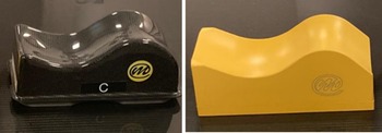
All patients had histologically confirmed squamous cell carcinoma of the oropharynx, hypopharynx, or larynx (T1–T2, N0–N1, M0). They were immobilised using a five-point thermoplastic mask and a knee support, with either a carbon fibre headrest in group 1 (n = 25) or a foam headrest in group 2 (n = 25). Headrests shown in Figure 1 were chosen based on mould room staff expertise. Their choice of either foam or carbon fibre headrests was determined based on suitability with the patient’s anatomy, as well as their compliance.
Carbon fibre headrest and foam headrest, photograph taken by the study’s primary author.

Imaging and data collection
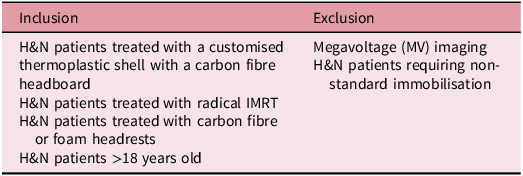
Patients received daily image guidance using three-dimensional kilovoltage cone-beam computed tomography (3D kV CBCT) for fractions 1 to 3, and then weekly thereafter. In between CBCTs, two-dimensional orthogonal kV (2D kV) imaging was used. The On Target 2 guidelines indicate that dual imaging is an acceptable approach to verification used within the limitations of the service. Reference McNair, Franks and van Herk1 For each of the 50 patients, the first 20 treatment fractions were reviewed. This pragmatic approach was to balance the different fractionations of radiotherapy between patients (20#vs?), in order to minimise factors that may influence patient positioning later in the radiotherapy course (>20#), such as weight loss or tumour volume change. A total of 1,000 image datasets were reviewed with inclusion and exclusion criteria outlined in Table 1. Imaging datasets in the form of CBCT images were used to assess displacements using bony anatomy and soft tissue relative to the isocentre. However, 2D kV images could only be assessed with bony anatomy relative to the isocentre position.
Imaging data inclusion and exclusion criteria

Translational (vertical, longitudinal, lateral) and rotational (pitch, yaw) displacements were recorded from the treatment verification system, based on deviations from the planning CT scan. Measurements were recorded to the nearest 0.01 cm or 0.1°. Data were collected in Microsoft Excel 2019. Then, transferred into IBM’s SPSS version 28.0 12 for statistical analysis.
Data analysis
Systematic error (Σ) was defined as the standard deviation of each patient’s mean displacements throughout treatment, as determined from imaging. Random error (σ) was defined as the root mean square of residual displacements throughout treatment, also confirmed from imaging. The Shapiro–Wilk test was used to assess data normality. Normally distributed variables were compared using independent-sample t-tests, whereas non-normally distributed variables were compared using Mann–Whitney U tests. Statistical significance was set at p < 0.05 with a 95% confidence interval. Differences of <0.1 cm or <1° were considered clinically negligible, in line with published guidance on acceptable set-up tolerances. Reference van Herk4,13
Therapeutic radiographer questionnaire
A structured, anonymous questionnaire was completed by ten experienced therapeutic radiographers (each with more than three years’ experience) involved in both pre-treatment and treatment delivery. They rated headrest preference across domains of:
-
• Setup reproducibility
-
• Perceived patient comfort
-
• Adaptability to patient anatomy
Results were aggregated and analysed descriptively. This survey aimed to incorporate staff-centred insights into headrest performance.
Dosimetric analysis
Supplementary dosimetric evaluations in the form of attenuation and conformity indices were derived from an Eclipse treatment planning system (TPS) using a Monte Carlo algorithm. A standard HNC dose of 2 Gy was prescribed to the centre of both the carbon fibre and foam headrests using a parallel opposed beam arrangement. This was done in an effort to compare both headrests dosimetrically.
Results
The Shapiro–Wilk test confirmed normality for all population means and systematic (Σ) errors in all directions. In contrast, the Shapiro-Wilk test rejected normality for all random (σ) errors in all directions. Population mean, systematic and random displacements for both headrest groups were well below clinical thresholds, with no statistically significant differences found in any translational direction. All the population mean errors, systematic errors and random errors for both headrests are outlined in Appendix A.
-
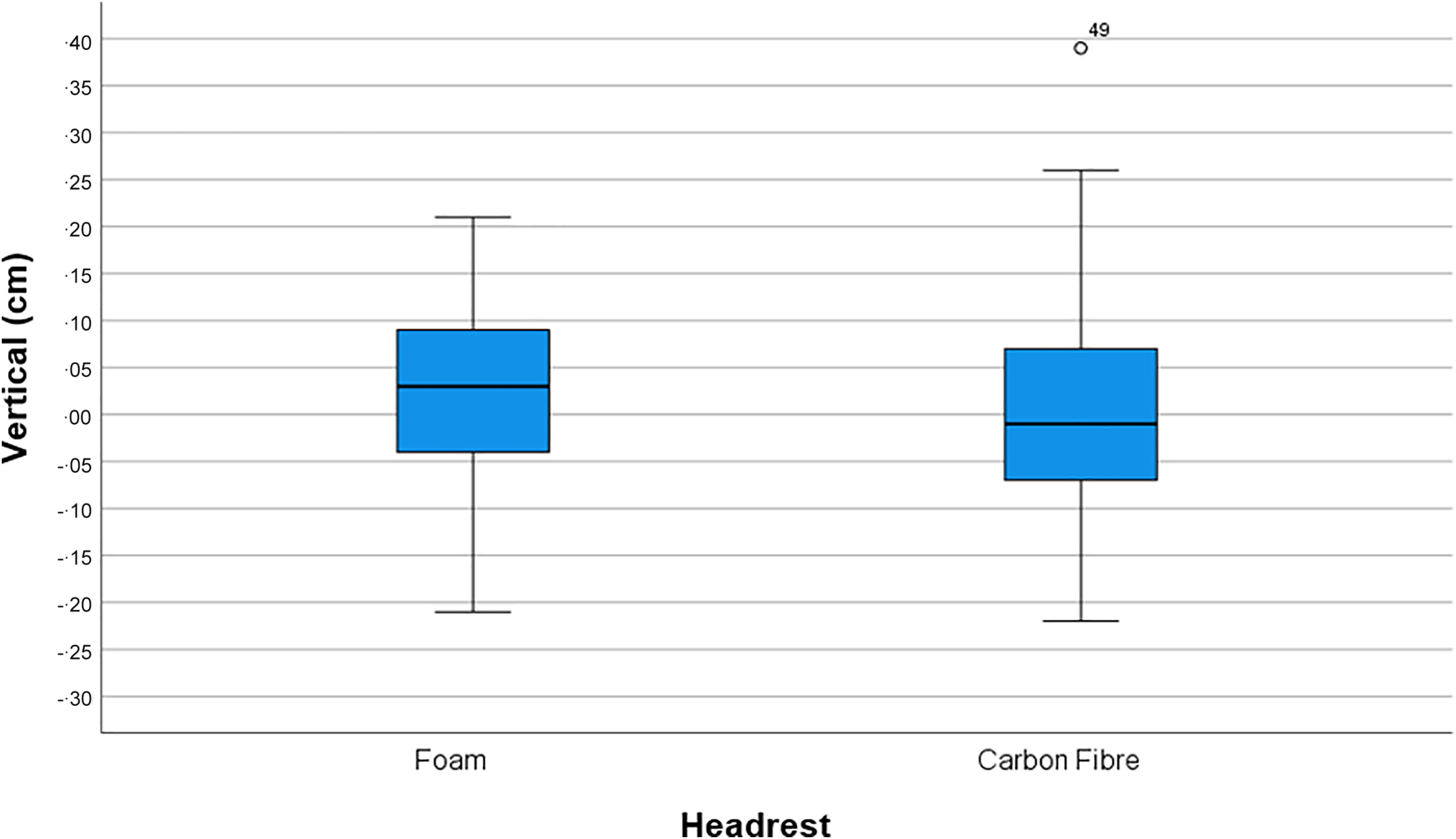
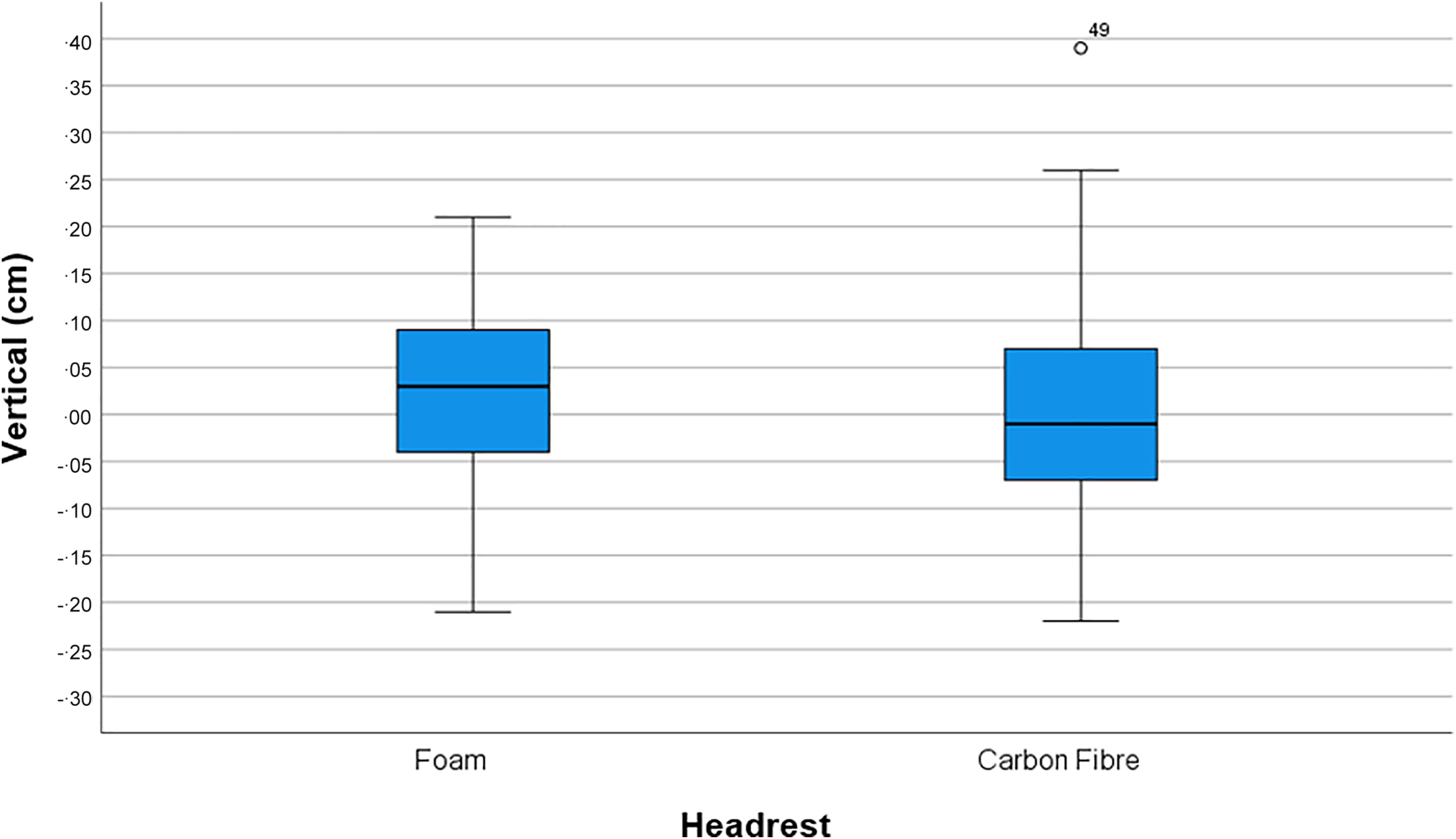
• Vertical errors (Figure 2 ): Mean headrest differences = 0.02 cm, clinically negligible. No statistically significant differences, population systematic error (Σ) (p = 0.68) and population random error (σ) (p = 0.53).
Figure 2.Boxplot chart of population mean vertical errors for both carbon fibre and foam headrests, measured in cm.

-
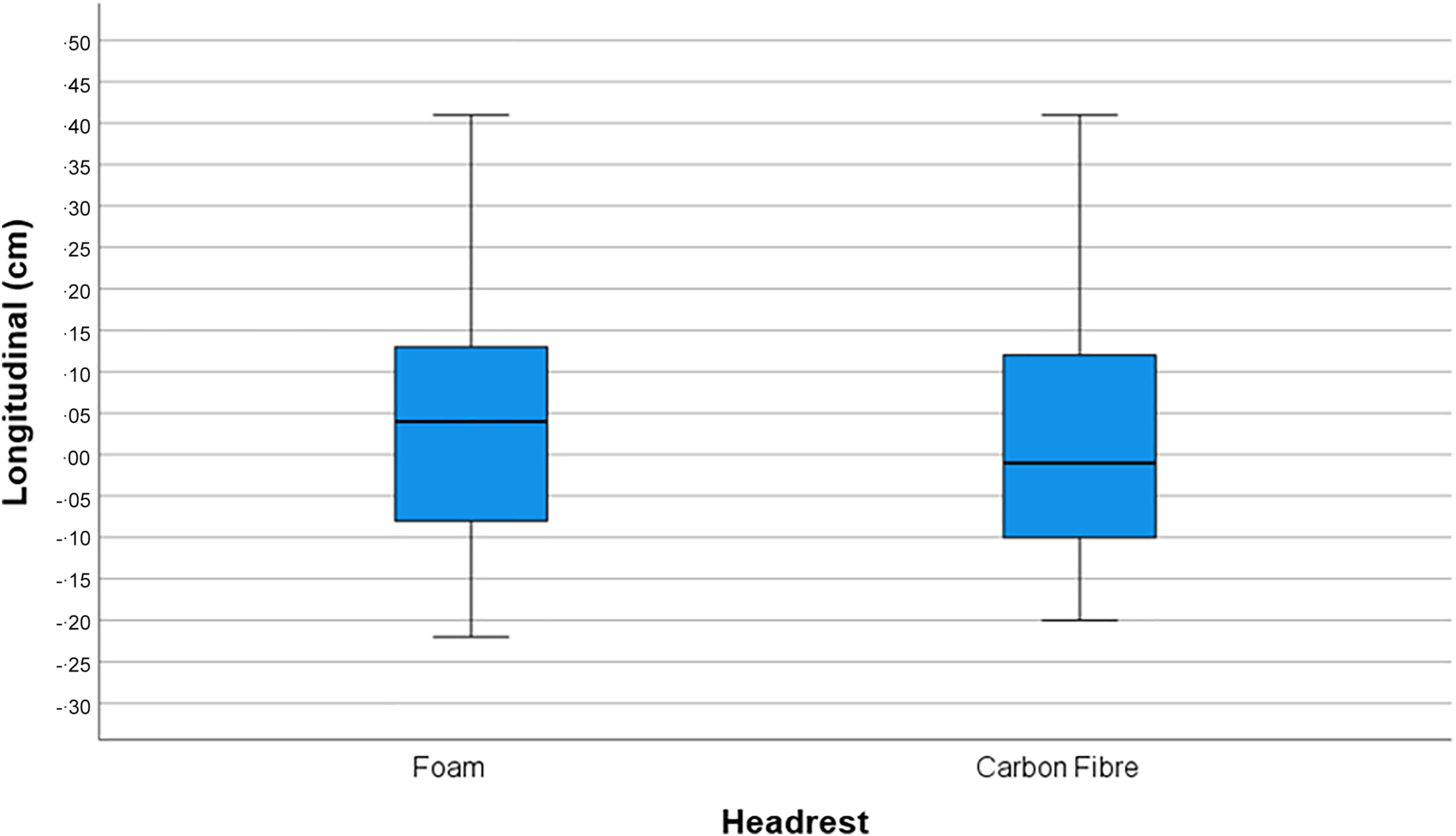
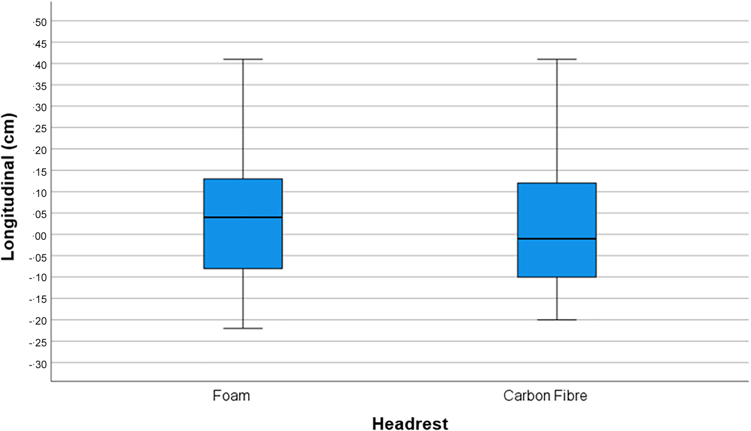
• Longitudinal errors (Figure 3 ): Mean headrest differences = 0.01 cm, clinically negligible. No statistically significant differences, population systematic error (Σ) (p = 0.77) and population random error (σ) (p = 0.88).
Figure 3.Boxplot chart of population mean longitudinal errors for both carbon fibre and foam headrests, measured in cm.
-
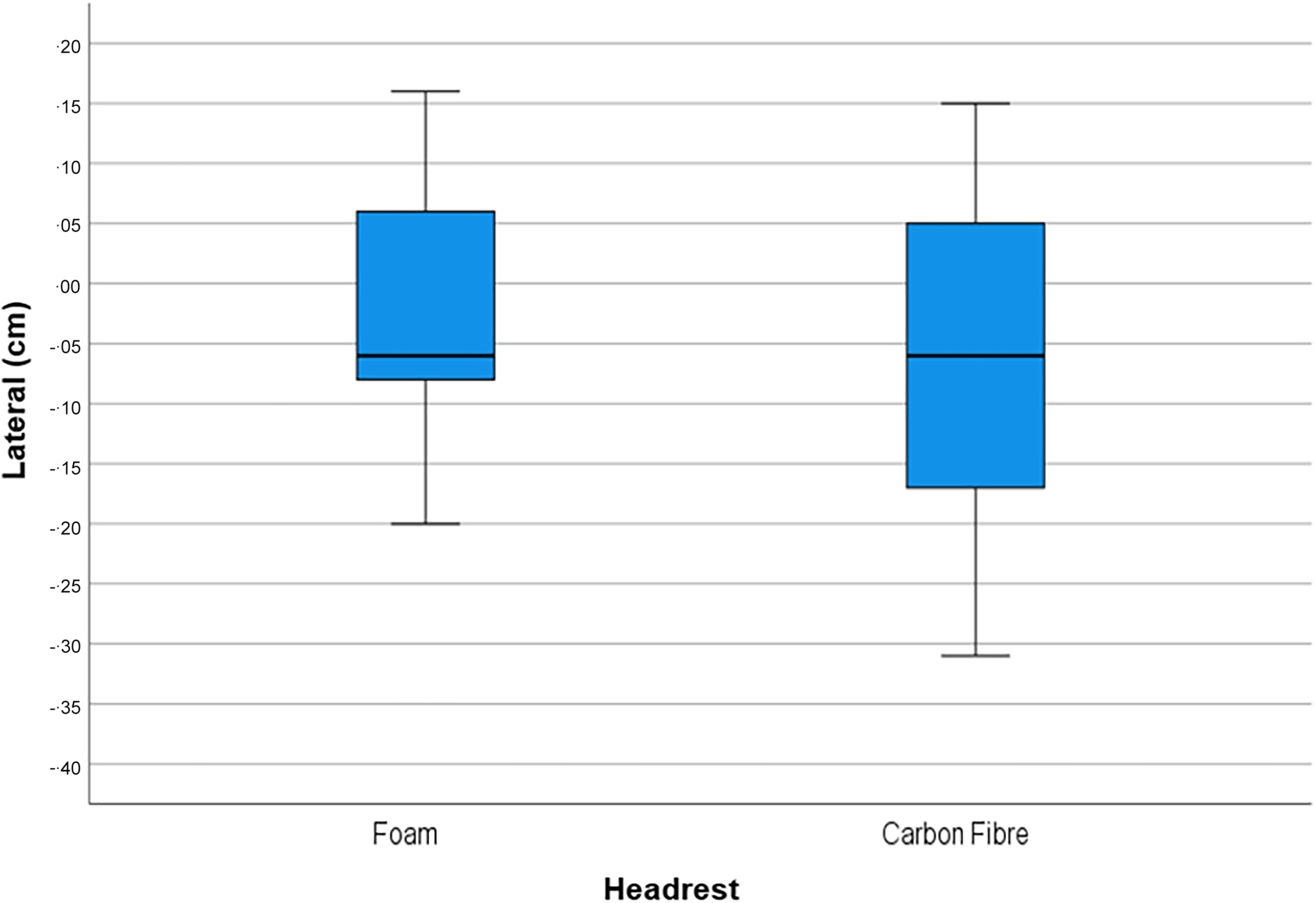
• Lateral errors (Figure 4 ): Mean headrest differences = 0.03 cm, clinically negligible. No statistically significant differences, population systematic error (Σ) (p = 0.38) and population random error (σ) (p = 0.88).
Figure 4.Boxplot chart of population mean lateral errors for both carbon fibre and foam headrests, measured in cm.

For rotational displacements:
-
• Pitch (Figure 5 ): Mean headrest differences = 0.1°, clinically negligible. No statistically significant differences, population systematic error (Σ) (p = 0.58) and population random error (σ) (p = 0.57).
Figure 5.Boxplot chart of population mean pitch errors for both carbon fibre and foam headrests, measured in °.
-
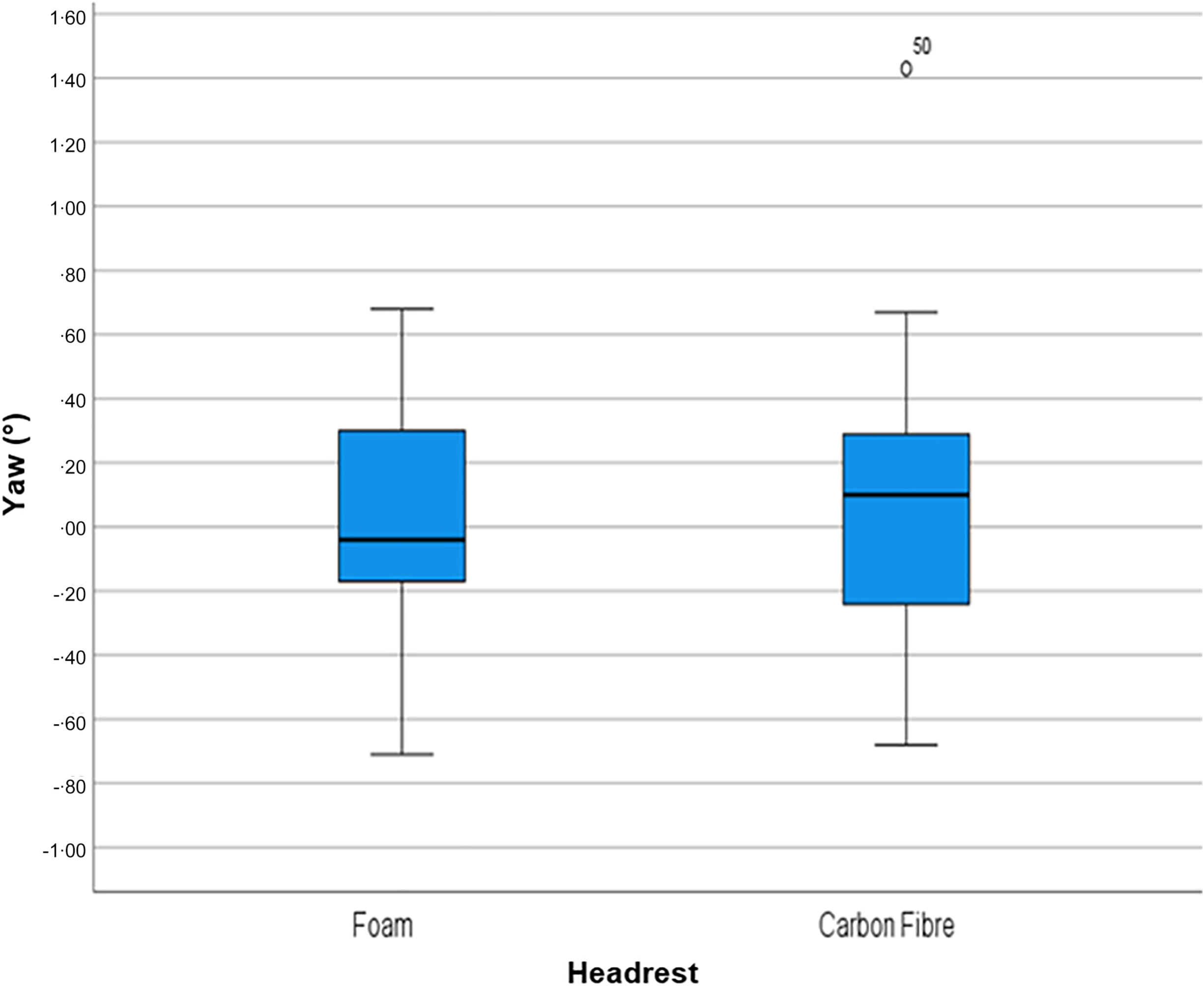
• Yaw (Figure 6 ): Mean headrest differences 0.05°, clinically negligible. No statistically significant differences for population systematic error (Σ) (p = 0.66). The only statistically significant difference (p = 0.028) was a 0.1° reduction in yaw population random error (σ) in the foam group. This was statistically but not clinically significant.
Figure 6.Boxplot chart of population mean yaw errors for both foam and carbon fibre headrest, measured in °.


Boxplot analyses (Figures 2–6) revealed tight clustering for both groups, confirming low variability. A few outliers were observed in the carbon fibre group in vertical, pitch and yaw directions. Supplementary dosimetric evaluation (Appendix B) demonstrated superior dose conformity with carbon fibre and fewer hotspots in high-dose regions.
Therapeutic radiographer feedback
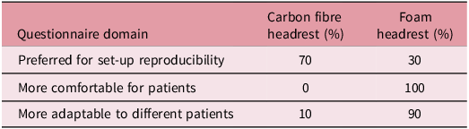
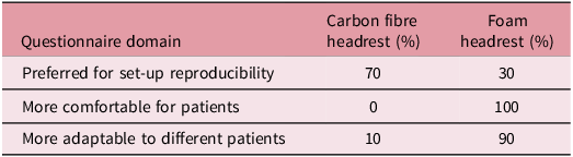
These findings (Table 2) show that from this cohort of therapeutic radiographers, carbon fibre headrests were favoured for operational consistency, and foam headrests were preferred in terms of comfort and adaptability for treating HNC radiotherapy patients.
Key findings from the staff questionnaire (n = 10)
Dosimetric findings
These findings (Appendix B) show that the carbon fibre headrest demonstrated greater dose conformity compared with the foam headrest. In addition, the TPS indicated that the carbon fibre headrest exhibited less attenuation, being 34 Hounsfield units less dense, and that the maximum isodose hotspot was 0.8% higher than for the foam headrest.
Discussion
This retrospective study compared the set-up accuracy of carbon fibre and foam headrests in patients undergoing IMRT for HNC. With 1,000 image datasets analysed across five degrees of freedom, both headrest types demonstrated equivalent performance in maintaining patient position within clinically acceptable tolerances. This was reflected in the results (Appendix A), with only one significant finding, and in the boxplot analyses (Figures 2–6), which demonstrated tight clustering in both groups, confirming low variability. The only statistically significant finding—a 0.1° reduction in yaw random error in the foam headrest group—was well below the 1° rotational threshold typically considered clinically meaningful in HNC radiotherapy. Reference van Herk4,13 It would have been valuable to calculate the difference in planning target volumes (PTV) margins between the headrests using Van Herk’s formula Reference van Herk4 ; however, due to limitations—absence of delineation, intrafraction and registration data, and no measurement of residual errors prior to treatment—this was not possible. Future studies could undertake a representative margin recipe to assess the potential impact of headrest choice on treatment outcomes.
These findings build on earlier studies examining headrest design. Howlin et al. Reference Howlin, O’Shea and Dunne9 and Houweling et al. Reference Houweling, van der Meer and van der Wal10 reported that while customised or vacuum-moulded supports may marginally reduce set-up errors, such benefits often fail to translate into meaningful clinical gains, particularly in the era of daily image guidance. Our study adds value by directly comparing standard carbon fibre and foam headrests, addressing a gap where most studies focused on bespoke or modified supports. Reference Howlin, O’Shea and Dunne9–Reference Rodrigues, Veen and van Egmond11,Reference Androjna, Marcius and Peterlin14
Carbon fibre has long been favoured in radiotherapy for its physical properties—low density, high strength and excellent radiotranslucency—leading to less beam attenuation and greater dosimetric consistency. Reference Njeh, Parker and Spurgin6,Reference Olson, Phillips, Eng, Lenards, Hunzeker and Lewis7,Reference Soriani, Strigari and Petrongari15 This is supported by our dosimetric findings (Appendix B), which showed the carbon fibre headrest demonstrated greater dose conformity and less attenuation than the foam headrest. These qualities are particularly important in high-precision techniques such as SABR and proton therapy, where dosimetric uncertainty can significantly affect clinical outcomes. Reference Billiet, Vingerhoed and Van Laere16,Reference Olch, Gerig and Li17
Despite these advantages, headrest comfort plays a crucial role in preventing intrafraction motion. Prolonged treatment times, often required in HNC radiotherapy due to complex dose modulation, can cause patient discomfort and increase motion during delivery. Reference Dhillon, Erler and Poon18,Reference Kang, Lee and Chan19 While this study did not use formal patient-reported outcome measures (PROMs), radiographer feedback indicated that foam headrests may be perceived as more comfortable—an observation supported in previous literature comparing immobilisation devices. Reference Houweling, van der Meer and van der Wal10,Reference Rodrigues, Veen and van Egmond11,Reference Lamprecht20 The literature indicates that patient comfort is sometimes overlooked in favour of accuracy and this is mirrored in other similar studies. Reference Cheng and Wang21–Reference Goldsworthy, Latour and Palmer23 Mask tightness may also affect comfort; carbon fibre headrests do not compress under the patient’s weight, potentially increasing mask tightness and discomfort. Future studies should incorporate comfort assessment when evaluating HNC immobilisation.
Operational and financial considerations are also relevant. Foam headrests require replacement approximately every three years due to deterioration from daily disinfectant cleaning, whereas carbon fibre products offer greater durability and resistance to deformation. Reference Leech, Coffey and Mast5,24 Suppliers quoted an average of £344 for a set of three foam headrests and £910 for a carbon fibre equivalent; brand names are not included, as the quotation is commercially sensitive. Radiographer feedback (Table 2) suggested carbon fibre headrests are favoured for operational consistency. Although foam may appear more cost-effective initially, carbon fibre could prove more cost and operationally effective long term, though the initial investment may challenge smaller centres.
The study’s findings—namely, the lack of clinically significant differences in set-up accuracy—raise an important practical consideration: is it justifiable to adopt carbon fibre headrests based solely on marginal dosimetric improvements? For standard fractionation with daily IGRT, the answer may be no. However, for stereotactic treatments, re-irradiation cases, or facilities exploring couch attenuation modelling, carbon fibre remains an appealing option.
There are limitations. The retrospective design introduces bias. Another potential bias is the mould room staff’s headrest selection; carbon fibre headrests were newly introduced in the department prior to data collection. The primary researcher (>3 years’ experience) examined the imaging datasets to reduce inter-observer variation as shown in numerous RT studies. Reference Huger, Graff and Harter25–Reference Devereux, Frantzis and Sisson27 However, intra-observer bias may exist, as the researcher was not blinded to headrest type. While intra-observer variation in image analysis has been evaluated in numerous RT studies, Reference Rodgers, Hales and Whiteside28–Reference Belshaw, Agnew and Irvine31 quantifying it was outside this study’s scope.
While the sample size was considerable, the absence of a formal power analysis limits definitive conclusions regarding headrest equivalence. Intrafraction motion was not evaluated—something that could influence outcomes with tighter PTV margins. Only the first 20 fractions were analysed, as weight loss and foam stability may affect displacements over time.
At the time, the host radiotherapy department used a combination of 3D CBCT and 2D kV imaging. Although dual image verification is acceptable in On Target 2 guidelines, Reference McNair, Franks and van Herk1 CBCT verification is superior for detecting anatomical and physiological changes for corrective action before radiotherapy is delivered. Reference Li, Zhu and Zhang32,Reference Kim, Pawlicki and Le33 All online displacements were corrected; offline checks corrected only systematic errors (>0.8 cm). No systematic errors were corrected via isocentre shifts. Future research should quantify differences between modalities, as this may skew set-up results. Future studies analysing foam and carbon fibre headrests or other immobilisation should use 3D CBCT verification to assess six-degrees-of-freedom, which may provide nuanced information on patient positioning.
Future research should include randomised controlled trials over the full course of treatment with PROMs, assessment of intrafraction stability through post-treatment imaging and long-term durability testing to quantify economic implications.
Conclusion
Carbon fibre and foam headrests demonstrated equivalent accuracy in immobilising patients undergoing HNC IMRT, both within acceptable set-up error thresholds. Where performance is equivalent, decision-making should factor in patient comfort, cost and departmental workflow. Carbon fibre’s radiological advantages may be beneficial in select cases, but foam remains a valid and adaptable choice. Future prospective studies should include PROMs, device longevity and economic analysis.
Supplementary material
The supplementary material for this article can be found at https://doi.org/10.1017/S1460396926100466.



 Open access

