


Introduction
Ollulanus tricuspis is a small nematode found worldwide (Dunn Reference Dunn1978; Hasslinger Reference Hasslinger1984). The taxonomic position of this genus has been the subject of significant debate over the years. In early classifications, it was placed within the families Heligosomidae and later Heligmosomatidae (Skrjabin et al Reference Skrjabin, Shikhobalova, Schulz, Popova, Boev and Delyamure1952), whilst Chitwood (Reference Chitwood1938) proposed the family Ollulaniidae. However, according to the cladistic classification system of Durette-Desset et al (Reference Durette-Desset, Hugot, Darlu and Chabaud1999), which is based on distinctive characters such as the morphology of the female nematode’s tail spines, groups lacking spines on the female tail are placed within the superfamily Trichostrongyloidea, whilst families possessing a tail spine, such as Ollulanus, have been placed within the superfamily Molineoidea (specifically the family Molineidae, subfamily Ollulaninae). This is the currently accepted classification in modern nematode systematics. This is the accepted placement in current modern nematode systematics. It is found in the stomachs of domestic and wild felids and, rarely, dogs, foxes, and pigs. (Hasslinger Reference Hasslinger1984; Kato Reference Kato, Oishi, Ohno, Nakashima, Wada, Morita and Tsujimoto2015; Rickard and Foreyt Reference Rickard and Foreyt1992). Adults and larvae live beneath the mucosal layer of the stomach wall, but in severe infections, they can reach the first part of the duodenum.
Ollulanus tricuspis has a direct life cycle and is transmitted when susceptible animals ingest the vomit of infected animals. O. tricuspis is usually non-pathogenic, causing increased mucus production, loss of appetite, and intermittent vomiting (Cameron Reference Cameron1927; Hargis et al Reference Hargis, Prieur and Blanchard1983). However, as the life cycle is completed endogenously, it can multiply in the host, causing severe chronic gastritis and, in rare cases, death (Collett et al Reference Collett, Pomroy, Guilford, Johnstone, Blanchard and Mirams2000; Hargis et al Reference Hargis, Prieur and Wescott1981; Wilson and Presnell Reference Wilson and Presnell1990). The infection is diagnosed by the presence of adults in vomit obtained by administering emetic drugs, by endoscopy or biopsy of the stomach, or by examination of stomach contents (Guy Reference Guy1984; Hargis et al Reference Hargis, Prieur and Blanchard1983). Most diagnoses have been made by examining vomit or stomach contents collected during necropsy (Guy Reference Guy1984; Hasslinger Reference Hasslinger1984; Poglayen et al Reference Poglayen, Traldi, Capelli and Genchi1985).
Females have a distinctly pointed tail and measure 0.8–1.0 × 0.04 mm. Their anterior ends are usually spirally curved, and there are usually two or three protrusions at the tip of the tail. Males measure 0.7–0.8 × 0.035–0.04 mm. They possess a small buccal capsule and spicules. The copulatory bursa is well developed, with spiculeae of equal length (approximately 0.05 mm) and distal ends that divide into two branches, one thick and one thin. One spicule terminates sharply, the other rounded (Umur Reference Umur, Köroğlu, Güçlü, Tınar and Tınar2011).
The first report in Türkiye was made in 1968 by Merdivenci following a necropsy performed on a wild cat (Felis sylvestris) (Merdivenci Reference Merdivenci1968). Following the initial report, the presence of the parasite has been reported in various studies on both cats and dogs in Türkiye (Burgu Reference Burgu1985; Dinçer et al Reference Dinçer, Cantoray and Taşan1980; Güralp Reference Güralp1984; Kaplan Reference Kaplan1995; Tınar et al Reference Tınar, Coşkun, Demir, Aydın, Kaplan and Çırak1993; Zeybek Reference Zeybek1988). Studies conducted in other countries have reported the parasite in other animal species, including tigers, red foxes, lions, cheetahs, and pigs (Chauvier and Chabaud Reference Chauvier and Chabaud1964; Collett et al Reference Collett, Pomroy, Guilford, Johnstone, Blanchard and Mirams2000; Hinaidy Reference Hinaidy1976; Lensink et al Reference Lensink, Rijpstra and Erken1979; Stockdale and Lautenslager Reference Stockdale and Lautenslager1973).
This study is based on the diagnosis of Ollulanus tricuspis infection in the vomit of an 8-month-old domestic cat belonging to one of our staff members, which had chronic vomiting complaints. Following this case, the prevalence of O. tricuspis was investigated in cats that died for various reasons in order to determine the situation in the province of Samsun. This study is the first report on the presence of O. tricuspis in cats in Samsun and is also the first molecular study conducted on this parasite in Türkiye.
Material and methods
Material collection
Our study was initiated following the diagnosis of Ollulanus tricuspis infection in the vomitus of an 8-month-old domestic cat, presenting with chronic vomiting, which then formed the basis for a subsequent study involving necropsy of 50 cats. The study material consisted of samples from a total of 50 cats. One sample was the vomit content of a cat presenting with a complaint of chronic vomiting. The remaining 49 subjects were deceased stray cats that had been brought to the Ondokuz Mayıs University Animal Hospital for various clinical reasons and were subsequently necropsied. The stomach, stomach contents, and duodenum of the 50 cats subjected to necropsy were examined under a binocular stereo microscope (Nikon SMZ-1500). The stomach and duodenal contents were examined under the stereo microscope after dilution. Adults were detected in the stomach contents of three cats. The adults were collected using fine eye forceps.
Parasitological examination of samples, morphological identification
The 860 adult specimens collected from three cats were morphologically examined under a light microscope (Nikon Eclipse 80i, Tokyo, Japan). Morphometric measurements were performed, and the specimens were identified as O. tricuspis with the help of the literature (Cameron Reference Cameron1927; Umur et al Reference Umur, Köroğlu, Güçlü, Tınar and Tınar2011). After morphological identification was completed, portions of the specimens were set aside for PCR and stored at +4 °C.
Molecular studies
The collected adults were subjected to genomic DNA (gDNA) extraction using a commercial DNA isolation kit (Invitrogen PureLink Genomic DNA Mini Kit). The obtained gDNA was stored at −20 °C until molecular analyses. PCR amplification of an approximately 1250 bp fragment covering the ITS-1, 5.8S, and ITS-2 gene regions was performed using the primers NC2 (5′-TTAGTTTCTTTTCCTCCGCT-3′) and NC5 (5′-GTAGGTGAACCTGCGGAAGGATCATT-3′) as described by Zhu et al (Reference Zhu, Gasser and Chilton1998).
The PCR mixture was prepared to a total volume of 50 μL by adding 10 mM Tris HCl, pH 8.3, 50 mM KCl, 2.5 mM MgCl₂, 250 mM dNTP, primers (100 pM NC2/NC5), and 1.25 U Taq polymerase. 4 μL of template DNA was used. The amplification protocol was performed on a thermal cycler (SimpliAmp, Applied Biosystems) at 94 °C for 5 minutes; 37 cycles of 94 °C for 30 seconds denaturation, 53 °C for 30 seconds annealing, 72 °C for 1 minute extension, followed by a final 5-minute extension at 72 °C. PCR products were run on a 1.5% agarose gel stained with ethidium bromide with a 100 bp DNA ladder and photographed using a UV transilluminator (ERBiyotek, UV-6L-2020). All positive amplicons were purified and directly sequenced in both directions using NC2 and NC5 primers on an automated sequencer from a commercial service.
Phylogenetic analyses
Forward and reverse sequences belonging to an adult isolate were assembled using Contig Express in the Vector NTI Advance 11.5 (Invitrogen) programme to obtain the consensus nucleotide sequence for each isolate, and the quality scores of their chromatograms were examined. BLASTn analysis was performed on the consensus sequences in the GenBank database (https://www.ncbi.nlm.nih.gov/genbank/). Subsequently, multiple alignments of the nucleotide sequences were performed using the Clustal W algorithm with BioEdit (Hall Reference Hall1999).
Following morphological diagnosis, a 1250 bp region covering the ITS-1, 5.8S, and ITS-2 gene regions was amplified using the NC2 and NC5 primers. For the phylogenetic tree, AB245044 Cooperia oncophora, AF194127 Nematodirus helvetianus (Nadler et al Reference Nadler, Hoberg, Hudspeth and Rickard2000), AF194128 Nematodirus spathiger (Nadler et al Reference Nadler, Hoberg, Hudspeth and Rickard2000), AF194138 Nematodirus battus (Nadler et al Reference Nadler, Hoberg, Hudspeth and Rickard2000), DQ408624 Heligmosomoides polygyrus bakeri (Cable et al Reference Cable, Harris, Lewis and Behnke2006), HQ844231 Haemonchus contortus, JF680984 Teladorsagia circumcincta, KT428384 Marshallagia marshalli (Kuchboev et al Reference Kuchboev, Sobirova, Karimova, Amirov, von Samson-Himmelstjerna and Krücken2020), KT428385 Ostertagia ostertagi, ON677953 Trichostrongylus axei (Buchmann et al Reference Buchmann, Christiansen, Kania and Thamsborg2022), Spiculopteragia houdemeri AB682696 (Sultan et al Reference Sultan, Omar, Makouloutou, Kaneshiro, Saita, Yokoyama, Suzuki, Hosoi and Sato2014) isolates were used to create the dataset. Phylogenetic relationships were inferred using both maximum likelihood (ML) and Bayesian inference (BI) methods (Figure 1).
Phylogenetic relationships of Ollulanus tricuspis inferred from ITS-1, 5.8S, and ITS-2 rRNA gene sequences using Maximum Likelihood (ML) and Bayesian Inference (BI) analyses. Spiculopteragia houdemeri (GenBank accession no. AB682696) was designated as the outgroup. Numbers at the nodes represent bootstrap support values (%) from ML analysis (left) and posterior probabilities from BI analysis (right); values below 70% bootstrap support are not shown. All sequences are identified by GenBank accession numbers with their geographic origins.

Figure 1. Long description
Panel A shows a full-body view of the nematode, which is curved in a C-shape with a tapered anterior and posterior. A scale bar is located at the bottom center. Panel B, located at the top right, is a high-magnification view of the male posterior end. It features two prominent, brownish, Y-shaped spicules within the translucent body and a complex, rounded bursa at the distal tip. A scale bar is in the bottom right corner. Panel C, at the bottom right, shows the posterior end of a female. The body tapers to a tail that terminates in three small, distinct points or cusps. A scale bar is located in the bottom right corner.
The ML tree was constructed under the GTR substitution model using PhyML, with branch support assessed via 1,000 bootstrap replicates (Guindon et al Reference Guindon, Dufayard, Lefort, Anisimova, Hordijk and Gascuel2010). Bayesian inference was performed using MrBayes v.3.2.7. Two independent Markov chain Monte Carlo (MCMC) simulations were run simultaneously for 10 million generations, with sampling every 200 generations, utilising four chains per run and a temperature parameter of 0.2. Before constructing a majority-rule consensus tree, the initial 25% of the sampled trees from each run were discarded as burn-in. Spiculopteragia houdemeri (GenBank accession no. AB682696) was designated as the outgroup to root the trees. The nucleotide sequence of O. tricuspis was deposited in GenBank under accession number PV855833.
Result
A vomit sample taken from a cat with chronic vomiting, being cared for in a professor’s garden, was sent to the parasitology department. The sample was placed in a Petri dish, diluted 1:1 with physiological saline, and examined under a stereomicroscope, revealing the presence of O. tricuspis. The infected cat, under clinical follow-up, was successfully treated with a single oral dose of 5 mg/kg Levamisole 2.5% solution (Sitrax®), after which the vomiting symptoms reportedly resolved.
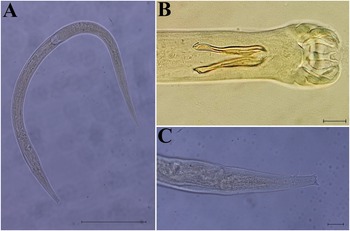
In total, Ollulanus tricuspis was detected in three of the 50 cats examined. One of these positive cases was clinically diagnosed based on the vomit contents of a cat suffering from chronic vomiting. The other two positive cases were identified among 49 stray cats that underwent necropsy. A total of 860 adult O. tricuspis worms were removed from the stomachs of these two cats. 854 adult worms were found in the first cat and 6 in the second. Of the 50 cats necropsied, a total of 860 adult O. tricuspis were collected in 3 (6%) cats, comprising 396 males and 464 females from the stomach and duodenum. The parasites were generally observed in a coiled form, with the anterior end of the females and the males almost completely coiled around themselves like a spring. The dimensions of the parasite were 702 (660–750) μm in length, 23.8 (21–29) μm in thickness, and 48 (45–51) μm in spicule length in males, while females measured 828 (760–900) μm in length, 26.25 (20.0–30.0) μm, and tail length 32.8 (31–36) μm (Figure 2). The dimensions of the parasites were determined by measuring 10 adult nematodes in both female and male nematodes.
Ollulanus tricuspis. A: Female (scale bar:100). B: Male rear end (scale bar:10). C: Female rear end (scale bar:10).

Figure 2. Long description
A horizontal phylogenetic tree with a scale bar of 0.07 at the bottom. The tree branches from a root on the left toward specific species labels on the right.
Top cluster:
- J F 6 8 0 9 8 4 Teladorsagia circumcincta, United Kingdom.
- K T 4 2 8 3 8 5 Ostertagia ostertagi, Uzbekistan.
- K T 4 2 8 3 8 4 Marshallagia marshalli, Uzbekistan.
Middle cluster:
- H Q 8 4 4 2 3 1 Haemonchus contortus, China.
- P V 8 5 5 8 3 3 Ollulanus tricuspis O M U P A R dot 11 dot 25 dot 08, Türkiye, highlighted in red text.
- A B 2 4 5 0 4 4 Cooperia oncophora, Japan.
- O N 6 7 7 9 5 3 Trichostrongylus axei, Denmark.
Bottom cluster:
- D Q 4 0 8 6 2 4 Heligmosomoides polygyrus, United Kingdom.
- A F 1 9 4 1 3 8 Nematodirus battus, United Kingdom.
- A F 1 9 4 1 2 8 Nematodirus spathiger, United States.
- A F 1 9 4 1 2 7 Nematodirus helvetianus, United States.
Outgroup at the bottom left:
- A B 6 8 2 6 9 6 Spiculopteragia houdemeri, Japan.
Nodes are labeled with support values such as 1 forward slash 99, 0.96 forward slash 95, and 1 forward slash 100.
Searching GenBank for O. tricuspis isolate records revealed only one isolate from Australia belonging to the 28S gene region. To date, no research has been found on the molecular identification and phylogenetic relationship of this species, and there are no isolate records in Türkiye. This study resulted in the acquisition of the first sequence data for the ITS-1, 5.8S, and ITS-2 gene regions of O. tricuspis from Türkiye, and its phylogenetic relationship with other closely related isolates worldwide was investigated.
In the present study, we attempted to clarify the phylogenetic and taxonomic status of O. tricuspis, which belongs to the subfamily Ollulaninae within the family Molineidae, using ribosomal DNA sequence data. Since there is only one species in this subfamily, a dataset comprising our novel sequence and 11 reference isolates (totalling 12 sequences) from families such as Cooperiidae, Haemonchidae, Molineidae, Trichostrongylidae, and Heligmosomidae was utilised. The obtained data show that O. tricuspis belongs to the same major clade as Haemonchus contortus (HQ844231), Cooperia oncophora (AB245044), and Trichostrongylus axei (ON677953). The analysis confirms the phylogenetic placement of O. tricuspis within the superfamily Trichostrongyloidea with a maximum Bayesian posterior probability (1), indicating a close evolutionary relationship with Haemonchus, Cooperia, and Trichostrongylus species (Chilton et al Reference Chilton, Huby-Chilton, Koehler, Gasser and Beveridge2015).
Discussion
This study presents a detailed investigation into Ollulanus tricuspis infection in domestic cats, initiated by the unique clinical observation of an infected feline belonging to a faculty member. The initial diagnosis of O. tricuspis in a cat presenting with chronic vomiting not only highlighted the presence of this parasitic nematode in our region but also underscored the necessity for a broader examination of its prevalence and pathology. Consequently, our findings from the subsequent necropsy study of 50 cats provide further insights into the morphometric characteristics and diagnostic challenges associated with O. tricuspis, thereby expanding the current understanding of this gastric nematode in feline populations. This combined approach, moving from a compelling case study to a more extensive necropsy series, allowed us to bridge individual clinical presentation with broader epidemiological and pathological patterns.
Chronic or intermittent vomiting is the main clinical finding associated with O. tricuspis. However, vomiting in cats and dogs can be a symptom of many diseases. Along with the medical history obtained from the owners, the focus is usually on diseases with a higher probability of occurrence, such as chronic enteropathies, chronic pancreatitis, chronic liver and biliary tract diseases, and kidney diseases. Limited knowledge about the epidemiology, biology, and systematics of the parasite further complicates diagnosis, identification, and treatment. Diagnosis of O. tricuspis is usually made morphologically during post-mortem examination. In the present study, a molecular phylogenetic analysis of O. tricuspis collected from domestic cats during post-mortem examination was performed.
Ollulanus tricuspis is widespread throughout the world, particularly in domestic and wild cats. After being reported in domestic cats by E. Aneurin Lewis in 1927, it has also been reported in dogs, red foxes, pumas, and cheetahs in addition to domestic and wild cats. O. tricuspis infection in animals is generally characterised by vomiting, weight loss, loss of appetite, and gastric lesions. Sometimes infected animals may be asymptomatic, making diagnosis difficult. Despite the gastrointestinal disturbances caused by the parasite, research on it is limited. Diagnosis is usually made by necropsy, stomach contents, or endoscopic biopsy. Lillis et al (Reference Lillis1967) reported finding O. tricuspis in faeces using the zinc sulphate flotation method. However, apart from this, there have been no other reports of O. tricuspis in faeces. Since O. tricuspis is expelled through vomiting, diagnostic methods using faeces do not yield accurate results. One study reported it in association with gastric adenocarcinoma, but the connection between the two could not be fully explained (Dennis et al Reference Dennis, Bennett and Ehrhart2006). The first record of O. tricuspis in Türkiye was established by Merdivenci (Reference Merdivenci1968) through necropsy examinations of wild cats (Felis sylvestris). Subsequent studies (Dinçer et al Reference Dinçer, Cantoray and Taşan1980; Güralp Reference Güralp1984) revealed that the parasite was also present in domestic carnivores. Later reports (Burgu Reference Burgu1985; Kaplan Reference Kaplan1995; Tınar et al Reference Tınar, Coşkun, Demir, Aydın, Kaplan and Çırak1993; Zeybek Reference Zeybek1988) further confirmed its broader host spectrum and indicated an endemic presence in the regions where these studies were conducted. The global occurrence of O. tricuspis spans across multiple continents, infecting a wide range of domestic and wild carnivores. The reported prevalence and distribution of this parasite in different geographical regions are summarised in Table 1.
Reported prevalence and distribution of Ollulanus tricuspis in domestic and wild carnivores across different geographical regions (+: case report)

Table 1. Long description
The table is organized into four columns: Animal species, Prevalence percent, Country, and References.
Domestic cat data includes:
* Wales: 12.9 percent (Lewis, 1927).
* Italy: 2 percent (Bronzini, 1956).
* Australia: Multiple reports ranging from 3 percent to 42 percent (1960 to 2015).
* U S A: 0.2 percent (Lillis, 1967) and 28 percent (Hargis et al, 1981).
* Türkiye: Case reports and prevalence of 17 percent (1968 to 1985).
* New Zealand: 4.3 percent and 13 percent (1973 to 1984).
* Germany: 18.3 percent and 38 percent (1977 to 1981).
* Belgium: 5.8 percent (Thienpoint et al, 1981).
* England and Spain: Case reports marked with a plus symbol.
Domestic dog data includes:
* Türkiye: Case report (Zeybek, 1988).
* Japan: Case report (Kato et al, 2015).
Wild carnivore data includes:
* Red fox in Austria: Case report (Hinaidy, 2010).
* Puma in U S A: Case report (Rickard and Foreyt, 1992).
* Cheetah in New Zealand: Case report (Collet et al, 2000).
A plus symbol indicates a case report. An asterisk indicates a single animal record.
* : Single animal record; USA: United States of America.
In this study, we aimed to clarify the phylogenetic and taxonomic position of O. tricuspis, a member of the Ollulaninae subfamily, the sole subfamily (monotypic) within the Molineidae family, using ribosomal DNA sequence data. To assess evolutionary relationships within a broader context, the newly obtained sequence data were analysed alongside 11 reference isolates (totalling 12 sequences) from the Cooperiidae, Haemonchidae, and Trichostrongylidae families, which constitute the core components of the Trichostrongyloidea superfamily. Furthermore, in our analysis, a nematode from the Heligmosomatidae family was selected as an outgroup to reflect the historical taxonomic affinity of the Ollulanus genus in the literature and its previously associated origins (Chitwood Reference Chitwood1938; Skrjabin et al Reference Skrjabin, Shikhobalova, Schulz, Popova, Boev and Delyamure1952). The molecular data obtained clearly place O. tricuspis within the Trichostrongyloidea superfamily. While our topology positions O. tricuspis within the same major clade as Haemonchus contortus (HQ844231), Cooperia oncophora (AB245044), and Trichostrongylus axei (ON677953), the statistical support for these specific internal branches is not sufficiently strong to resolve its exact familial associations. Therefore, although this analysis robustly confirms the evolutionary relationship of O. tricuspis with the superfamily Trichostrongyloidea, our conclusions regarding its direct phylogenetic proximity to the Haemonchidae, Cooperiidae, or Trichostrongylidae must remain restrained. This aligns with the previous findings of Chilton et al (Reference Chilton, Huby-Chilton, Koehler, Gasser and Beveridge2015), who also confidently assigned Ollulanus to the Trichostrongyloidea using 28S rRNA sequences, but similarly noted a lack of strong support for any further sub-familial or familial associations.
In conclusion, this study describes the diagnosis of O. tricuspis in a domestic cat from Türkiye, which prompted a subsequent necropsy investigation. Although morphological diagnoses of O. tricuspis have been previously reported in Türkiye, our findings represent the first genetic record of this parasite from the region and demonstrate that the phylogenetic position of O. tricuspis is consistent across different geographic populations and gene regions. Furthermore, the 50-cat necropsy series contributes to the understanding of its prevalence and morphological characteristics. However, further studies involving larger populations are required to comprehensively assess the full extent of genetic variation and epidemiological patterns of O. tricuspis within Türkiye.
Data availability statement
Data available on GenBank accession number PV855833.
Author contribution
Tuğçe Tuygun, Mehmet Öztürk, Rübeyda Dinç, Cenk Soner Bölükbaş, Ali Tümay Gürler and Şinasi Umur
Conceptualisation, data curation, formal analysis, funding acquisition, investigation, methodology, project administration, software, visualisation, writing – original draft, writing – review and editing.
Financial support
This study is funded by TÜBİTAK (Türkiye Bilimsel ve Teknolojik Araştırma Kurumu) under project number 123C415.
Competing interests
The authors declare they have no financial or non-financial conflicts of interest.
Consent to participate
All authors confirm their participation in the study.
Ethics declarations
Institutional ethical approval was not required for the post-mortem material used in this study. However, informed consent was obtained from the owner for the animal that received clinical treatment.
Ethics approval
The authors confirm that the journal’s ethical policies, as noted on the journal’s author guidelines page, have been adhered to. Specific Ethics and Welfare Committee approval was not required as naturally dead material was used.


 Open access

