





Introduction
Idiopathic ventricular arrhythmias, covering premature ventricular complexes and ventricular tachycardias, can be treated by radiofrequency catheter ablation. Reference Shen, Hu and Shao1 These arrhythmias rarely originate from the epicardial surface of the left ventricle. The predominant origin of epicardial ventricular arrhythmias is the left ventricular summit, located superior to the aortic segment of the left ventricular ostium. Ventricular arrhythmias originating from this region can be eradicated with radiofrequency catheter ablation performed within the great cardiac vein or via epicardial technique. Reference Yamada, McElderry and Doppalapudi2 Ablation from a close anatomical point, such as the right ventricular outflow tract, aortic cusps, and aortic mitral continuity, is also feasible. Reference Shirai, Santangeli and Liang3 Nevertheless, catheter ablation of ventricular arrhythmias that originated from the basal portion of the left ventricular summit near the left main coronary artery is difficult due to the thick epicardial fat pad that envelops the region and the proximity to the coronary arteries. Reference Yamada and Kay4 Ventricular arrhythmia originating from the basal section of the summit is infrequently observed in paediatric patients. Our paper highlighted the clinical use of radiofrequency catheter ablation in two patients with this exceptional tachyarrhythmia.
Case report
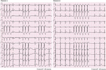
Patient 1 is a 9-year-old male, weighing 27 kg, who presented with intermittent palpitations, and no structural heart disease was detected in his echocardiographic evaluation. The patient had non-sustained ventricular tachycardia on his ECG and triplet and couplet beats. A 24-hour rhythm Holter evaluation revealed 21% of monomorphic ventricular extrasystoles (Figure1). Patient 2 is a 13-year-old male weighing 67 kg, presenting with palpitations, and was referred for ablation due to non-sustained ventricular tachycardia observed during an exercise test. Both patients experienced sustained ventricular tachycardia episodes despite one year of combined medical treatments; hence, ablation was contemplated as a therapy option for them.
12-lead ECG recordings featuring non-sustained ventricular tachycardia.

All the procedures were performed under general anaesthesia. The EnSite Precision and WorkMate Claris™ System was used for activation mapping. The RF Marinr™ 7 Fr steerable multi-curve ablation catheter is used for mapping and RF ablation. Point-by-point mapping was performed using the ablation catheter in both patients. The process started with the mapping of the right ventricular outflow tract. EnSite 3D mapping and fluoroscopy were both used sequentially for intracardiac imaging during the procedure. Prior to administering energy, the safety of the ablation site is verified using angiography. 3D mapping was conducted during sinus rhythm with the ventricular extrasystoles. Activation mapping revealed the left ventricular outflow tract as the earliest ventricular activation of extrasystoles.
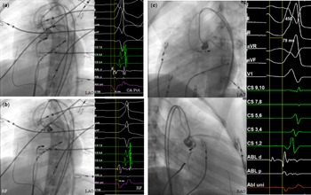
During the ablation procedure of patient 1, the mapping catheter advanced to the left coronary artery by the retro-aortic approach. The earliest spot was detected in the left coronary artery with a –43 msn early activation compared to surface ECG, with deep and sharp QS unipolar recordings. It was a very risky point for ablation. So the left coronary cusp region was mapped due to proximity to the origin of the ventricular activation. The ablation point was determined with late unipolar recordings compared to −35 msn early bipolar signals within the left coronary cusp. Contrast injection was simultaneously performed into the left coronary artery and the left coronary cusp. The distance from the ablation site to the origin of the left coronary artery was measured as 5.2 mm (Figure 2A-B). Radiofrequency catheter ablation was applied to this region with 35 W and 55 degrees for 30 s. Automaticity was observed with the beginning of ablation, and it was seen that the extrasystoles disappeared. After the procedure, the coronary artery patency was evaluated with selective left coronary artery injection, and the procedure was terminated. The duration of the procedure was 150 min, with fluoroscopy exposure lasting 40 min.
A: The left anterior oblique fluoroscopy image shows the position and potential (CA Pot) of the ablation catheter in the left coronary artery. (Patient 1) B: The left anterior oblique view shows the radiofrequency ablation site on the left coronary cusp. (Patient 1) C: The position of the ablation catheter on the aortic-mitral continuity under the aortic valve and the ablation signal are shown in the left anterior oblique and right anterior oblique view with left coronary artery injection. (Patient 2).

Six months post-procedure, contrast-enhanced computed CT angiography verified the absence of stenosis or thrombus in the coronary arteries. No recurrence was noted at 20 months post-procedure.
The ablation procedure of patient 2 was also conducted with the same methods as in patient 1. After the determination of LVOT as the primary target, the mapping catheter advanced to the coronary cusp and the ablation point by the retrograde path. During the evaluation of the aortic cusps and subvalvular structures, it was observed that the earliest activation zone was located in the aorto-mitral continuity region. This region was targeted for ablation. Figure 2C displays the ablation signal alongside the fluoroscopy image. It was determined with –79 msn early activation compared to surface ECG, with deep and sharp QS unipolar recordings, which are also late compared to the bipolar signal that is revealing a far-field signal. This indicates that the lesion area is relatively remote from the tachycardia focus. The anatomical correlation was the subvalvular region close proximity to the aortic-mitral continuity. Radiofrequency catheter ablation was administered to this location, resulting in the disappearance of extrasystole. No recurrence was observed in the patient’s 3-month follow-up.
Discussion
The basal left ventricular summit ventricular arrhythmias exhibited a predominance of a left bundle branch block pattern, increased R-wave amplitude in the inferior leads, decreased III/II and aVL/aVR ratios, and late precordial transition relative to the apical left ventricular summit ventricular arrhythmias. Reference Yamada, Doppalapudi, Litovsky, McElderry and Kay5 Treatment of ventricular arrhythmias arising from the basal left ventricular summit is tough due to the difficulties of accessing this region and its proximity to the left coronary artery. Reference Yamada, McElderry and Doppalapudi2
The successful ablation of the left ventricular summit was influenced by several parameters, including the diminutive size of the coronary sinus vessel lumen, elevated impedance within the cardiovascular system, the confusing anatomy of the coronary venous system, and proximity to the coronary artery. Reference Li, Lin, Li, Ji and Lin6 The communicating branch of the great cardiac veins traverses the epicardial surface, with minimal adipose tissue present between the branch and the epicardial surface. So radiofrequency catheter ablation eliminates arrhythmias at the basal left ventricular summits. Conversely, the distal segment of the great cardiac veins and the anterior interventricular vein pass or lie upon the epicardial fat, which surrounds the epicardial surface; thus, radiofrequency catheter ablation within these veins may be constrained by elevated impedance. Reference Yamada, Doppalapudi, Litovsky, McElderry and Kay5 One of the ablation strategies was ablation from the adjacent endocardial site, the earliest of which had the earliest activation from the aortic root/cusp, the adjacent RV outflow tract, and the left atrial appendix, including great cardiac veins. Reference Das, Hawson and Koh7 For the paediatric patients with smaller body sizes, the coronary sinus is even smaller and is not the preferred route to reach the summit region. Therefore, ablation through aortic mitral continuity or aortic cusp may be preferred due to its close anatomical location to the summit region, thanks to its minimal fat pad presence. However, the small anatomical structures in paediatric patients, along with their proximity to coronary arteries, may elevate the risk of procedural complications.
Ablation targets the area with the earliest activation. The inability to directly ablate the earliest site within the left ventricular summit leads to a lower success rate of ablation procedures and a higher recurrence rate in adults. Reference Futyma and Sauer8 Radiofrequency catheter ablation can be effectively applied in managing ventricular arrhythmia originating from the left ventricular summit in children with no recurrence observed so far. This experience has been limited to individual reports due to the extremely low incidence of cases. Relatively late unipolar signals compared to bipolar signals taken from successful ablation targets in all procedures reflect distance from the ectopic focus.
Left coronary cusp PVCs originate from the endocardial aspect of the left ventricular outflow tract and typically show earlier activation in the coronary cusp compared to other sites. These PVCs can often be successfully ablated from within the aortic cusp. In contrast, left ventricular summit PVCs arise from the epicardial region, often near the coronary arteries, and may show earlier activation in the coronary venous system. For Patient 1, since we do not have such recordings, we cannot state with 100% certainty that the origin was within the left ventricular summit. However, the earlier signals recorded within the coronary arteries, compared to the cusp region, along with unipolar signals appearing slightly later than bipolar signals within the coronary arteries, and the ECG findings, suggest that the true origin may have been deeper or closer to an epicardial site rather than the classical left ventricular summit. Overall, these findings indicate that the origin is likely closer to the summit region rather than the cusp.
Conclusion
Ventricular tachycardia originating from the basal region of the left ventricular summit is rarely seen in paediatric patients. Applying radiofrequency catheter ablation to the area closest to the source can successfully and safely terminate the tachycardia.
Acknowledgements
Acknowledgements should be extended to those individuals or institutions whose contributions to the study were limited or minimal.
Financial support
The authors declare that the study received no funding.
Competing interests
The authors declare that there is no conflict of interest to disclose.
Ethical standard
Informed consent was obtained from the patient and his family to share patient clinical information and imaging photographs anonymously for scientific purposes.
Declaration of generative AI and AI-assisted technologies in the writing process
There is no usage of generative AI and AI-assisted technologies in writing.

 Open access

