





Introduction
Seabirds are among the most threatened vertebrate groups globally [Reference Butchart1–Reference Dias3]. While climate change and overfishing have long been recognized as major drivers of their population declines, emerging infectious diseases have recently gained prominence as additional and potentially severe threats. Among these, highly pathogenic avian influenza virus (HPAIV) A/H5 clade 2.3.4.4b has emerged as a major conservation concern due to its capacity to cause mass mortality events in wild bird populations [4–Reference Garcês and Pires6].
This clade was first reported in Eurasia in 2016 causing sporadic mortality events in poultry and wild birds. In 2020, a genomic reassortment enabled an expansion of both its geographic and host range, triggering large-scale mortality events across Europe, Africa, and North America [Reference Bellido-Martín7]. Between 2021 and 2022, the virus spread southward through the Pacific and Mississippi migratory flyways, eventually reaching South America and causing unprecedented mass mortality events in seabirds and marine mammals [Reference Azat8–Reference Pardo-Roa11]. By 2023, mass mortalities had also been documented in the Subantarctic region, affecting multiple seabird species including Gentoo Penguins (Pygoscelis papua), Black-browed Albatross (Thalasarche melanophris), and Chilean Skuas (Stercorarius chilensis) among others [Reference Banyard12].
At the western Antarctic Peninsula (WAP), the first suspected cases of HPAIV A/H5 were reported in Adélie Penguins (Pygoscelis adeliae) and Antarctic Shags (Leucocarbo bransfieldensis) [Reference León13], followed by confirmed infection of HPAIV A/H5 clade 2.3.4.4b in a Brown Skua (Stercorarius antarcticus) in early 2024 [Reference Bennett-Laso14]. Since then, additional seabird carcasses, primarily in the northern WAP, have raised concerns that HPAIV outbreaks may exacerbate ongoing declines in seabird populations breeding in this region [Reference Tiwari15]. These areas are of high conservation importance, supporting large fractions of global seabird populations and several endemic or threatened species [Reference Harris16].
Seabirds appear particularly susceptible to HPAIV, with mortality rates in wild populations reaching up to 90% during outbreaks [Reference Garcês and Pires6]. The demographic consequences can be especially severe during breeding seasons affected by adult mortality [Reference Furness and Tasker17, Reference Lewison18] and the high density of colonies further facilitates viral transmission [Reference Boulinier19]. Nevertheless, species vary in their susceptibility. Evidence from the Northern Hemisphere suggests that skuas may be disproportionately affected [Reference Bellido-Martín7]. During the 2021–2022 outbreak of HPAIV A5/H5N1 clade 2.3.4.4b, Great Skua (Stercorarius skua) experienced severe mortality (69 to 76% population decline) along with drastic reduction in reproductive success [Reference Camphuysen, Gear and Furness20]. At the Antarctic Peninsula, current evidence suggests that species of the same genus such as Brown and South Polar Skuas (Stercorarius maccormicki) appear to be similarly impacted [Reference Dewar, Alcami and Wille21]. Their scavenging behaviour and extensive migratory ranges may also facilitate viral spread across regions, underscoring the need for active HPAIV surveillance in these species [Reference Dewar, Alcami and Wille21, Reference Dewar22].
Recent genomic analyses indicate that many skuas in the Antarctic Peninsula are admixed individuals resulting from hybridization between South Polar and Brown Skuas, forming two dominant genomic clusters: one with higher South Polar ancestry (70–96%) towards the southern Peninsula and another with higher Brown Skua ancestry (56–75%) towards the northern Peninsula, South Georgia, and Bouvet Island [Reference Jorquera23]. Because hybrids often exhibit intermediate morphologies, reliance on phenotypic traits alone may lead to misidentifications [Reference Ritz, Hahn, Janicke and Peter24], increasing the risk of inaccurate species classifications in field surveys.
Given the substantial demographic impact of HPAIV A5/H5N1 clade 2.3.4.4b elsewhere [Reference Gamarra-Toledo9, Reference Pardo-Roa11] and the similar vulnerability observed in Stercorarius species across both hemispheres [Reference Camphuysen, Gear and Furness20, Reference Banyard25], there is an urgent need to document and understand outbreak dynamics in Antarctic populations.
Here, we present a systematic survey of HPAIV-associated mortality in seabirds along the WAP during the 2024–2025 breeding season, with a particular focus on skuas. Our surveillance, conducted between November 2024 and January 2025, included behavioural assessments to identify signs consistent with HPAIV infection and field necropsies of all recovered carcasses, from which samples were collected for viral detection. By integrating field observations, mortality records, H5 antibodies detection, and phylogenetic viral analyses, our study provides urgently needed baseline data for evaluating the unfolding outbreak in Antarctica. These results are critical not only for understanding the immediate conservation impacts on key seabird species, but also for informing early-warning systems, biosecurity protocols, and long-term monitoring strategies under a rapidly changing disease landscape.
Materials and methods
Ecological surveillance strategy, site selection, and sampling effort
Three field expeditions were conducted during the 2024–2025 austral summer season. First, a long-term survey has been performed at Harmony Point, Nelson Island (62.305650 S°, 59195120 W°), between 23 November 2024 and 11 January 2025. Additionally, two vessel-based surveys with landings at multiple sites were performed, one aboard the Betanzos vessel from 3 January 2025 to 16 January 2025, and another aboard the Karpuj vessel from 14 January 2025 to 15 January 2025. For this timing, most of the seabirds at the Antarctic Peninsula are expected to be at incubation – hatching or early chick rearing.
For the stationary survey, annual research activities have been performed in this area, which allowed us to include the HPAIV surveillance activities. For the vessel-based surveys, landings were focused on areas along the WAP where a medium to high risk of HPAIV introduction had been previously assessed [Reference Dewar, Alcami and Wille21, Reference Dewar22] as well as those that were accessible for the vessel. Priority was given to skua populations near areas hosting high-density breeding colonies including Pygoscelis penguins and Antarctic Shags. Skuas near nesting colonies of Southern Giant Petrels (Macronectes giganteus) were also included. At each location, efforts were made to maximize area coverage, considering available sampling time, accessibility, and weather conditions.
In total, 16 geographic areas were surveyed along the WAP between 62.132683 S°–67882431 S° latitude and 58122040 W°–67399990 W° longitude. The surveyed sites included several Antarctic Specially Protected Areas (ASPA) such as Biscoe Point (ASPA No. 139), Harmony Point (ASPA No. 133), Litchfield Island (IBA criteria A4i; ASPA No. 115), and Avian Island (IBA criteria A1, A4i, A4ii, A4iii; ASPA No. 117). Additional locations between King George Island and the WAP (Table 1 and Figure 1).
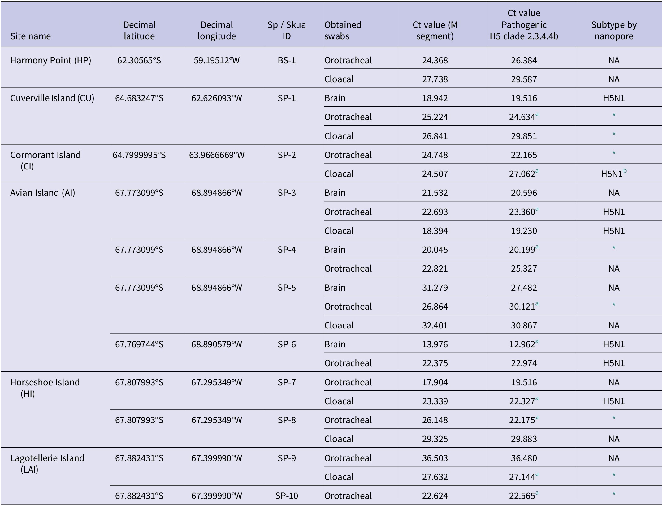
Sampling sites with positive cases of HPAIV H5N1 clade 2.3.4.4b in dead adult skuas

Table 1 Long description
This is a structured data table containing 8 columns and 20 rows of data organized by geographic sampling locations. The columns from left to right are: Site name with abbreviations, decimal latitude, decimal longitude, Species and Skua ID, swab sample obtained, Ct value for M gene segment, Ct value for the pathogenic clade H5 2.3.4.4b, and influenza subtype determined by nanopore sequencing (results include H5N1, NA, or an asterisk).
BS, Brown Skua; NA, sample was not sequenced; SP, South Polar Skua. Notice that not all carcasses had sufficient tissue for the collection of all swab types.
a Sample was confirmed by SAG.
b Incomplete.
* No genomic amplification at multi-segment PCR, not positive to amplify the viral genome, or only M and NS segments are available.
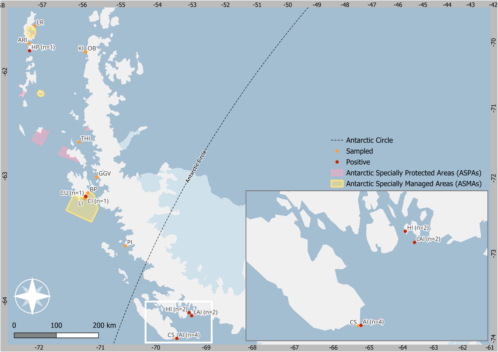
Sampling sites across the Antarctic Peninsula. Sampling sites from north to south: Lions Rump (LR), Ardley Island (ARI), Harmony Point (HP), Kopaitic Island (KI), Bernardo O’Higgins Base (OB), Two Hummock (THI), Cuverville Island (CU), Lichtfield Island (LI), Cormorant Island (CI), Biscoe Point (BP), Gabriel Gonzalez Videla Base (GGV), Perch Island (PI), Carvajal Station (CS), Avian Island (AI), Horseshoe Island (HI), Lagotellerie Island (LAI). Mortalities and positive HPAIV cases are denoted with red stars, while no mortalities or suspected cases are indicated with yellow circles. Six of the localities visited, Ardley Island (ARI), Harmony Point (HP), Litchfield Island (LI), Biscoe Point (BP), Avian Island (AI) and Lagotellerie (LAI) have ASPA or ASMA categories and all of them are Important Bird Areas (IBA). Full list of abbreviations in Supplementary Table 1.

Figure 1 Long description
This figure is a geographic map showing the monitoring locations along the Antarctic Peninsula included in this study. The map includes all sampled or visited sites, as well as the locations where HPAIV H5N1-positive dead skuas were found. It also identifies Antarctic Specially Protected Areas (ASPAs) and Antarctic Specially Managed Areas (ASMAs). The map includes coordinate grid around its borders, with latitude ranging from −70° to −74° and longitudes from −57° to 72°. The Antarctic Circle is represented by a dashed diagonal line, showing that some positive locations were situated south of the Antarctic Circle. A large rectangular inset in the lower right corner provides a magnified view of the southernmost area outlined by a white square on the main map. The map legend includes: Dashed Line: Antarctic Circle; Orange Dots: Sampled locations; Red Dots: HPAIV-positive locations; Pink shaded Areas: ASPAs; Yellow Shaded Areas with Outlines: ASMAs.
Since all individuals from the Antarctic Peninsula are introgressed among the three species of skuas, here we refer to the species according to the higher percentage of hybridization, South Polar and Brown Skuas. Therefore, the individuals were classified according to the higher genomic proportion reported in the same localities or nearby areas included in this study [Reference Jorquera23]. At each site, visual inspection of seabirds and colonies was conducted to assess the nesting activity, detect mass mortality events, or dead individuals with evidence of rapid death progression. Also, clinical signs consistent with HPAIV infection were assessed in alive seabirds such as lack of coordination, lethargy, loss of balance, opisthotonus, and respiratory distress [Reference Camphuysen, Gear and Furness20].
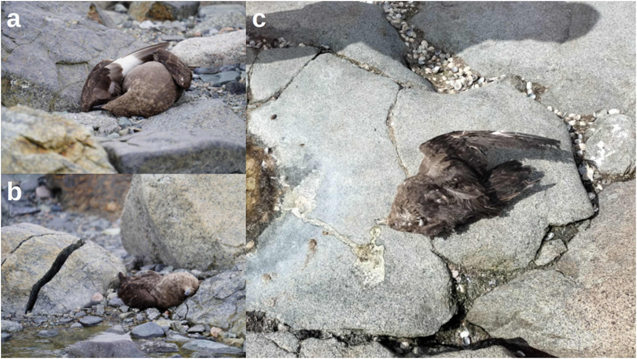
For dead birds, orotracheal, cloacal, brain, or lung swabs were collected for HPAIV detection. Swabs were stored in microtubes, preserved in the Copan eNAT® System, DNA/RNA Shield™, or InhibiSURE™, and then stored in 4 °C, −20 °C, or in liquid nitrogen until analysis. Carcasses were observed to detect abnormal postures suggestive of a death caused by HPAIV infection, such as torticollis posture (twisted) (Figure 2a,b). Necropsies were conducted only on fresh, intact avian carcasses that showed no signs of scavenging or advanced decomposition, to identify macroscopic lesions consistent with HPAIV infection. Body condition was assessed for all carcasses using a five-point fat scoring system to estimate the subcutaneous fat [Reference Brown, Nolan and Ketterson26]. External palpation and visual inspection were used to detect traumatic injuries and to infer if the bird had recently died. Severely ill agonizing birds were not handled until their death.
Several dead skuas were observed in pairs near the reported nesting sites in the breeding and rearing areas of the IBA. (a–b) Both sampled birds in Horseshoe Island showed neck position indicating a possible opisthotonus sign at the time of death. Birds had no sign of predation and had well-preserved plumage. (c) Some animals exhibited dry vomit around the bill.

Figure 2 Long description
This figure is a composite of three photographs of dead skuas, labeled a, b and c, illustrating the positions and external appearance of the carcasses at the time they were found. Photograph a (Top-left): A dead skua lying on its side on grey rocks. The wings are partially arched or folded over its back. The carcass is in a dorsal recumbency position, with well-preserved plumage and no apparent traumatic lesions. Photograph b (Bottom left): A skua carcass in a compact or curled position. The position of the neck suggests possible opisthotonus sign at the time of dead. The plumage is also well-preserved, and there are no sign of predation or external trauma. Photograph c (Right): A carcass resting directly on a large, flat, grey boulder. A discharge or vomitus is visible on the rock immediately in the front of the head.
Biosecurity measures
All procedures followed strict biosafety protocols in accordance with the Antarctic Treaty guidelines as well as recommendations outlined in the Scientific Committee on Antarctic Research (SCAR) Biological Risk Assessment for HPAIV in the Southern Ocean [Reference Dewar, Alcami and Wille21]. All samples collected were obtained under animal capture permits and Millennium Institute Biodiversity of Antarctic and Subantarctic Ecosystem 4/CBSCUA/2022. All procedures were approved by Instituto Antártico Chileno (INACH) (permits N° 00624/2024, 00623/2024, 00693/2024).
Molecular detection and phylogenetic analyses
Molecular detection of influenza A virus (IAV) was performed on 24 samples from 11 skuas at the Laboratory of Molecular Virology, Pontificia Universidad Católica de Chile (LVM-UC) Although more skuas were found dead, not all of them were accessible for a safe sampling and some lacked sufficient tissue for sampling. First, sample lysis was performed with TRIzol™ reagent (Invitrogen TM 15596018) followed by RNA viral extraction with E.Z.N.A Viral RNA Kit (R6874, Omega Biotek) [Reference Pardo-Roa11]. Then, the highly conserved matrix (M) gene of IAV was detected by RT-qPCR assay according to WHO recommendations employing the primers InfA Forward 5′ GACCRATCCTGTCACCTCTGAC 3′, InfA Reverse 5′ AGGGCATTYTGGACAAAKCGTCTA3′ Primer, and InfA Probe1 5′ FAM-TGC AGT CCT CGC TCA CTG GGC ACG-BHQ1–3′ [27]. All positive samples were subsequently tested to determine the influenza A subtype H5 clade 2.3.4.4b by RT-qPCR with the primers H5.2344-1673F TACCAAATAYTGTCAATTTATTCAAC, H5.2344-1749R GTAAYGACCCRTTRGARCACATCC, and H5.2344-1718P FAM-CTGGCAATCATDRTGGCTGGTCT-BHQ1 [27]. All One-Step RT-qPCRs were performed with AgPath-ID™ One-Step RT-PCR Reagents (4387391 Applied Biosystems™) following manufacturer’s instructions. Samples with Ct values < 37 were considered positives.
According to national guidelines and regulations, a subset of positive samples, one per individual, was submitted to the Virology and Biotechnology Laboratories of the Agricultural and Livestock Service of Chile (SAG, Lo Aguirre) for confirmation of the results. First, general detection of IAV was performed using the VetMAX™-Gold AIV Detection Kit (Applied Biosystems™, Cat No. 4485261) and subsequently, RT-qPCR assays were conducted to determine the specific lineage of H5 within clade 2.3.4.4b, following the standard operating procedures NVSL-WI-1732.02 and NVSL-WI-1767.01 from the National Veterinary Services Laboratories (NVSL) of the USDA (NVSL, 2023a; NVSL2023b).
Genome sequencing and phylogenetic analyses
Samples with a diagnostic Ct value < 37 were selected for whole-genome amplification. The viral genome was amplified using a multi-segment One-Step RT-PCR genome amplification employing SuperScript™ III One-Step with DNA Platinum™ Taq polymerase, (12574026 Invitrogen™) with the influenza primers, Opti1-F1 5′:GTTACGCGCCAGCAAAAGCAGG-3′, Opti1-F25′:GTTACGCGCCAGCGAAAGCAGG-3′, and Opti1-R1:5′:GTTACGCGCCAGTAGAAA-CAAGG-3′ [Reference Mena and Neher28]. Next-generation sequencing was performed using the Oxford Nanopore Technology (ONT) platform with the Native Barcoding Kit (SQK-NBD114.96) for library preparation at LVM-UC. End-prep was performed using the NEBNext Ultra II End Repair/dA-tailing Module (E7546, NEB). Native barcodes were ligated using the Native Barcoding Expansion 96 (EXP-NBD196) and the NEB Blunt/TA Ligase Master Mix (M0367, NEB), assigning a unique barcode per sample. The barcoded libraries were pooled and purified using SPRISelect beads. Native adapters were ligated to the library using the NEBNext Quick Ligation Module (E6056, NEB), followed by purification with SPRISelect beads (B23318, Beckman Coulter) and quantification using a Qubit Fluorometer (Invitrogen™). The library was loaded onto the sequencer using the ligation sequencing kit (SQK-LSK109) according to ONT instructions for R.10.4 flow cells. Sequencing was carried out for 72 h. According to LMV-UC quality criteria, only complete sequences were submitted to GenBank.
The dataset used for phylogenetic analysis was generated from AIV sequences from avian hosts from South America and Antarctica obtained from GenBank and GISAID databases with a collection date between 1 January 2023 and 31 December 2024. MAFFT (Linux v7.526) [Reference Katoh29] was used to align all sequences with Antarctic sequences generated in this study using only one sequence per individual. We also included some sequences from North America to provide a global context for the sequences.
The phylogenetic trees were inferred for the segments (Hemagglutinin (HA) and Neuraminidase (NA)) separately using the maximum likelihood (ML) method available in IQ-Tree3 (https://ecoevorxiv.org/repository/view/8916) with a general-time reversible (GTR) model of nucleotide substitution, incorporating a gamma-distributed rate variation among sites, selected by the BIC criterion with ModelFinder in IQ-tree software. An ultrafast bootstrap resampling process was performed with 1000 replicates to assess the reliability of each node. The result was visualized in FigTree (v1.4.4).
Antibodies detection against influenza
First, the detection of influenza antibodies was performed for all skuas’ serum or plasma samples employing the commercial kit ID Screen® influenza A Antibody Competition Multi-species (ID.vet Cat No. FLUACA-5P) and for all positive samples, a second detection for antibodies against H5 was performed using the commercial kit ID Screen® influenza H5 Antibody Competition 3.0 Multi species (ID.vet #FLUACH5-2P). ELISA assays were performed following the manufacturer’s instructions. Positives results were identified according to the kit instructions, based on the ratio of ELISA optical densities between the specimen and the negative control (S/N). An internal positive control was included in each assay.
Results
Summer 2024–2025 monitoring efforts
During stationary surveillance at Harmony Point, eight adult Brown Skuas were found dead. Six carcasses lacked sufficient tissue for sampling, while the remaining two were seen before dying and exhibited neurological signs consistent with HPAIV infection, including lethargy, tremors, opisthotonus, and loss of equilibrium (Supplementary Video 1). Only one of these birds was later confirmed dead and samples were collected. This individual remained in a snowmelt stream, where its condition progressively deteriorated until its death, which occurred approximately two days after the first observation of signs. The second skua showing signs compatible with influenza was not seen again in subsequent days. Although the carcass was not retrieved, the bird was presumed dead (Supplementary Table 1).
During itinerant expeditions aboard the Betanzos and the Karpuj vessels, a total of 27 dead adult skuas were found at 50% of the visited sites, including, Cuverville (n = 1), Cormorant Island (n = 4), Biscoe Point (n = 2), Carvajal Station (n = 2), Avian Island (n = 6), Horseshoe Island (n = 6), and Lagotellerie Island (n = 6) (Supplementary Table 1). However, due to logistical constraints and adverse weather conditions that prevented prolonged stays at the sites, dead birds from Carvajal Station and Biscoe Point were not sampled. No skua mortality was observed at the remaining eight sites.
Most dead skua carcasses exhibited well-preserved plumage and showed no visible signs of trauma or injury. Most of them (20/35) were found in a ventral position, often with neck postures suggestive of opisthotonus at the time of death (Figure 2a,b). Two individuals had dried vomit around the beaks (Figure 2c). Carcasses without signs of predation had estimated body condition scores of 2/5 and 3/5, consistent with acute death progression. While most dead skuas appeared to have died recently, some carcasses showed evidence of scavenging or were too decomposed for sampling.
Although adults and chicks of Pygoscelis penguin and Antarctic Shag were observed dead, the numbers were consistent with natural mortalities regularly observed in the area, and no other evidence related with HPAIV was detected. Other live bird species, including Antarctic Terns (Sterna vittata) and Snowy Sheathbill (Chionis albus), were observed across sites. None displayed clinical signs compatible with HPAIV infection. All these birds at breeding colonies or active nests exhibited typical territorial behaviour for the breeding season.
HPAIV A/H5 clade 2.3.4.4b is associated with skua mortality
A total of 24 samples from 11 carcasses were collected for viral detection, representing 31% of detected individuals. Samples from other carcasses [Reference Ritz, Hahn, Janicke and Peter24] were either inaccessible for a safe sampling or lacked sufficient tissue. Of these, 33% were cloacal (n = 8), 41% orotracheal (n = 10), and 25% were obtained from the brain (n = 6).
All swabs were positive for influenza A gene M and were also positive for HPAIV A/H5 clade 2.3.4.4b lineage. All samples derived to the SAG National Reference Laboratory were confirmed according to their standardized protocols, supporting the circulation of the clade in Antarctic seabirds (Table 1). Ct values for clade 2.3.4.4b were between 12,9 in a brain sample from SP-6 (Avian Island) to 36,4 in a cloacal sample from SP-9 (Lagotellerie Island). No clear differences in Ct values were detected among sampling sites or tissue types (Table 1).
Skuas positive to HPAIV A/H5 clade 2.3.4.4b were detected in six localities, including Harmony Point (n = 1), Cuverville (n = 1), Cormorant Island (n = 1), Avian Island (n = 4), Horseshoe Island (n = 2), and Lagotellerie Island (n = 2) (Supplementary Table 1 and Figure 1). Skua population across these locations have shown to differ in admixture proportions based on genomic analyses by Jorquera et al., (2025). Specifically, individuals from most of those sampled sites exhibit admixed ancestry with a predominant genomic contribution from South Polar Skuas, whereas individuals from Harmony Point are primarily assigned to an admixed cluster with a higher Brown Skua genomic component.
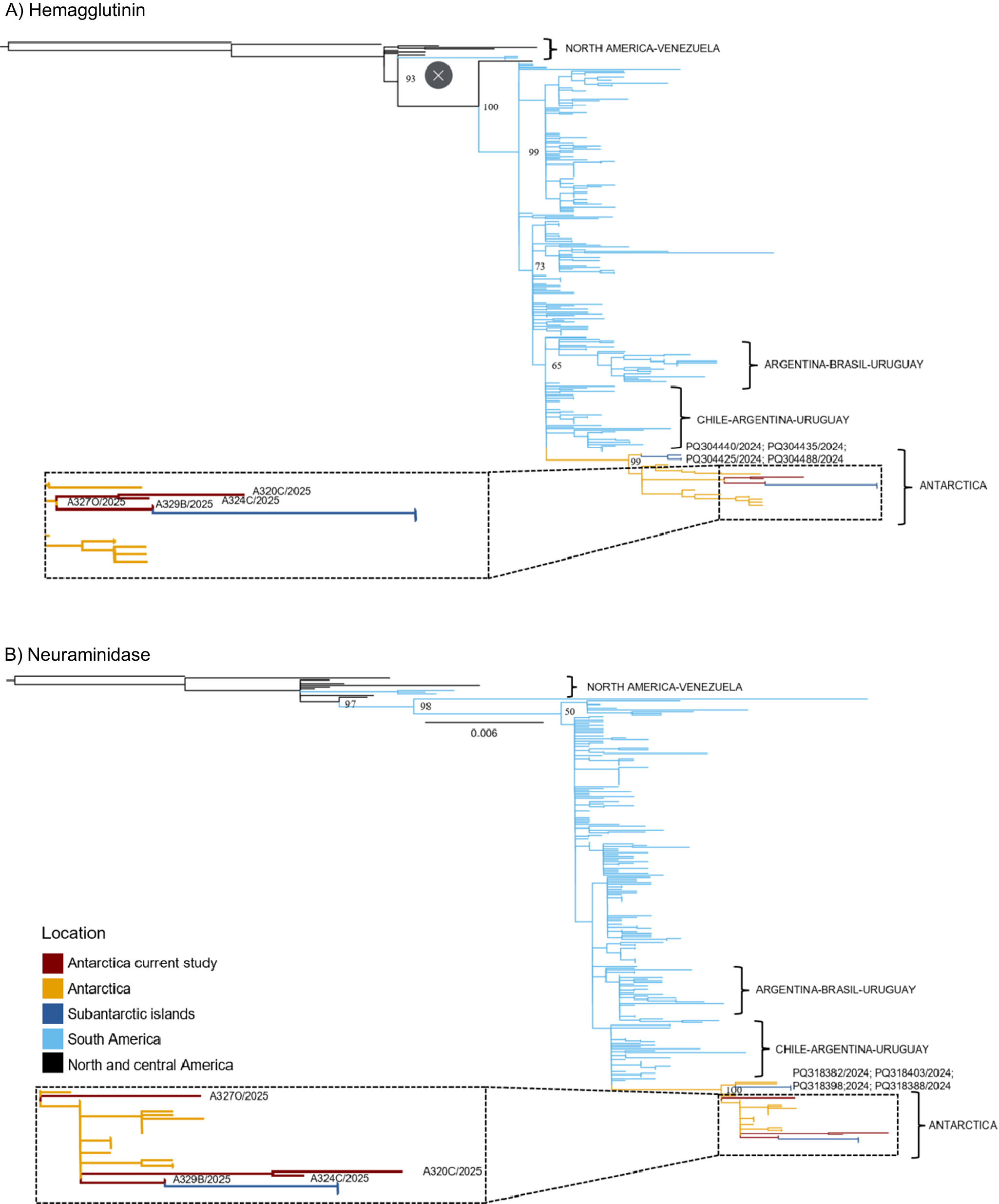
Six complete influenza virus genomes were generated from four individuals. One sequence from Cuverville and Horseshoe Island, and two per Avian Island, were used for phylogenetic reconstruction of the HA and NA segments, ensuring one per individual (Figure 3a,b). Phylogenetic trees showed that all sequences clustered within a monophyletic clade comprising all Antarctic sequences. The sequences from this study were most closely related to viruses reported from Fildes Bay (King George Island) during the same period. Consistent with other studies, the phylogenetic structure indicates a single introduction of the influenza virus into Antarctica, likely associated with South American sources.
Maximum-likelihood (ML) trees were inferred for each surface protein. (a) Hemagglutinin (HA) with 296 sequences. (b) Neuraminidase (NA) with 290 sequences. The datasets include sequences from North and South America, as well as the Antarctic region, with collection dates ranging from 1 January 2023 to 31 December 2024. Ultrafast bootstrap values are provided for key nodes.

Figure 3 Long description
This figure presents maximum-likelihood phylogenetic trees of two influenza virus surface glycoproteins: A) Hemagglutinin and B) Neuroaminidase. Both trees illustrate the evolutionary relationships and geographical clustering of viral isolates. Branch colors correspond to the sampling locations defined in the legend: Dark Red: Antarctica from the current study; Orange: Antarctica (previously published sequences); Dark Blue: Subantarctic islands; Light Blue: South America; Black: North and Central America. The basal linages are composed primarily of sequences from North America and Venezuela. Withing the South American lineage, two major sub-clades are evident: one comprising isolates from Argentina-Brazil-Uruguay, and another including Chile-Argentina-Uruguay. The Antarctic clades contain all sequences generated in the present study.
Serology assays to detect antibodies against influenza A/H5 were performed on serum collected from two healthy Brown Skuas captured at Harmony Point. Both individuals were seropositive for A/H5, however, none tested positive for influenza A in cloacal swabs.
Discussion
Our surveillance identified 35 dead skuas, 11 of which tested positive for HPAIV A/H5 clade 2.3.4.4b, representing the first documented evidence of skua mortality associated with this virus beyond the Antarctic Circle (AC). These individuals were found in areas where recent reports had indicated suspected HPAIV-related mortalities. The first PCR-confirmed case in skua during the 2023–2024 season was reported on Lagoon Island by WAHIS (SCAR database, https://scar.org/library-data/avian-flu). Subsequently, several suspected skua cases were reported in the SCAR database by the IAATO during the 2024–2025 breeding season. To our knowledge, the first detailed report confirming a positive case during the 2024–2025 season was provided by this study [Reference León13], and this was later supported by additional confirmed cases in Marguerite Bay [Reference Gorta30] and Cuverville Island [Reference Wille31]. These findings suggest that the current circulation of this viral clade in Antarctic seabird breeding sites may be disproportionately affecting skuas, compared with other sympatric species.
At Harmony Point, neurological signs and rapid clinical deterioration preceding death closely resemble signs described in other wild birds infected with HPAIV [Reference Banyard25, Reference Rijks32]. In carcasses, the head and wing position are signs of acute death, and together with the low RT-qPCR Ct values, further support HPAIV as the most likely cause of death [Reference Dewar, Alcami and Wille21, Reference Bennet33, Reference Muñoz34]. Although we did not perform differential diagnoses for other infectious agents such as avian cholera, influenza infection is known to cause fatal outcomes in closely related species such as the Great Skua [Reference Camphuysen, Gear and Furness20, Reference Banyard25], and has also been documented in skuas from Antarctica [Reference Bennett-Laso14]. The molecular detection of HPAIV A/H5 2.3.4.4b in skuas across Avian, Horseshoe, and Lagotellerie Islands highlights the need for geographically extensive surveillance throughout the region.
In contrast, no other sympatric seabird exhibited evident mortality or clinical signs of influenza infection, despite co-occurrence with infected skuas. No unusual behaviour or deaths were recorded for Antarctic Tern, even when it is a potential candidate for transpolar viral transmission. The same pattern was observed in Southern Giant Petrel or Snowy Sheathbill even though both species are scavengers and could plausibly become infected through consumption of contaminated carcasses [Reference Gorta35].
The observed interspecific differences in mortality suggest that species within the genus Stercorarius may exhibit higher susceptibility to HPAIV infection, potentially due to the acute manifestation of the disease. No live skuas were sampled, as very few solitary individuals were observed alive and nesting. This pattern highlights the importance of prioritizing skuas for active monitoring to clarify their role in Antarctic outbreaks.
South Polar and Brown Skuas belong to the order Charadriiformes, a taxonomic group considered a major reservoir of AIVs and a key vector for long-distance viral dispersal [Reference Gorta35]. Both species undertake seasonal migrations, but their strategies differ markedly. South Polar Skuas migrate extensively to the North Atlantic and North Pacific [Reference Rijks32], whereas Brown Skuas generally remain within the Southern Hemisphere [Reference Bennet33, Reference Muñoz34]. These differences in migratory behaviour may influence their exposure to viral strains circulating at lower latitudes between the two species.
The HPAIV A/H5 infection patterns observed here parallel those recently reported for Great Skuas in the Northern Hemisphere, where severe outbreaks disproportionately affected this group. These similarities may reflect shared ecological traits, such as predation and scavenging, which increase exposure to infected birds or carcasses. Differences in trophic niche may further influence exposure risk. For example, Brown Skua predominantly preys on penguin chicks and eggs during the breeding season, whereas South Polar Skua feeds mainly on marine prey such as fish and krill [Reference Phillips36]. These dietary distinctions likely shape their interactions with infected hosts or contaminated carcasses, potentially mediating species-specific disease outcomes [Reference Carneiro, Manica and Phillips37, Reference Santa Cruz and Krüger38].
The HPAIV A/H5N1 clade 2.3.4.4b in the Subantarctic region was first detected in September 2023 on South Georgia Island and subsequently reported in multiple species, including kelp gulls, albatrosses, penguins, and marine mammals such as Antarctic Fur Seals (Arctocephalus gazella) and Southern Elephant Seals (Mirounga leonina) [Reference Pardo-Roa11, 39]. Reports of this viral clade in penguins, other wild birds, and marine mammals were later documented in the Antarctic Peninsula and the South Shetland Islands [Reference León13, Reference Bennett-Laso14, Reference Aguado40]. However, genomic and serological data for skuas, as well as for other Antarctic hosts, remain insufficient to determine the transmission routes or to clarify the role of skuas in viral dispersal and propagation. In this study, we detected antibodies against A/H5 in two surviving individuals without detectable viral load, indicating prior exposure to the influenza virus. These findings highlight the need for long-term surveillance, including integrated serological and phylogeographical analyses, to better understand the pathways by which skuas become infected in the Antarctic environment.
If species within the genus Stercorarius are indeed highly susceptible to HPAIV infection, the demographic consequences could be detrimental. Although both species are currently categorized as ‘Least Concern’ by the International Union for Conservation of Nature (IUCN), their population trends differ. South Polar Skua remains globally stable, whereas Brown Skuas populations have shown declines that predate the HPAIV A/H5 outbreak [39]. Some efforts reveal that regional nesting dynamics across the Antarctic Peninsula exhibit heterogeneous patterns. At Ryder Bay, one of the southernmost breeding sites on the Antarctic Peninsula, long-term monitoring has evidenced a sustained increase in breeding pairs and occupied territories of South Polar Skua [Reference Phillips36]. In contrast, the breeding population of both species has remained relatively stable at Harmony Point over the last 25 years [Reference Santa Cruz and Krüger38]. Further north, at Signy Island (South Orkney Islands) the breeding population of Brown Skua has increased, whereas South Polar Skua has declined from 10 breeding pairs to just one [Reference Carneiro, Manica and Phillips37]. Similarly, recent declines of South Polar Skua have been reported at Stinker Point, Elephant Island [Reference Petry41]. These fluctuations likely result from local ecological drivers such as prey availability and interspecific competition [Reference Ritz, Hahn, Janicke and Peter24, Reference Carneiro, Manica and Phillips37, Reference Reinhardt42]. Unfortunately, no population estimates are available for skuas in Antarctic locations associated with reports of HPAIV A/H5. Moreover, although the devastating findings reported here, characterized by skuas mortalities and presence of solitary nesting individuals, and supported by other reports across the Antarctic Region [Reference Gorta30, Reference Wille31], suggest potential population-level impacts, the current evidence remains insufficient to confidently assess the effects of the outbreak on this species. Given that many breeding niches may already be saturated [Reference Sasan43], additional mortality associated with HPAIV could exacerbate pressures on vulnerable populations. All these characteristics suggest that skuas should be considered a sentinel species for evaluating the introduction and evolution of influenza viruses in the Antarctic Continent.
Interspecific variation in HPAIV responses among bird species are likely the result of complex interactions among environmental factors, host characteristics, and viral traits [Reference Sasan43]. Prior exposure to low pathogenicity AIV, for instance, may confer partial immunity and enhance host survival [Reference Fereidouni44]. Moreover, although Anseriformes and Charadriiformes orders have been considered as reservoirs to AIV, substantial interspecific susceptibility variation exists. Such differences could stem not only from ecological or behavioural traits but also from underlying genomic variation influencing host immune responses [Reference Idoko-Akoh45, Reference Morris46]. Comparative approaches integrating ecological and genomic data represent a valuable approach to better understanding species-specific susceptibility.
Conclusions
The detection of HPAIV A/H5 clade 2.3.4.4b in adult skuas and the apparently higher susceptibility of Antarctic species from the genus Stercorarius enhances the necessity of maintaining and strengthening active surveillance across Antarctic seabird populations. Although these findings must be interpreted cautiously, enhanced monitoring, including long-term colonies censuses and population monitoring of skuas, is essential to determine the epidemiological role of skuas and to evaluate the potential demographic consequences of ongoing viral circulation.
Supplementary material
The supplementary material for this article can be found at http://doi.org/10.1017/S0950268826101733.
Data availability statement
The data that supports the findings of this study will be openly available in GenBank from January 2026 at https://www.ncbi.nlm.nih.gov/genbank/, accession numbers for each segment are at Supplementary Table 2.
Acknowledgements
We are very grateful to the entire team of ICM-ANID ICN2021_002 Millennium Institute BASE. We deeply thank the Betanzos crew, the Captain José Reyes, Edgardo barrios Villouta; the INACH logistic staff, in particular Alejandro Font and Pablo Espinoza; Gaspar Mejías, Carolina Márquez, and Constanza Barrientos for their help during fieldwork. A preprint has been published and additional analyses were provided in this research [47]. Magdalena Johow, Christian Mathieu, and Carolina Aguayo were added due to their contribution to the molecular confirmation of the clade at the SAG.
Author contribution
Conceptualization: F.L., C.U., E.J.P., L.K., C.P., J.A.V.; Data curation: F.L., C.U., E.J.P., E.G., F.C.T., C.P., J.A.V.; Formal analysis: F.L., C.U., E.J.P., L.K., C.P., J.A.V.; Funding acquisition: F.L., J.A.V.; Investigation: F.L., C.U., E.J.P., P.N.C., K.B.D., E.G., A.C.F., F.C.T., M.L.C., M.J., C.M., C.A., H.v.B., Z.K., L.K., E.P., C.P., J.A.V.; Methodology: F.L., C.U., E.J.P., P.N.C., K.B.D., E.G., A.C.F., M.L.C., M.J., C.M., Z.K., L.K., E.P., C.P., J.A.V.; Project administration: F.L., J.A.V.; Resources: J.A.V.; Software: F.L., C.U., E.J.P., P.N.C., K.B.D., E.G., J.A.V.; Supervision: E.J.P., C.P., J.A.V.; Validation: F.L., C.U., E.J.P., P.N.C., K.B.D., E.G., A.C.F., F.C.T., M.L.C., C.M., H.v.B., Z.K., E.P., C.P., J.A.V.; Visualization: F.L., E.J.P., P.N.C., K.B.D., E.G., F.C.T., M.L.C., M.J., C.M., L.K., C.P., J.A.V.; Writing - original draft: F.L., C.U., E.J.P., L.K., C.P., J.A.V.; Writing - review & editing: F.L., C.U., E.J.P., P.N.C., K.B.D., E.G., A.C.F., F.C.T., M.L.C., M.J., C.M., C.A., H.v.B., Z.K., L.K., E.P., C.P., J.A.V.
Funding statement
This study was funded by Agencia Nacional de Investigación y Desarrollo (ANID) CM-ANID ICN2021_002 Millennium Institute BASE and ANID Becas/Doctorado Nacional 21241517; Instituto Antártico Chileno (INACH): Programa de Areas Marinas Protegidas (AMP24 03052) and INACH RT-30-22; The Oceanographic Institute Albert I, ICN2021_044-CGR; National Institute of Allergy and Infectious Diseases; National Institutes of Health, Department of Health and Human Services; Centers of Excellence for Influenza Research and Response (CEIRR) under Contact No. 75N93021C0017 Option 18A; Brazilian National Council for Scientific and Technological Development (CNPq) under the Projects number 440901/2023–5.
Competing interests
The authors declare that they have no competing interests or conflicts of interest related to this research, authorship, or publication of this manuscript.


 Open access

