




Introduction
Consumption of a high-fat diet (HFD) combined with a sedentary lifestyle has been associated with various metabolic disorders. Reference Molteni, Barnard, Ying, Roberts and Gómez-Pinilla1 In addition to its well-established role in the development of obesity-related health problems, maternal obesity has been linked to adverse pregnancy outcomes, including fetal and neonatal mortality, maternal hypertension, and gestational diabetes. Reference Lu, Rouse, DuBard, Cliver, Kimberlin and Hauth2–Reference Thompson4 Prenatal exposure to an HFD may induce epigenetic modifications that lead to long-term alterations in physiological and metabolic regulation, thereby increasing disease susceptibility in offspring. Reference Barker5,Reference Lucas6
High-fat or high-calorie diets have been shown to increase the production of reactive oxygen species (ROS) Reference Zhang, Dong, Ren, Driscoll and Culver7 and promote protein oxidation, Reference Souza, Moreira and Siqueira8 resulting in oxidative cellular damage. Reference Gutteridge and Halliwell9 In this context, inflammatory and oxidative mediators such as interleukin-6 (IL-6), interleukin-8 (IL-8), tumor necrosis factor-alpha (TNF-α), C-reactive protein, and malondialdehyde (MDA) play a critical role in the development of metabolic dysfunction. Reference Sahebkar, Serban, Ursoniu and Banach10
Oxidative stress is commonly assessed using parameters including MDA, paraoxonase 1 (PON1), arylesterase (ARES), total oxidant status (TOS), and total antioxidant status (TAS). MDA is widely used as a marker of oxidative damage to lipids, proteins, and DNA, Reference Erturk, Bozkurt and Sahin11 while PON1 exerts a protective effect against the pro-oxidant activity of oxidized low-density lipoproteins. Reference Thomàs-Moyà, Gianotti, Proenza and Lladó12 Given the synergistic interaction among plasma antioxidants, overall oxidative balance is more accurately evaluated by measuring TAS and TOS rather than individual antioxidant components. Reference Erel13
One of the most sensitive and stable biomarkers of oxidative DNA damage is 8-hydroxydeoxyguanosine (8-OHdG), which results from ROS-induced oxidation of deoxyguanosine. Reference Fenga, Gangemi, Di Salvatore, Falzone and Libra14 Elevated levels of 8-OHdG in body fluids have been associated with increased oxidative stress and DNA damage, particularly in overweight individuals, as indicated by body mass index. Reference Dede, Ozden, Avci, Kural and Kantarci15
Although the precise molecular mechanisms underlying obesity-related metabolic dysfunction are not yet fully elucidated, strong evidence supports a close association among adipogenesis, chronic inflammation, and oxidative stress. Reference Monteiro and Azevedo16,Reference Manna and Jain17 Consequently, there is growing interest in therapeutic strategies that not only reduce adiposity but also ameliorate inflammation and oxidative stress. Due to the limitations and potential adverse effects of many pharmacological agents, nutritional and plant-based interventions have gained increasing attention, with N-acetylcysteine (NAC) emerging as a promising candidate.
NAC is a thiol-containing dietary supplement with well-documented antioxidant and anti-inflammatory properties. Reference De Andrade, Moura and dos Santos18 As an acetylated derivative of the sulfur-containing amino acid cysteine, NAC plays a critical role in glutathione biosynthesis and free radical scavenging. Reference Zhang, Ju, Ma and Wang19–Reference Mokhtari, Afsharian, Shahhoseini, Kalantar and Moini21 It is widely used in clinical practice, including the treatment of acetaminophen overdose, Reference Skvarc, Dean and Byrne22 respiratory diseases, and acute intoxications. 23–Reference Samuni, Goldstein, Dean and Berk25 Moreover, NAC has demonstrated therapeutic potential in a range of conditions, such as polycystic ovary syndrome, pregnancy-related complications, neurodegenerative diseases, and metabolic disorders. Reference Mokhtari, Afsharian, Shahhoseini, Kalantar and Moini21
Experimental studies have shown that NAC supplementation can enhance intracellular antioxidant capacity, reduce oxidative damage, and attenuate inflammatory responses in obesity-related conditions. Reference Talebi, Dehairs and Rambow26 During pregnancy, NAC administration has been reported to increase glutathione levels, reduce mitochondrial dysfunction, and protect against ROS-induced DNA damage. Reference Amin, Shaaban and Bediawy27 These findings suggest that NAC may mitigate the detrimental effects of maternal obesity and HFD consumption on metabolic and oxidative outcomes.
Based on this evidence, the present study aims to investigate the effects of prenatal N-acetylcysteine administration on glucose metabolism and oxidative status in HFD exposed pregnant rats and their offspring. It is hypothesized that prenatal NAC treatment may prevent HFD-induced impairments in glucose metabolism and excessive ROSs production in both dams and their offspring.
Methods
Experimental design
A total of sixteen 25-day-old Wistar albino female rats, weighing approximately 30 g, were used in this study. The animals were randomly assigned to four groups: Control, HFD, NAC, and HFD + NAC, with four rats in each group (n = 4 dams per group). The Control and NAC groups were fed a standard diet (10% k/cal from fat), while the HFD and HFD + NAC groups received a HFD (60% k/cal from fat) (Arden Research and Experiment Company, Ankara, Turkey).
Female rats were fed their respective diets starting from postnatal day 25 and continuing throughout the preconception period, pregnancy, and lactation. At 12 weeks of age, females were housed overnight with proven male breeders. Vaginal smear samples were collected the following morning, and the presence of sperm was designated as gestational day 0. Pregnant rats were then housed individually.
NAC (Sigma-Aldrich, Merck) was administered only to the dams in the NAC and HFD + NAC groups via intragastric gavage at a dose of 150 mg/kg/day, starting from the beginning of pregnancy and continuing throughout lactation. Reference Gonçalves, Benvegnú and Bonfanti28 Offspring did not receive NAC directly at any stage of the experiment. Therefore, the observed effects in the offspring reflect indirect exposure mediated by alterations in the maternal metabolic and intrauterine environment, consistent with the concept of maternal nutritional programming.
Approximately 21 days after mating, the dams gave birth and were housed with their respective litters in individual cages. In each group, male offspring (n = 8 per group; total n = 32) were selected for further evaluation. The average litter size ranged between 6 and 8 pups per dam, with no significant differences among groups. To minimize litter effects, no more than two male offspring from the same dam were included in each experimental group. Male offspring were selected to avoid the confounding influence of sex hormones, as female rodents are reported to be partially protected against HFD-induced metabolic and hepatic alterations. Reference Peng, Xu and Li31 Liver tissue samples were collected at the end of the 25th postnatal day. All animals were euthanized under ketamine (50 mg/kg) and xylazine (10 mg/kg) anesthesia. Flow chart of the study is presented in Figure 1.
Flow chart of the study.

Figure 1. Long description
The flowchart begins with sixteen Wistar albino female rats weighing 30 grams and 25 days old. The rats are divided into four groups: Control (n=4), NAC (n=4), HFD (n=4), and HFD+NAC (n=4). The Control and NAC groups are fed a standard diet (10% kcal) for 12 weeks, while the HFD and HFD+NAC groups are fed a high-fat diet (60% kcal) for the same duration. After 12 weeks, the rats mate with male breeders for one day. Vaginal smear checks for sperm presence indicate day 0 of pregnancy. The pregnant rats are then divided into four groups: Control pregnant (n=4), NAC pregnant (n=4), HFD pregnant (n=4), and HFD+NAC pregnant (n=4). The Control pregnant group continues with the standard diet, the NAC pregnant group receives a standard diet with NAC administration (150 mg/kg intragastric), the HFD pregnant group continues with the high-fat diet, and the HFD+NAC pregnant group receives a high-fat diet with NAC administration (150 mg/kg intragastric) for 28 days. Twenty-one days later, births occur. Each dam with their respective litters (dams n=4, offspring n=8) is housed in separate cages. On postnatal day 28, all animals are euthanized.
Histological procedure
Ten percent neutral buffered formalin solution was used to fix the liver tissues. For histological tissue processing, tissues were washed overnight in running water and embedded in paraffin after dehydration and xylene clearing. Five-micrometer sections were taken from the tissues in the paraffin blocks by means of a microtome (Leica, Multicut, Germany) and stained with hematoxylin and eosin. Finally, histopathological changes in the liver were evaluated and photographed under light microscopy (Zeiss Axiolab 5, Jena, Germany).
Glucose tolerance test (GTT)
The intraperitoneal (IP) glucose tolerance test (GTT) was performed following a 15-hour overnight fast prior to the termination of the experiment. The animals received an IP injection of 2 g/kg of 10% D-glucose solution. Reference Satapathy, Ochani and Dancho29 Glycemic status was assessed using a commercial glucometer (Accu-Chek Active, Roche) with blood samples collected from the tail vein at 0, 30, 60, 90, and 120 min post-injection. Reference Ranieri, Fusco and Panieri30
Insulin tolerance test (ITT)
Prior to the end of the experiment, rats fasted for 5 hours were subjected to an intraperitoneal injection of recombinant human insulin at a dose of 0.4 U/kg. Blood glucose levels were measured using a commercial glucometer immediately before (0 min) and at 15, 30, and 60 min after the IP injection. Reference Titta, Trinei and Stendardo32
Immunohistochemistry procedure
Serial sections of 5 µm thickness were obtained from paraffin-embedded tissue blocks and mounted on positively charged slides. The immunoreactivity of 8-OHdG (Dilution rate: 1:250, Cat number: BS-1278R, Bioss USA) was evaluated using the streptavidin-biotin complex (sABC) method. After deparaffinization in xylene and rehydration through graded alcohols, antigen retrieval was performed by heating the slides in citrate buffer (pH 6.0) using a microwave oven. After the cooled slides were washed in PBS, endogenous peroxidase activity was blocked with 3% hydrogen peroxide. Permeabilization was achieved using 0.2% Triton X-100 in PBS, followed by incubation with a protein blocking solution to prevent nonspecific antibody binding. The sections were then incubated overnight at +4°C with appropriately diluted primary antibody. After PBS washes, secondary antibody reagents (TP-125-HL, Thermo Fisher Scientific, LabVision Corporation, Fremont, CA, USA) were applied according to kit instructions, and the antigen-antibody reaction was visualized using AEC chromogen. Counterstaining was performed with Gill’s hematoxylin, and the slides were mounted with an aqueous mounting medium before microscopic examination. In negative controls, PBS was used instead of the primary antibody.
Liver tissue homogenization
Approximately 0.5 g of liver tissue was rinsed with 10% phosphate-buffered saline (PBS, 150 mM, pH 7.4). After removing excess buffer from the tissue surface, the samples were homogenized in 5 mL of PBS using a Teflon-glass homogenizer at 2000 rpm for 1 minute. The homogenates were then transferred into microcentrifuge tubes and centrifuged at 11,300 rpm for 10 minutes at +4°C. The resulting supernatants were collected into fresh tubes and stored at +4°C until biochemical analyses were performed.
Total antioxidant status (TAS) analysis
TAS was analyzed in tissue samples collected at the end of the experimental procedure using commercial kits developed by Erel (Relassay, Turkey) on a Beckman Coulter AU680 autoanalyzer. Reference Erel13
Total oxidant status (TOS) analysis
TOS was analyzed in blood samples collected at the end of the experimental procedure using commercial kits developed by Erel (Rel Assay Diagnostics, Gaziantep, Turkey) on a Beckman Coulter AU680 autoanalyzer. Reference Erel33
Analysis of paraoxonase and arylesterase activities
Paraoxonase (PON1) and arylesterase (ARES) activities were measured using commercial kits developed by Erel (Rel Assay Diagnostics, Gaziantep, Turkey) on a Beckman Coulter AU680 autoanalyzer. Reference Suchocka, Swatowska, Pachecka and Suchocki34
MDA analysis
MDA levels were determined based on the measurement of the optical density at 532 nm of the pink chromogen formed by the reaction of MDA with thiobarbituric acid under acidic conditions. Reference Ohkawa, Ohishi and Yagi35
Statistical analysis
Statistical analyses were performed using SPSS (Version XX, IBM Corp., Armonk, NY, USA). Data were expressed as mean ± standard deviation. The normality of distribution was assessed using the Shapiro–Wilk test, and homogeneity of variances was evaluated with Levene’s test. A two-way analysis of variance (ANOVA) was conducted to determine the main effects of diet (control vs. HFD), treatment (±N-acetylcysteine, NAC), and their interaction (diet × treatment) on oxidative stress parameters (TOS, OSI, MDA), antioxidant enzyme activities (PON1, ARES), and metabolic indices (AUC ITT and AUC GTT). When significant main effects were detected, pairwise comparisons were performed using Bonferroni post hoc correction. Effect sizes were calculated using partial eta squared (η 2), and interpreted as small (η 2 ≥ 0.01), medium (η 2 ≥ 0.06), and large (η 2 ≥ 0.14). A p-value < 0.05 was considered statistically significant.
Results
Histopathological results
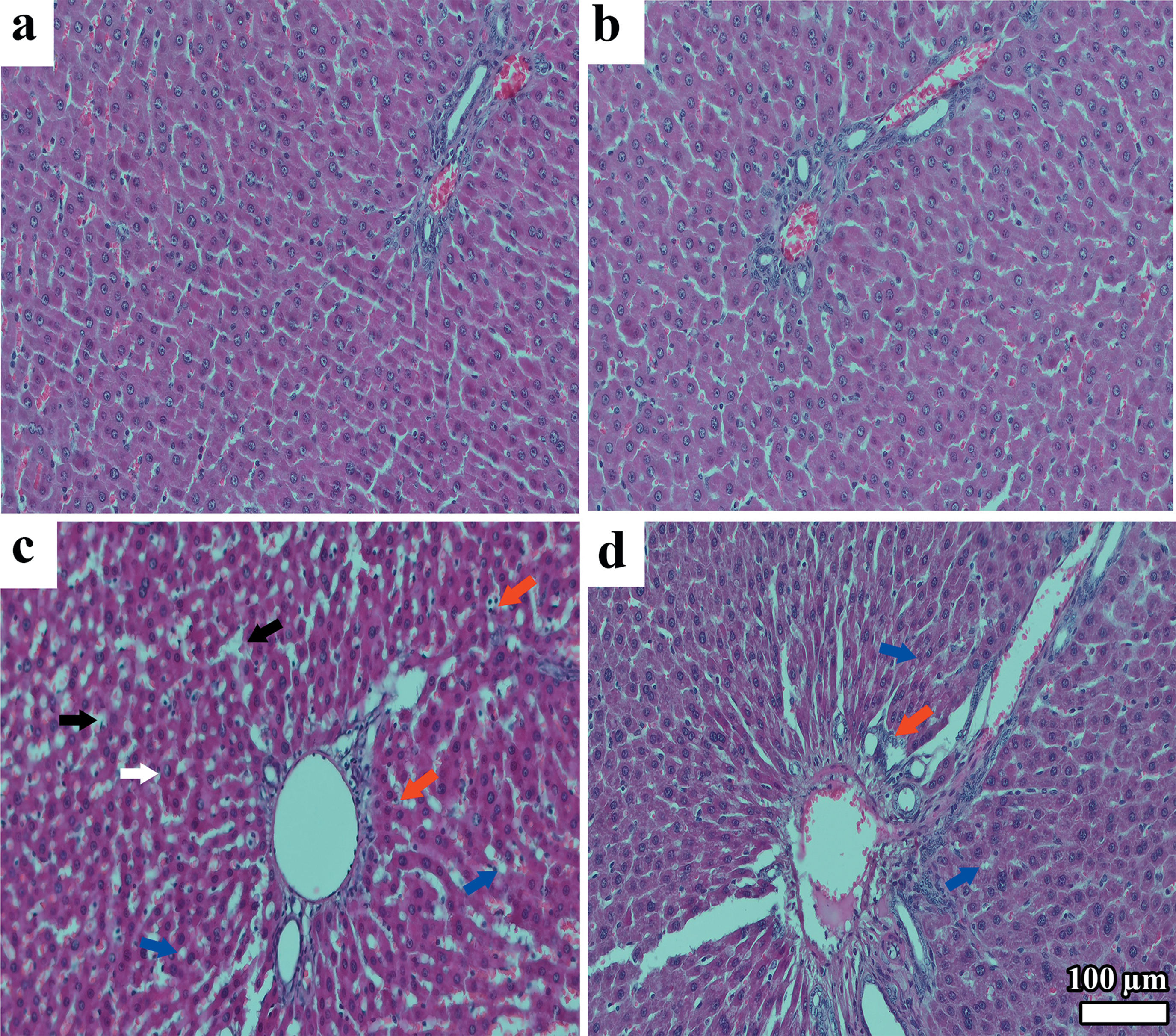
Histopathological examination of maternal liver tissues revealed that the sections from the control group displayed normal morphology (Figure 2a). Similarly, no histopathological alterations were observed in the NAC group, resembling the control group (Figure 2b). In the HFD group, sinusoidal dilation, ballooning of hepatocytes, inflammation, and microvesicular steatosis were noted. (Figure 2c). In the HFD+NAC group, less hepatocyte ballooning and microvesicular steatosis were noted compared to the HFD group (Figure 2d).
H&E staining of maternal liver sections. Control group (a), NAC group (b), HFD group (c), HFD+NAC group (d); Sinusoidal dilation (black arrow), Ballooning of hepatocytes (white arrow), Microvesicular steatosis (blue arrow), İnflammation (red arrow), H&E stain, X20.
Histopathological examination of liver tissues from male offspring revealed that the control group exhibited normal histological appearance (Figure 3a). Similarly, no histopathological alterations were observed in the NAC group, comparable to the control group (Figure 3b). In the HFD group, diffuse degeneration within the liver parenchyma, and hepatocyte swelling and microvesicular steatosis were detected (Figure 3c). In contrast, in the HFD+NAC group, hepatocytes mainly appeared normal. However, compared to the HFD offspring, much less hepatocyte swelling and microvesicular steatosis were detected (Figure 3d).
H&E staining of liver sections from male offspring. Control group (a), NAC group (b), HFD group (c), HFD+NAC group (d); Ballooning of hepatocytes (white arrow), Microvesicular steatosis (blue arrow), H&E stain, X20.

Immunohistochemical results
Immunohistochemistry for 8-hydroxy-2’-deoxyguanosine (8-OHdG), a marker of oxidative damage, was performed on liver sections from both mothers and male offspring (Figures 4 and 5). The H-score analysis of 8-OHdG immunostaining revealed a statistically significant difference among the groups (p < 0.05) (Figure 6).
Immunohistochemical staining of maternal liver sections. Control group (a), NAC group (b), strong 8-OHdG positive reactions in hepatocytes of the HFD group (c), moderate positive reactions in hepatocytes of the HFD+NAC group (d); Positive IHC staining with 8-OHdG (black arrow), IHC staining (sABC method), X20.

Immunohistochemical staining of liver sections from male offspring. Control group (a), NAC group (b), strong 8-OHdG positive reactions in hepatocytes of the HFD group (c), moderate positive reactions in hepatocytes of the HFD+NAC group (d); Positive IHC staining with 8-OHdG (black arrow), IHC staining (sABC method), X20.

H-score results for 8-OHdG immunohistochemical staining in dams and male offspring liver tissues (p < 0.05), n = 4 dams and n = 8 offspring per group. **p < 0.01, ***p < 0.001, ****p < 0.0001.

Immunohistochemical staining of maternal liver sections are presented in Figure 4. No immunoreactivity was observed in the liver sections from the control and NAC groups, and their staining patterns were similar. In contrast, increased 8-OHdG expression was observed in hepatocytes of the HFD groups. Compared to the control group, the HFD maternal group showed intense immunopositive staining for 8-OHdG in the cytoplasm of hepatocytes. NAC treatment in the HFD group led to a reduction in the immunopositivity of 8-OHdG in hepatocytes.
Immunohistochemical staining of liver sections from male offspring is presented Figure 5. In liver sections from male offspring, no immunoreactivity was detected in the control and NAC-treated groups, with similar staining profiles. Compared to the control group, the HFD offspring group exhibited intense and strong 8-OHdG immunopositivity in hepatocyte cytoplasm. In the HFD-NAC group, 8-OHdG expression was reduced to a moderate level compared to the HFD group.
Biochemical analyses
Following the termination of the experiment, various biochemical parameters, including ITT, GTT, TAS, TOS, PON1, ARES, and MDA, were assessed in blood samples. Additionally, values such as ITT AUC, GTT AUC, and OSI were calculated.
Graphs were used to compare the responses of different maternal and offspring groups to the GTT and insulin tolerance test (ITT).
Figure 7 demonstrates the effects of maternal obesity on both glucose and insulin tolerance. The HFD dam group exhibited higher glucose levels in both GTT and ITT compared to the other groups. In the NAC-treated groups, a significant reduction in glucose levels was observed in both non-HFD and HFD dams. Notably, glucose levels in the HFD + NAC group were significantly lower than those in the HFD group alone, with this effect being particularly pronounced in the GTT.
Maternal glucose responses to ITT and GTT.

Offspring Glucose Responses to ITT and GTT are presented in Figure 8. Offspring born to HFD dams exhibited significantly higher glucose levels in both GTT and ITT compared to the control group. According to the ITT results, insulin responsiveness was impaired in the offspring of HFD mothers. Offspring in the NAC-treated group showed a metabolic profile closely resembling that of the control group in terms of both insulin and glucose tolerance. Notably, the GTT and ITT outcomes of the HFD + NAC offspring group were improved compared to those of the HFD group alone.
Offspring glucose responses to ITT and GTT.

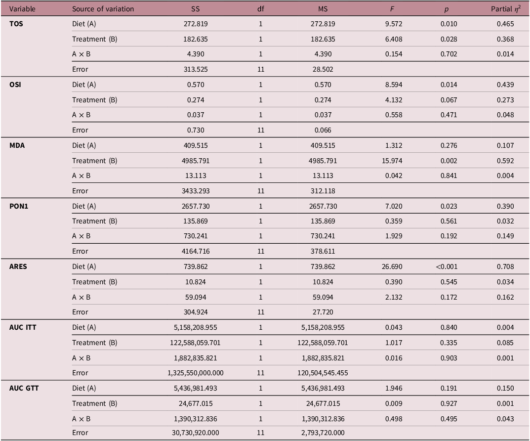
The effects of HFD and NAC on oxidative stress markers, antioxidant enzyme activities, and glucose metabolism parameters were evaluated using two-way ANOVA in Table 1. For oxidative stress parameters, both diet and treatment had significant main effects on total oxidant status (TOS) (diet: p = 0.010, η 2 = 0.465; treatment: p = 0.028, η 2 = 0.368). Similarly, oxidative stress index (OSI) was significantly affected by diet (p = 0.014, η 2 = 0.439), whereas the effect of treatment did not reach statistical significance (p = 0.067). In contrast, malondialdehyde (MDA) levels were significantly influenced by treatment (p = 0.002, η 2 = 0.592), but not by diet (p = 0.276). Regarding antioxidant enzymes, PON1 activity was significantly affected by diet (p = 0.023, η 2 = 0.390), with no significant effect of treatment (p = 0.561). ARES activity showed a highly significant effect of diet (p < 0.001, η 2 = 0.708), while treatment had no significant impact (p = 0.545). For glucose metabolism parameters, neither diet nor treatment had significant effects on AUC for insulin tolerance test (AUC ITT) or glucose tolerance test (AUC GTT). No significant interaction effects (diet × treatment) were observed for any of the measured parameters.
Evaluation of the effects of HFD and NAC on oxidative stress, antioxidant enzymes, and glucose metabolism parameters in dams

Table 1. Long description
The table presents the effects of diet and treatment on oxidative stress markers, antioxidant enzyme activities, and glucose metabolism parameters using two-way ANOVA. It includes eight variables: TOS, OSI, MDA, PON1, ARES, AUC ITT, and AUC GTT. The table has 8 rows and 7 columns. Column headers are Variable, Source of variation, SS, df, MS, F, p, and Partial n squared. Row labels include TOS, OSI, MDA, PON1, ARES, AUC ITT, and AUC GTT. Each row provides data for Diet (A), Treatment (B), and A x B interactions, along with Error values. Notable trends include significant effects of diet on TOS, OSI, and PON1, and significant effects of treatment on TOS, MDA, and PON1. No significant interaction effects were observed for any of the measured parameters.
Data were analyzed using two-way ANOVA with diet (control vs HFD) and treatment (±NAC) as fixed factors. Partial eta squared (η 2) was used as a measure of effect size. ITT: insulin tolerance test, GTT: glucose tolerance test, TAS: total antioxidant status, TOS: total oxidant status, OSI: oxidative stress index, PON1: paraoxonase 1, ARES: arylesterase, MDA: malondialdehyde (p < 0.05).
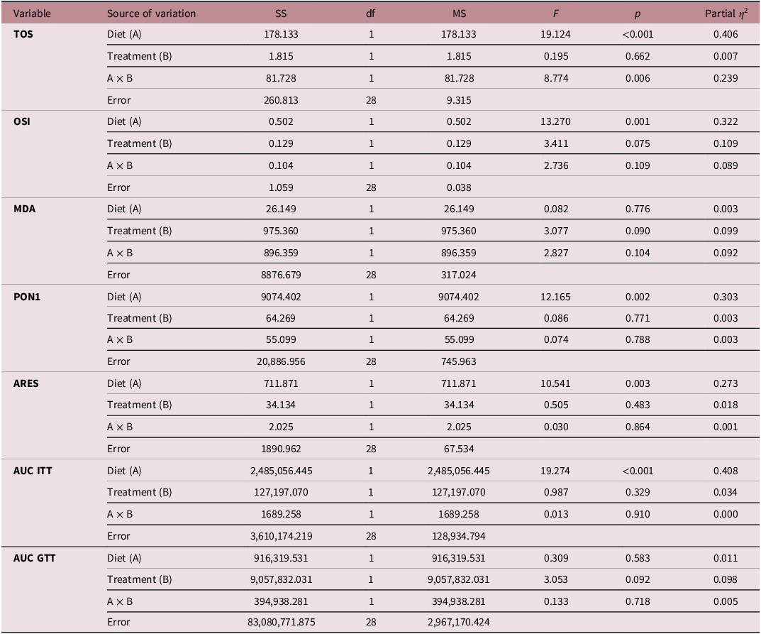
The effects of HFD and NAC on oxidative stress markers, antioxidant enzyme activities, and glucose metabolism parameters in offspring were assessed using two-way ANOVA in Table 2. For oxidative stress parameters, diet had a significant main effect on TOS (p < 0.001, η 2 = 0.406), whereas treatment had no significant effect (p = 0.662). Notably, a significant diet × treatment interaction was observed (p = 0.006, η 2 = 0.239). Similarly, OSI was significantly influenced by diet (p = 0.001, η 2 = 0.322), while treatment (p = 0.075) and the interaction term (p = 0.109) were not statistically significant. MDA levels were not significantly affected by diet (p = 0.776), treatment (p = 0.090), or their interaction (p = 0.104), although treatment and interaction effects approached significance. Regarding antioxidant enzymes, PON1 activity was significantly affected by diet (p = 0.002, η 2 = 0.303), with no significant effects of treatment (p = 0.771) or interaction (p = 0.788). Similarly, ARES activity showed a significant main effect of diet (p = 0.003, η 2 = 0.273), whereas treatment (p = 0.483) and the interaction (p = 0.864) were not significant. For glucose metabolism parameters, diet had a significant effect on AUC ITT (p < 0.001, η 2 = 0.408). However, treatment (p = 0.329) and the interaction (p = 0.910) were not significant. In contrast, no significant effects of diet (p = 0.583), treatment (p = 0.092), or their interaction (p = 0.718) were observed for AUC GTT.
Evaluation of the effects of HFD and NAC on oxidative stress, antioxidant enzymes, and glucose metabolism parameters in offsprings
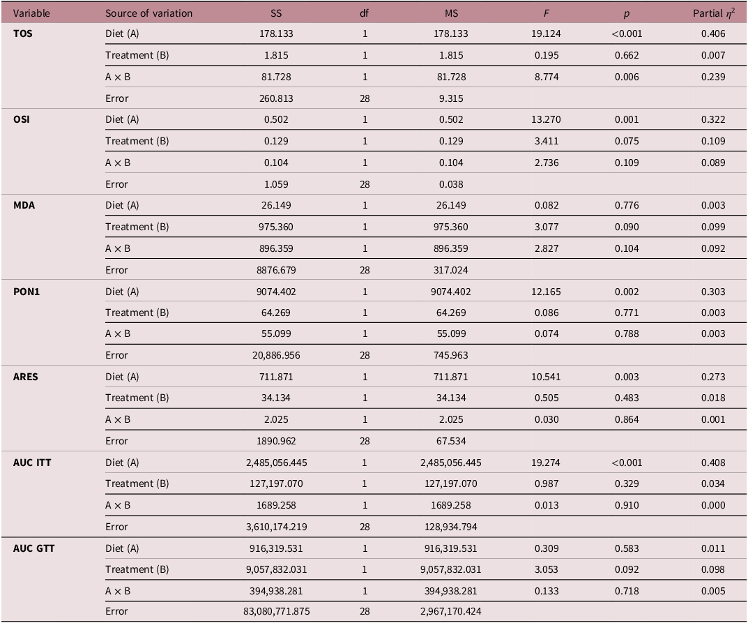
Table 2. Long description
The table presents the effects of diet and treatment on oxidative stress markers, antioxidant enzyme activities, and glucose metabolism parameters in offspring. It includes data for TOS, OSI, MDA, PON1, ARES, AUC ITT, and AUC GTT. The table has 8 rows and 7 columns. Column headers are Variable, Source of variation, SS, df, MS, F, p, and Partial n2. Row labels include TOS, OSI, MDA, PON1, ARES, AUC ITT, and AUC GTT. Each row provides data for Diet (A), Treatment (B), and A x B interactions. Notable trends include significant effects of diet on TOS, OSI, PON1, ARES, and AUC ITT, while treatment and interaction effects vary across parameters.
Data were analyzed using two-way ANOVA with diet (control vs HFD) and treatment (±NAC) as fixed factors. Partial eta squared (η 2) was used as a measure of effect size. ITT: insulin tolerance test, GTT: glucose tolerance test, TAS: total antioxidant status, TOS: total oxidant status, OSI: oxidative stress index, PON1: paraoxonase 1, ARES: arylesterase, MDA: malondialdehyde (p < 0.05).
Discussion
In our study, both histopathological evaluations using hematoxylin-eosin staining and semi-quantitative immunohistochemical assessment of 8-OHdG were performed on liver tissues of dams and male offspring.
Biochemical analyses included the ITT, GTT, total antioxidant status (TAS), total oxidant status (TOS), oxidative stress index (OSI), paraoxonase 1 (PON1), arylesterase (ARES), and malondialdehyde (MDA) levels.
Histopathological evaluation
In the present study, the livers of the control and NAC-only groups exhibited preserved hepatic architecture without any histopathological abnormalities and showed no or minimal 8-hydroxy-2′-deoxyguanosine (8-OHdG) immunoreactivity. These findings indicate that NAC administration alone does not adversely affect hepatic morphology or induce oxidative DNA damage, supporting its hepatic safety during gestation.
In contrast, maternal exposure to a HFD resulted in marked hepatic structural alterations, including sinusoidal dilatation, hepatocellular ballooning degeneration, microvesicular steatosis, and inflammatory cell infiltration. Concurrently, increased 8-OHdG immunopositivity was observed in hepatocytes of HFD dams, indicating significant oxidative DNA damage. These findings are consistent with previous studies demonstrating that maternal HFD intake or obesity induces hepatic steatosis, sinusoidal dysfunction, inflammation, and oxidative stress in both dams and their offspring. Reference Bruce, Cagampang and Argenton36–Reference Monks, Orlicky and Stefanski38 The hepatocellular ballooning degeneration and microvesicular steatosis observed in the HFD-fed groups represent hallmark histopathological features of non-alcoholic fatty liver disease, which is commonly associated with obesity and insulin resistance. These alterations are likely driven by excessive accumulation of free fatty acids in hepatocytes, leading to lipotoxicity and mitochondrial dysfunction. Impaired β-oxidation of free fatty acids promotes the generation ofROS, which exacerbates cellular injury through lipid peroxidation and DNA damage. Reference Cao, Liu and Zhang37 The accompanying sinusoidal dilatation may reflect vascular remodeling or portal alterations secondary to chronic inflammatory stress.
Maternal NAC supplementation partially attenuated both histopathological damage and oxidative DNA injury induced by the HFD. In the HFD + NAC group, hepatocytes exhibited sharply demarcated, non-staining vacuoles indicative of residual intracellular lipid accumulation; however, the overall severity of ballooning degeneration, steatosis, and 8-OHdG immunoreactivity was reduced compared to the HFD group. NAC is a well-known precursor of glutathione and has been reported to reduce oxidative stress-induced liver injury in experimental models of steatosis and non-alcoholic fatty liver disease. Reference Monks, Orlicky and Stefanski38,Reference Begriche, Igoudjil, Pessayre and Fromenty39 The attenuation of hepatocyte ballooning and steatosis observed in NAC-treated groups supports its anti-inflammatory and cytoprotective properties, potentially mediated through restoration of redox balance and modulation of pro-inflammatory signaling pathways such as NF-κB. Reference Samuni, Goldstein, Dean and Berk25
In male offspring of HFD dams, diffuse parenchymal degeneration and degeneration predominantly localized to the periacinar region were observed, accompanied by increased 8-OHdG immunoreactivity. The periacinar zone is particularly vulnerable to hypoxic and toxic insults due to its relative distance from the portal blood supply and lower oxygen tension. These findings suggest that intrauterine exposure to maternal obesity imposes metabolic and oxidative stress on the developing liver, predisposing offspring to hepatic injury. Similar observations have been reported in offspring of HFD or HFD–fed dams, where fetal programming mechanisms increase susceptibility to hepatic steatosis, insulin resistance, and oxidative damage later in life. Reference Bruce, Cagampang and Argenton36
Conversely, male offspring from the HFD + NAC group exhibited largely preserved hepatocyte morphology with reduced 8-OHdG staining intensity compared to the HFD group, indicating partial protection against oxidative DNA damage. However, the presence of extramedullary hematopoietic foci in portal areas suggests that NAC administration did not completely reverse all aspects of hepatic adaptation or developmental remodeling. A previous study investigating the effects of NAC in a rat model of non-alcoholic steatohepatitis similarly reported that NAC treatment improved steatosis and necroinflammatory scores but did not fully normalize liver histology. Reference Thong-Ngam, Samuhasaneeto, Kulaputana and Klaikeaw40
Taken together, these findings indicate that maternal HFD disrupts hepatic homeostasis in both dams and their offspring through mechanisms involving lipid accumulation, oxidative stress, and inflammatory injury. Maternal NAC supplementation appears to ameliorate several of these histopathological and oxidative alterations, supporting its potential role in mitigating the adverse intrauterine effects of maternal HFD. Nevertheless, the persistence of certain histological features, even in NAC-treated groups, underscores the complexity of obesity-induced hepatic programming and suggests that additional mechanisms beyond oxidative stress contribute to long-term liver pathology.
Biochemical evaluation
Higher glucose levels in both GTT and ITT tests in HFD dams compared to other groups were observed. The rapid decrease in glucose levels at the 30th minute in the ITT test especially indicates an effective insulin response. When evaluating the effects of NAC treatment, the significant reduction in glucose levels in both non-HFD and HFD dams suggests that NAC may improve insulin tolerance and glucose homeostasis through its regulatory effects on oxidative stress and inflammation. The markedly lower glucose levels in the NAC + HFD group compared to the HFD group were observed.
Offspring born to HFD dams showed significantly higher glucose levels in both GTT and ITT tests compared to the control group, supporting the influence of the intrauterine environment on metabolic programming. The glucose peak observed at the 30th minute during GTT and the slower decline thereafter indicate impaired glucose tolerance in these offspring. The poor insulin response was observed in offspring of HFD dams. Offspring in the NAC group exhibited insulin and glucose tolerance comparable to the control group, indicating that prenatal antioxidant support may positively influence fetal metabolic programming and offer protection against future metabolic disorders. Notably, the improved GTT and ITT results in the HFD + NAC offspring compared to the HFD-only group suggest that NAC may partially counteract the negative effects of maternal obesity. However, since glucose levels in this group did not reach control values, the protective effect of the treatment may be limited. NAC treatment during the prenatal period may improve both insulin and glucose tolerance in offspring. These findings underscore the critical role of prenatal interventions in shaping transgenerational metabolic health.
Sex may also play a role in glucose regulation. While differences among groups in GTT and ITT were less pronounced in mothers at various time points, they were more evident in male offspring. A similar outcome was reported by Berry et al. who observed more pronounced changes in sensitivity among males, while females exhibited less change. Reference Berry, Bellisario and Panetta41 A study associates maternal obesity with increased oxidant production and oxidative damage in various systems, particularly placental tissue. Reference Aye, Powell and Jansson42 This supports studies suggesting that NAC’s antioxidant effects may be limited in high oxidative stress environments. Reference Jorde, Brattelid and Haug43
Firstly, significant dietary effects on TOS and OSI clearly demonstrate that HFD increases the oxidative load in the maternal organism. The significant effect of both diet and NAC treatment on TOS suggests that NAC may be effective in reducing oxidant load. However, the lack of statistically significant effect of NAC on OSI (p = 0.067) indicates that NAC may not modulate all components of oxidative stress equally. This suggests that NAC may act primarily through specific oxidant types or specific biochemical pathways.
The fact that MDA levels, a key indicator of lipid peroxidation, were affected only by treatment (with a high effect size) supports the idea that NAC exhibits a strong antioxidant effect, particularly in suppressing lipid peroxidation. Reference Faghfouri, Zarezadeh and Tavakoli-Rouzbehani44 The lack of a significant effect of diet on MDA suggests that the oxidative damage caused by HFD arises primarily through other oxidative parameters, or that MDA may have lower sensitivity in this model.
When the antioxidant defense system is examined, the significant effect of diet on both PON1 and ARES activities indicates that HFD not only increases oxidant production but also suppresses antioxidant enzyme systems. The particularly high effect size (η 2 = 0.708) for ARES reveals that this enzyme is highly sensitive to diet-induced oxidative stress changes. Reference Sarhat, Wadi and Mahmood45 In contrast, the lack of a significant effect of NAC treatment on these enzyme activities suggests that NAC’s antioxidant effect occurs primarily through direct free radical scavenging mechanisms and that it does not sufficiently modulate enzymatic antioxidant defense. Reference Aldini, Altomare and Baron46
There was no statistically significant difference in insulin tolerance (ITT AUC) and glucose tolerance (GTT AUC) parameters among maternal groups. The study shows that the HFD applied during the study period was sufficient to trigger oxidative stress, but had not yet reached the threshold to develop significant insulin resistance or glucose intolerance. Oxidative stress is known in the literature to be a precursor to metabolic disorders. Reference Raut and Khullar47 Therefore, the current situation may represent a premetabolic stage.
The lack of a significant diet × treatment interaction in any parameter in the study indicates that the effects of NAC occur independently of dietary status and are limited in modifying HFD-specific effects. This finding suggests that the protective effect of NAC is more related to reducing overall oxidative stress levels, but its interaction with diet-induced metabolic changes may be weak.
Analysis of the TOS data of the offspring revealed that diet (p < 0.001, η 2 = 0.406) and especially the Diet × Treatment interaction (p = 0.006) were significant. This interaction, not observed in mothers, indicates that the effect of NAC on the oxidant load of offspring varies depending on the type of diet the mother is exposed to. This provides strong evidence that maternal NAC supplementation can modulate the pro-oxidant load caused by HFD in the offspring at a programming level. Furthermore, the significant dietary effect on the OSI (p = 0.001) confirms that high fat intake pushes offspring into a background of chronic oxidative stress from an early age. The lack of significant treatment and interaction effects suggests that NAC does not systematically modulate all components of oxidative stress. This finding suggests that NAC may act primarily through specific oxidant mechanisms and have a weaker effect on composite indices reflecting overall oxidative balance.
When antioxidant enzyme activities were examined, significant effects of diet were observed on both PON1 and ARES. This indicates that HFD not only increases oxidant production but also disrupts antioxidant defense systems. The moderate effect sizes (η 2 ≈ 0.27–0.30) support the idea that these enzymes are biomarkers sensitive to diet-induced oxidative stress changes. Reference Kim, Maden and Burt48 The continued decrease in the activity of these enzymes in offspring born from the HFD group suggests that maternal nutritional deficiency leaves a lasting epigenetic imprint or functional suppression in the liver and serum antioxidant capacity of the offspring. In contrast, the lack of a significant effect of NAC treatment on these enzymes suggests that the antioxidant effect of NAC occurs primarily through direct free radical scavenging, and that its capacity to modulate enzymatic antioxidant systems may be limited. Reference Aldini, Altomare and Baron46 This incomplete restoration underscores that overcoming obesity-induced metabolic and hepatic programming requires multi-targeted interventions beyond single-pathway antioxidant support.
One of the most striking findings of the study is the AUC ITT results of the offspring. While no significant effect of diet on insulin tolerance was found in mothers, maternal HFD had a very strong negative effect on the offspring (p < 0.001, η 2 = 0.408). This, consistent with the Barker Hypothesis, proves that maternal metabolic stress predisposes to insulin resistance in the peripheral tissues of the offspring rather than the mother. Reference Gluckman and Hanson49 Interestingly, the insignificant AUC GTT results indicate that the offspring do not yet exhibit an overt diabetic picture, but are entering a prediabetic phase with decreased insulin tolerance.
Conclusion
A HFD induces adverse histopathological changes in tissues. While NAC-treated animals showed tissue morphology similar to controls, complete recovery to control levels was not achieved in HFD + NAC groups. This study demonstrates that maternal HFD negatively reprograms the metabolic and redox profiles of offspring, leading to significant impairments, particularly in insulin tolerance. The persistent decrease in PON1 and ARES enzyme activities observed in the offspring proves that maternal nutritional stress leaves a functional suppression and a lasting epigenetic imprint on the antioxidant defense systems of the offspring. Although prenatal NAC supplementation partially improves metabolic parameters by reducing oxidative load through a direct free radical scavenging mechanism, its inability to completely reverse enzymatic systems and epigenetic programming underscores that dietary modification, not just antioxidant support, is of primary importance in maintaining transgenerational health.
Limitations
This study is limited by the lack of comprehensive systemic obesity assessments; therefore, the term “rats fed a HFD” is used instead of “obese rats.” Only male offspring were analyzed to avoid hormonal confounding, and NAC effects in offspring reflect indirect maternal exposure rather than direct treatment. A limitation of this study is the relatively small number of dams per group (n = 4), which may limit the generalizability of the findings. Therefore, the results should be interpreted with caution and confirmed in larger studies. Since NAC was administered during both gestation and lactation, the current design could not distinguish between prenatal and postnatal effects. Future studies using cross-fostering may help clarify the contribution of these exposure periods.
Financial support
This research project was financed by Kastamonu University Scientific Research Projects Unit with project reference [KÜBAP-01/2022-16].
Competing interests
The authors declare no conflict of interest.
Ethical standard
This study was performed in line with the principles of the Declaration of Helsinki. Approval was granted by the Kastamonu University Animal Experiments Local Ethics Committee (24.06.2022/10).



 Open access

