





Introduction
Right ventricular outflow tract stenting is a palliative treatment modality in symptomatic newborns and infants with tetralogy of Fallot or with pulmonary atresia and a ventricular septal defect. Reference Goldstein, Petit and Qureshi1 This procedure is performed mainly in patients who exhibit additional severe comorbidities and hypoplastic pulmonary arteries Reference Pizzuto, Cuman and Assanta2–Reference Abumehdi, Sasikumar and Chaudhari5 and, therefore, are not candidates for primary surgical correction and need an improvement in oxygen saturation. In the current era, some centres advocate for right ventricular outflow tract stenting as a first-line palliative treatment for these conditions. Reference Bertram, Emmel and Ewert6,Reference Quandt, Ramchandani and Penford7 In the majority of cases, bare metal coronary stents or other bare metal stents are used for this procedure. Reference Pizzuto, Cuman and Assanta2,Reference Ghaderian, Ahmadi and Sabri4,Reference Quandt, Penford, Ramchandani, Bhole, Mehta and Stumper8 Tissue ingrowth due to endothelium hyperplasia may cause stent narrowing, Reference Prabhu, Mehra, Kanaparthi, Maiya and Rao9,Reference Nokhrin, Tarasov and Mukhamadiyarov10 and it may be difficult or impossible to surgically remove these stents without causing significant harm to the native pulmonary valve tissues or to the right ventricular outflow tract myocardium. Reference Pizzuto, Cuman and Assanta2,Reference Barron, Ramchandani and Murala11–Reference McGovern, Morgan, Oslizlok, Kenny, Walsh and McMahon13 For these reasons, some cardiac surgeons object to using right ventricular outflow tract stenting as a palliative strategy. These complications may potentially be avoided by the implantation of a covered stent into the right ventricular outflow tract. Reference Nishi, Nakayama, Ueda, Ishikawa and Matsuda14,Reference Kwa, Dawson and Laird15 Here, we present our experience with patients who were treated using covered coronary stent-grafts in the right ventricular outflow tract.
Methods
Patients
From November 2017 to July 2021, all consecutive patients treated in the German Heart Centre, Munich, Germany (n = 20, 14 males), for tetralogy of Fallot and pulmonary atresia with ventricular septal defect with coronary covered stent-grafts in the right ventricular outflow tract were evaluated. The median patient age was 32 (4–306) days, the median body weight was 3.0 (1.26–6.5) kg, and the median height was 51.0 (40.0–74.0) cm. Nine patients were newborns, and the rest were treated in the first year of life. This study was approved by the institutional ethics committee.
Prior to any intervention, the treatment plan for each patient was based on an individual case discussion by the Heart Team. Patients selected for right ventricular outflow tract stenting with a covered stent were unanimously considered unsuitable candidates for alternative surgical or interventional palliative strategies. The primary reason was the presence of severely hypoplastic pulmonary arteries, which rendered the placement of a modified Blalock-Thomas-Taussig shunt technically unfeasible and unlikely to result in meaningful haemodynamic improvement. Ductal stenting was considered technically challenging in cases with unfavourable anatomy or not feasible when the duct had already closed by the time of the scheduled catheter-based intervention. Additionally, the decision-making process placed particular emphasis on the potential physiological benefits of preserving antegrade pulsatile flow across the right ventricular outflow tract, which may support pulmonary artery growth and promote more favourable overall haemodynamics. The patient’s caregivers also gave informed consent for each treatment.
Interventional technique
The BeGraft stent graft system (Bentley InnoMed, Germany) is used for the treatment of acute coronary artery perforations and ruptures, coronary artery aneurysms, and coronary bypass-vein graft aneurysms. The cobalt-chrome stent platform is highly flexible and covered with a microporous expanded polytetrafluoroethylene membrane. The available diameters are from 2.5 to 5.0 mm, and the lengths range from 8 to 24 mm. The BeGraft is implanted through a 5F guiding catheter.
All procedures were performed using a biplane angiographic platform. Cardiac catheterisations were conducted under intravenous sedation in spontaneously breathing patients. A femoral vein was punctured, and a 5 French Glidesheath Slender sheath (Terumo, Japan) was introduced in order to maintain a low outer profile as previously described. Reference Gendera, Cleuziou and Tanase16 Heparin (100 units/kg, intravenously) was administered unless contraindicated. The “telescopic technique” with a short 5 French Judkins Right 4 coronary guide catheter (55 cm, Cordis, USA) was used. In patients with an atretic pulmonary valve, radiofrequency perforation using the Baylis Radiofrequency Puncture Perforation System (BVM Medical, UK) was utilised to open the right ventricular outflow tract. After a right ventricular angiography and depiction of the right ventricular outflow tract, the diameter and length of the right ventricular outflow tract and pulmonary valve were carefully assessed. The stent diameter was selected to be 1–2 mm wider than the right ventricular outflow tract and to cover the entire length. In cases of incomplete coverage of the right ventricular outflow tract by the stent, additional stents were implanted. If possible, it was tried not to cover the pulmonary valve; however, if the pulmonary valve was severely hypoplastic (Z-score < −3), the stent was placed over the pulmonary valve. Control angiography was performed to confirm the required result and to exclude any complications. Patients were observed for a short period in the hospital, with an echocardiography examination performed to assess the results of the procedure. Anticoagulation was started with intravenous heparin and continued later with oral acetylsalicylic acid for 6 months or until the next surgical step. Clinical and echocardiographic reevaluations were carried out at regular intervals. To evaluate the growth of the pulmonary arteries after the stent implantation, we calculated the Nakata Index and Z-scores for the diameters of the main pulmonary artery, the left pulmonary artery, and the right pulmonary artery before and after treatment. Reference Dyar17,Reference Nakata, Imai and Takanashi18
Two explanted overlapping stents were placed in formalin for a histological work-up, which was achieved by infiltration in hard resin and applying the sawing-and-grinding technique, as described previously. Reference Sigler, Paul and Grabitz19
Statistical analysis
Statistical analysis was performed with PQStat 1.8.2.238 (PQStat Software, Poland). Data that were skewed are presented as medians, minimum, and maximal values. Categorical data are expressed as counts and percentages where appropriate. Comparisons between groups were performed with parametric or non-parametric tests. A p-value < 0.05 was considered statistically significant.
Results
All covered stents were implanted successfully. Median follow-up time was 156 (43–1578) days. Detailed demographic and clinical data are presented in Table, and a flowchart of the course of patient treatment is depicted in Figure 1.
The course of treatment for patients with RVOT stenting using the beGraft. BV: biventricle circulation, PDA: arterial duct, RVOT: right ventricle outflow tract.

Indications
There were two main indications for stenting the right ventricular outflow tract in our cohort. One group was comprised of patients (n = 5) with pulmonary atresia, a ventricular septal defect, major aortopulmonary collaterals, and very diminutive central and branch pulmonary arteries. In these patients, the atretic pulmonary valve was perforated before implanting the covered stent. The right ventricular outflow tract was stented immediately after the perforation of the valve in all but two patients (described below). In this indication, the diameter of the implanted stent was 3.5–4.5 mm, and it had a length of 12–16 mm.
The second group of patients consisted of symptomatic (cyanotic or duct-dependent) patients (n = 15) with tetralogy of Fallot. In these patients, stenting was done to avoid surgical palliation (aortopulmonary shunt or palliative right ventricular outflow tract patch). Patients with tetralogy of Fallot had their right ventricular outflow tract stented with stent grafts of larger diameters, ranging from 3.5 to 5.0 mm.
Additional stenting during the initial procedure and follow-up
Four patients had an additional stent(s) implanted during the initial procedure either to stabilise the stent graft or to cover the whole length of the stenosed right ventricular outflow tract. Patient no. 9 had a coronary stent (3.5 x 18 mm) implanted into the arterial duct due to echocardiographic signs of stenosis of the origin of the left pulmonary artery two days after the BeGraft implantation. To treat stenosis of the right ventricular outflow tract that developed proximal to the implanted BeGraft stent after initial palliation, 11 patients had an additional stent implanted at a median time of 40 (3–834) days after initial palliation.
Stent redilation
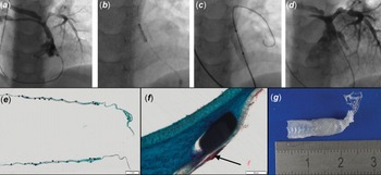
Ten patients (50%) with recurrent cyanosis needed a redilation of the implanted stent to adapt to somatic growth [with 4.0–8.0 mm percutaneous transluminal coronary angioplasty balloons]. Redilation was necessary for four patients from the pulmonary atresia with ventricular septal defect group and for six patients from the tetralogy of Fallot group. Median follow-up time for patients requiring stent redilation was 226 (43–835) days compared with 129 (50–531) days for the rest of the group, but the difference was not statistically significant. Four patients (nos. 1, 4, 6 and 15) needed more than one redilation (Figure 2 A–D). Patients no. 4 and 6 had a very resistant stenosis that required dilation with an ultra-high-pressure balloon.
Upper row: reintervention in a patient with pulmonary atresia, 28 months after implantation of a covered Bentley coronary stent in the RVOT (Patient No. 1). A: initial angiography showing diminutive pulmonary arteries and the previously implanted RVOT stent. B: implantation of a formula stent (6×12mm) in the RVOT. C: high-pressure balloon dilatation to intentionally fracture the stent. D: final angiography demonstrating a satisfactory procedural result. Lower row: E: histology (Movat pentachrome staining) showing two overlapping beGraft stents without inner or outer tissue appositions. F: minor fibrin condensations on the stent (red colour, black arrow). G: explanted stents.

Size of pulmonary arteries and arterial oxygen saturation
Complete angiographic data to compare the size of pulmonary arteries were available in 12 patients. In these patients, Z-scores for the main pulmonary artery (median from −7.3 to −3.2, p < 0.05), left pulmonary artery (median from −3.4 to 0.2, p < 0.05), and right pulmonary artery (median from −4.0 to 1.4, p < 0.05) increased significantly over a median time of 117 (63–394) days. We also observed a significant increase in the Nakata Index in these patients from a median of 51.1 to 189.9 mm/m2 (p < 0.05) over the same time period.
There was an immediate increase in oxygen saturation in our patients, from a median value of 81% before the procedure to 90% post-intervention (p < 0.05).
Complications
Three complications occurred in our cohort. One patient (no. 4 - pulmonary atresia with ventricular septal defect, major aortopulmonary collaterals, hypoplastic pulmonary arteries) suffered right ventricular outflow tract perforation, which resulted in the accumulation of blood in the pericardium and the left pleural cavity without haemodynamic compromise. This was managed with an aspiration of the blood from the pericardium, and the bleeding ceased spontaneously. The procedure was postponed, and the atretic pulmonary valve again was perforated 35 days later, and a 3.5 x 12 mm BeGraft was implanted without complications. In patient no. 3, after the perforation of the valve, we noticed an aneurysm of the pulmonary trunk. This was completely excluded by stent-graft implantation six days after the radiofrequency perforation. One serious complication occurred in the cohort of patients with tetralogy of Fallot. In patient no. 5, three stents (BeGraft 3.5 x 12 mm, 3.5 x 8 mm, and a Formula stent 4.0 x 12 mm [Cook, USA]) were utilised to cover the whole length of the right ventricular outflow tract. The last stent embolised to the right ventricle and through the ventricular septal defect to the descending aorta. It was not possible to retrieve it by interventional methods, and, finally, the stent was removed surgically.
Midterm results and surgical correction
All stent grafts were patent during the study period, examined by echocardiography or angiography; we did not detect any stent fractures. To date, sixteen patients have undergone surgical correction of their heart defect, with complete surgical removal of implanted BeGrafts.
Two patients needed an additional surgical shunt preceding correction due to low saturation. Twelve patients were surgically corrected at a median age of 6 (4.8–15.5) months. One patient died two months after surgical correction. In this patient, a take-down of the correction was performed (reopening of the ventricular septal defect, right-ventricle-pulmonary conduit removal, central shunt to the left and right pulmonary arteries) due to unsatisfactory oxygenation. The patient needed extracorporeal membrane oxygenation support and died due to multi-organ failure. Two additional patients needed extracorporeal membrane oxygenation support after surgery. Both needed support due to right ventricular failure and were successfully weaned 1 and 4 days after extracorporeal membrane oxygenation implantation.
Histological examination of the removed stent graft
Histological evaluation of the explanted stents showed no adjacent tissue from the surrounding right ventricular outflow tract muscular tissue (Figure 2 E–G). Accordingly, no significant tissue proliferation of pseudo-intima was found on the inner surface of the stents. However, very few fibrin depositions had formed. Tissue that had formed in between the two stents demonstrated regular fibromuscular cells embedded in an extracellular matrix without inflammatory cells or atypical cells.
Discussion
This study shows that catheter palliation in cyanotic patients with diminutive pulmonary arteries using covered coronary stents is feasible, safe, and effective. At our institution, right ventricular outflow tract stenting was performed in patients for whom other palliative strategies were deemed unsuitable, technically challenging, or potentially associated with a higher risk of periprocedural complications. Covered coronary stents were chosen because of the potential ease of surgical removal during the corrective operation.
If primary surgical correction is not feasible, right ventricular outflow tract stenting is one possible treatment option for the rehabilitation of pulmonary arteries. Reference Goldstein, Petit and Qureshi1,Reference Bertram, Emmel and Ewert6–Reference Quandt, Penford, Ramchandani, Bhole, Mehta and Stumper8,Reference Dohlen, Chaturvedi and Benson20,Reference Qureshi, Caldarone and Wilder21 An augmentation of pulmonary blood flow should result in an increase in vascular diameter in the small central pulmonary arteries to enable later surgical correction. The main advantages of this type of palliation compared to surgical aortopulmonary shunt or arterial duct stenting are avoiding distortion of pulmonary arteries, lower ICU admission rate, and the promotion of better, more symmetrical pulmonary arterial growth. Reference Pizzuto, Cuman and Assanta2,Reference Quandt, Ramchandani and Penford7,Reference Quandt, Ramchandani and Stickley22 However, the evidence for improved survival and less morbidity, especially in larger groups of patients and meta-analyses, is less clear. Reference Goldstein, Petit and Qureshi1,Reference Pizzuto, Cuman and Assanta2,Reference Ghaderian, Ahmadi and Sabri4
A known disadvantage of right ventricular outflow tract stenting is the early and common need for reintervention. Reference Goldstein, Petit and Qureshi1,Reference Pizzuto, Cuman and Assanta2,Reference Quandt, Ramchandani and Penford7,Reference Quandt, Penford, Ramchandani, Bhole, Mehta and Stumper8,Reference Nokhrin, Tarasov and Mukhamadiyarov10–Reference McGovern, Morgan, Oslizlok, Kenny, Walsh and McMahon13 One of the causes may be tissue ingrowth into the implanted stent. Progressive vessel narrowing after bare metal stent implantation is a recognised complication, Reference Quandt, Ramchandani and Penford7,Reference McMahon, El-Said, Grifka, Fraley, Nihill and Mullins23 necessitating reintervention to relieve the obstruction. Neointimal proliferation is one of the causes of stent restenosis in a non-coronary setting that is also observed in stents implanted in the right ventricular outflow tract. Reference Prabhu, Mehra, Kanaparthi, Maiya and Rao9,Reference Nokhrin, Tarasov and Mukhamadiyarov10,Reference Happel, Bertram, Kretschmar, Eildermann, Schmiady and Sigler24 The formation of neointimal tissue is a response of the vascular structures to several forms of injury and is compounded by the low biocompatibility and hemocompatibility of bare metal stents. Reference Newby and Zaltsman25 The use of a covered stent may limit tissue ingrowth into the stent struts, thus limiting the neointimal proliferation observed for stents implanted in other locations of the cardiovascular system. Reference Chang, Goodney, Baek, Nolan, Rzucidlo and Powell26–Reference Mwipatayi, Thomas and Wong27 The results of the histological examinations of the explanted stents confirm this hypothesis. No adjacent tissue from the surrounding right ventricular outflow tract muscular tissue, and no significant pseudo-intima was found on the inner surface of the stents. How this observation translates into clinical results is yet to be determined, but all implanted BeGraft stents remained patent, and no stenosis of a stent lumen was observed.
The indication for additional stent placement in the follow-up was a muscular stenosis of the right ventricular outflow tract proximal to the implanted BeGraft. None of the patients with the right ventricular outflow tract primarily stented with more than one stent (n = 4) needed an additional stent in follow-up. Contrary to this, 11 out of 16 patients primarily palliated with one stent required additional stenting during the observation period. Despite the intention to cover the whole right ventricular outflow tract during the primary intervention, this may indicate that the primary stent was too short.
The need for additional balloon redilation of the BeGraft resulted from prolonged palliation, with a median time of more than 7 months. This time of palliation is similar to that reported by Quandt Reference Quandt, Ramchandani and Penford7 and Castleberry Reference Castleberry, Gudausky, Berger, Tweddell and Pelech29 but longer than that reported in other work. Reference Sandoval, Chaturvedi and Benson28 Patients’ growth, not the stent lumen stenosis, was the indication for balloon redilation. The covered Bentley coronary stent can be safely expanded to a diameter of 5mm. Further expansion leads to a stent fracture. The initial stent diameter does not influence the maximum achievable expansion. Reference Gendera, Bambul Heck and Eicken30 Our results show that BeGraft stents can be safely expanded beyond the diameters provided by the manufacturer.
A similar, high burden of reinterventions has been reported by others, Reference Abumehdi, Sasikumar and Chaudhari5,Reference Quandt, Ramchandani and Penford7,Reference Quandt, Penford, Ramchandani, Bhole, Mehta and Stumper8,Reference Dohlen, Chaturvedi and Benson20,Reference Sandoval, Chaturvedi and Benson28 although the proportion of patients requiring additional stenting in our cohort is higher. This difference may be partially explained by a high proportion of patients with pulmonary atresia, severely hypoplastic pulmonary arteries, and low body weight.
The relatively small number of surgically corrected patients, long time of palliation, and numerous reinterventions reflect the complexity of the cardiac condition in our group of patients. This is especially true in regard to the five patients with an atretic pulmonary valve. Reference Walsh, Lee, Chaturvedi, Van Arsdell and Benson31 All of these patients, after the radiofrequency perforation, had a BeGraft stent implanted in the right ventricular outflow tract, similar to the technique described by Aurigemma et al. Reference Aurigemma, Moore, Vaughn, Moiduddin and El-Said32 Although an aneurysm of the main pulmonary artery may be clinically silent, it can carry the risk of severe complications (rupture or clot formation). In one of our patients, we were able to exclude such an aneurysm with the BeGraft, adding some safety for the patient.
The dimensions of the main pulmonary artery and its branches (expressed as Z-scores) were extremely small in our patients and smaller than those reported by other groups. Reference Abumehdi, Sasikumar and Chaudhari5,Reference Barron, Ramchandani and Murala11,Reference Bigdelian, Ghaderian and Sedighi12,Reference Nishi, Nakayama, Ueda, Ishikawa and Matsuda14,Reference Dohlen, Chaturvedi and Benson20 A large proportion of patients with atretic pulmonary valve and major aortopulmonary collaterals in our cohort may explain this difference. This haemodynamic configuration may cause severe underdevelopment of the central pulmonary artery and its branches. Despite this, we showed that right ventricular outflow tract stenting creates a possibility for the pulmonary artery and its main branches to grow. Similar results have been reported by others. Reference Abumehdi, Sasikumar and Chaudhari5,Reference Quandt, Ramchandani and Penford7,Reference Quandt, Penford, Ramchandani, Bhole, Mehta and Stumper8,Reference Barron, Ramchandani and Murala11,Reference McGovern, Morgan, Oslizlok, Kenny, Walsh and McMahon13,Reference Dohlen, Chaturvedi and Benson20,Reference Castleberry, Gudausky, Berger, Tweddell and Pelech29 BeGraft stent implantation adds to the variety of possible pulmonary artery rehabilitation procedures. Reference Goldstein, Petit and Qureshi1,Reference Barron, Ramchandani and Murala11
Surgical removal of an implanted stent from the right ventricular outflow tract may be difficult and technically challenging, with different mechanisms involved and different numbers of entirely excised stents reported. Reference Quandt, Penford, Ramchandani, Bhole, Mehta and Stumper8–Reference Bigdelian, Ghaderian and Sedighi12,Reference Nishi, Nakayama, Ueda, Ishikawa and Matsuda14,Reference Castleberry, Gudausky, Berger, Tweddell and Pelech29 Stent removal can result in destruction of the native pulmonary valve Reference Quandt, Penford, Ramchandani, Bhole, Mehta and Stumper8,Reference Barron, Ramchandani and Murala11,Reference Bigdelian, Ghaderian and Sedighi12,Reference Nishi, Nakayama, Ueda, Ishikawa and Matsuda14 and the right ventricular outflow tract myocardium. This can sometimes also endanger the coronary arteries on the surface (especially the left anterior descending artery), but there is evidence that right ventricular outflow tract stenting and at least partial stent removal are achievable even in patients with tetralogy of Fallot and anomalous coronary arteries. Reference Afifi, Mehta and Bhole33 In all of the sixteen patients who underwent surgical correction to date, the BeGraft stents were removed entirely and easily. This contrasts with the removal of bare metal stents from the right ventricular outflow tract, as our surgical team reported previously.
Complications
Overall, in our series, three patients experienced serious complications that were not directly related to covered stent usage. Perforation of the atretic pulmonary valve may be accompanied by an inadvertent perforation of the right ventricular outflow tract and a risk of aneurysm formation in a large portion of patients. Reference Walsh, Lee, Chaturvedi, Van Arsdell and Benson31 However, we believe that the use of a covered stent may help to manage these complications. Embolisation of an implanted stent is not common during right ventricular outflow tract stenting but, if present, usually carries a high risk of morbidity for the patient. Reference Quandt, Ramchandani and Penford7 In the case of a small coronary stent, the embolised stent can be retrieved in some patients, Reference Quandt, Ramchandani and Penford7 but this is more difficult if other stents are used. If unsuccessful, surgical removal must be performed, as in our case. There were no deaths related to right ventricular outflow tract stenting during the procedure or in the follow-up period.
Limitations
The group of patients is small, and the follow-up time is short. In addition, the BeGraft stent was used for right ventricular outflow tract stenting in very symptomatic young patients (mostly newborns or infants) unsuitable for or at high risk for other methods of palliation. The study cohort is also heterogeneous, comprised of patients with tetralogy of Fallot, pulmonary atresia with ventricular septal defect, and major aortopulmonary collaterals. The decision for right ventricular outflow tract stenting was based on an individual case discussion by the Heart Team and not upon predefined criteria. Additionally, inclusion of a control group receiving bare-metal stents for direct comparison was not feasible due to the surgical team’s reluctance to implant bare-metal stents, given concerns regarding their surgical removal and long-term outcomes. Finally, this study is retrospective, with all the known limitations.
Conclusions
Stenting of the right ventricular outflow tract with covered stents in young infants with diminutive pulmonary arteries that were not amenable for primary surgical correction was safe and effective in a small cohort of patients. This stent-graft system provides a durable method of palliation with a possibility of further redilation and limits the potential of neointimal proliferation. Utilisation of covered stents may facilitate surgical removal of the implanted stent during the surgical correction.
Demographic and clinical data of patients with the RVOT stented using BeGraft

AVSD = atrioventricular septal defect, MAPCAs = major aortopulmonary collaterals, MPA = main pulmonary artery, PAs = branches of the pulmonary artery, PaV = pulmonary valve, PA + VSD = pulmonary atresia with ventricular septal defect, PDA = arterial duct, PS = pulmonary valve stenosis, RVOT = right ventricle outflow tract, ToF = tetralogy of Fallot.
Acknowledgements
None
Financial support
None.
Competing interests
None.



 Open access

