





Introduction
CHD affects approximately 10 per 1000 live births. Reference Xu, Li, Deng, Xiong, Cheng and Ye1 Although advances in medical and surgical care have reduced global CHD mortality by over 75% in recent decades, neonates undergoing cardiac surgery with cardiopulmonary bypass remain at high risk for neurological injury and long-term neurodevelopmental impairment. Reference Xu, Li, Deng, Xiong, Cheng and Ye1–Reference Claessens, Algra and Ouwehand4 Up to 35–50% of neonates with critical CHD have evidence of brain injury prior to surgery, and as many as 50% acquire new structural brain injury following cardiopulmonary bypass. Reference Stegeman, Feldmann and Claessens5–Reference Lee, Sun and van Amerom8 Although often clinically silent, brain injury may manifest as seizures, affecting up to 20% of neonates postoperatively. Reference Andropoulos, Mizrahi and Hrachovy9–Reference Clancy, Sharif and Ichord11 These injuries result from a complex interplay of perinatal factors, altered central nervous system development, genetic conditions, and perioperative haemodynamic and metabolic disturbances. Reference Andropoulos, Hunter and Nelson6,Reference Lee, Sun and van Amerom8
Neuroimaging and neuromonitoring tools, such as brain magnetic resonance imaging (MRI), electroencephalography, and near-infrared spectroscopy, may aid detection of neurological injury and guide perioperative management, with emerging roles in neurological and neurodevelopmental prognostication. Reference Sood, Newburger and Anixt12,Reference Massey, Weinerman and Naim13 Despite their importance, standardised guidelines describing optimal perioperative neuroimaging and neuromonitoring are lacking, resulting in substantial practice variation across centres. We therefore surveyed Pediatric Cardiac Intensive Care Society-affiliated centres to characterise current neuroimaging and neuromonitoring practices and provider perceptions regarding their utility, sufficiency, and barriers to broader implementation.
Materials and methods
Survey
An online cross-sectional survey was developed by the Neuro-Cardiac Special Interest Group of the Pediatric Cardiac Intensive Care Society, comprised of physicians, nurses, and advanced practice providers, from cardiology, critical care, psychology, and neurology. It was reviewed by all authors for clarity, relevance, and completion time prior to distribution. The study was approved by the Pediatric Cardiac Intensive Care Society scientific review and research committee and by the University of Texas Southwestern institutional review board (STU-2024-0494) with a waiver of informed consent. The manuscript follows the Consensus-Based Checklist of Reporting of Survey Studies checklist. Reference Massey, Weinerman and Naim13 The survey was distributed to Pediatric Cardiac Intensive Care Society members via the society listserv using a secure REDCap© link. Reference Harris, Taylor and Minor15,Reference Harris, Taylor and Thielke16
Participants
At the time of the study, Pediatric Cardiac Intensive Care Society membership included 1035 individual members: 413 physicians, 368 advanced practice providers, 180 nurses, and 74 trainees affiliated with 153 institutions (109 in the United States and 44 international). Respondents provided information on their clinical role, title, subspeciality, and centre affiliation; no additional identifiers were recorded. To enhance response rates among medical directors, a targeted email invitation containing the survey link was sent to 137 medical directors identified in the Pediatric Cardiac Intensive Care Society directory.
Part 1 of the survey, completed by self-identified medical directors, evaluated institutional characteristics, neurodevelopmental and neurocritical care resources, and perioperative neuromonitoring and neuroimaging practices. Neuroimaging modalities assessed included brain MRI, head ultrasound (HUS), computerized tomography (CT), and transcranial doppler, while neuromonitoring modalities included near-infrared spectroscopy, electroencephalography (EEG), bispectral index, automated pupillometry, and diffuse correlation spectroscopy. Part 2, distributed to all respondents, evaluated perceptions of the utility and sufficiency of these practices. Perceived utility was rated using a 5-point Likert scale, while sufficiency of institutional resources and practices was assessed as a binary yes/no response. Additional survey details are shown in Supplementary Appendix 1.
Data analysis
Survey responses were summarised descriptively using proportions, with comparisons performed using chi-square or Fisher’s exact tests, as appropriate. If multiple medical directors from the same institution completed Part 1, one response was randomly selected for centre-level analysis (this occurred at one institution). In Part 2, all responses were analysed, with denominators varying by completed items. Likert-scale responses were dichotomised for regression analyses, with the responses “often” and “sometimes useful” categorised as “useful,” and the responses “rarely,” “neutral,” and “not applicable” were categorised as “not useful.” Mixed-effects logistic regression was used to evaluate differences in perceptions by respondent role while accounting for clustering by institution. Responses from individuals at institutions lacking a corresponding medical director survey were excluded from analyses that linked individual perceptions to institutional practices. We used either profile likelihood or bootstrapping to estimate standard errors. Odds ratios with their 95% confidence intervals were presented as OR (95% CI). For missing answers, we used available case analysis, i.e., maximising the available data by only excluding cases on a variable-by-variable basis. Analyses were performed using available responses without imputation in R version 4.4.1 (R Foundation for Statistical Computing).
Results
Respondent and centre characteristics
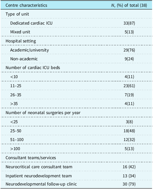
Thirty-eight medical directors representing unique institutions completed Part 1 (Table 1). Of these, 37 programmes were in the United States and one in Argentina. Most centres had a dedicated CICU (33/38, 87%) and were academically affiliated (76%). Neurodevelopmental follow-up clinics were common (79%), but fewer centres had dedicated neurocritical care consulting teams (42%) and inpatient neurodevelopmental teams (34%). Neurocritical care teams were more frequently reported in academic centres compared to non-academic centres (14/29 [48%] versus 2/9 [22%]; p = 0.25).
Respondent heart centre characteristics

Part 2 of the survey included 143 respondents from 60 distinct heart centres: 56 located within the United States and one each from Mexico, Canada, Brazil, and Argentina. This yielded an individual response rate of 13.8% (143/1035) and a centre response rate of 39.2% (60/153). Respondents included 95 physicians (66%), 29 advanced practice providers (20%), 18 nurses (13%), and 1 trainee. Physician subspecialties included 64 critical care (67%), 17 cardiology/critical care (18%), 9 cardiology (9%), 3 neurology (3%), 1 anaesthesia/critical care (1%), and 1 anaesthesia (1%). The median number of respondents per centre was 1 (interquartile range 1–2; range 1–23). Seventeen centres had responses from both a medical director and other members, 21 from a medical director only, and 22 from non-medical directors only.
Neuroimaging practices
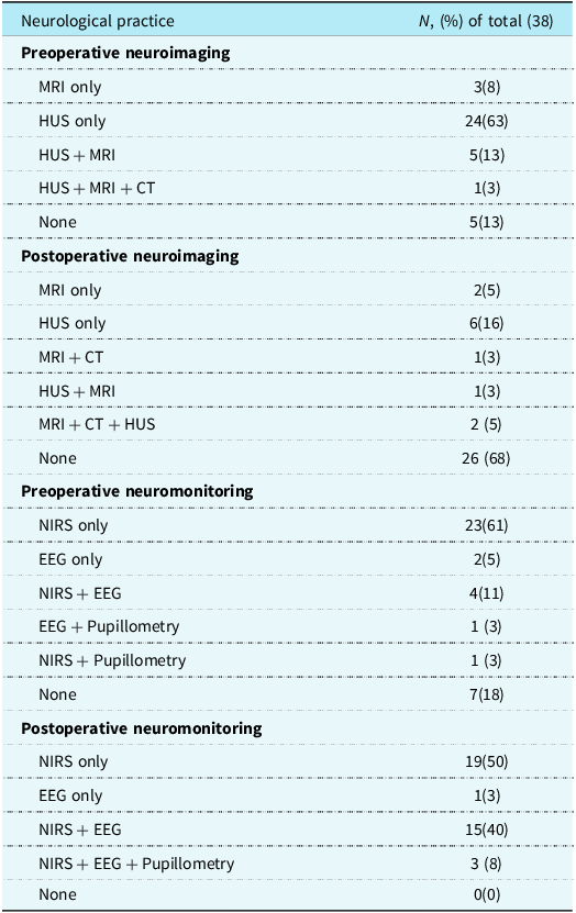
Most centres (33/38, 87%) reported performing preoperative neurological imaging, most commonly HUS alone (63%) or a combination of HUS and MRI (13%) (Table 2). Routine preoperative MRI, alone or combined with other modalities, was reported in 9 centres (24%), and utilisation did not differ by the presence of an neurocritical care team (19% vs. 27%).
Perioperative neuroimaging and neuromonitoring practices
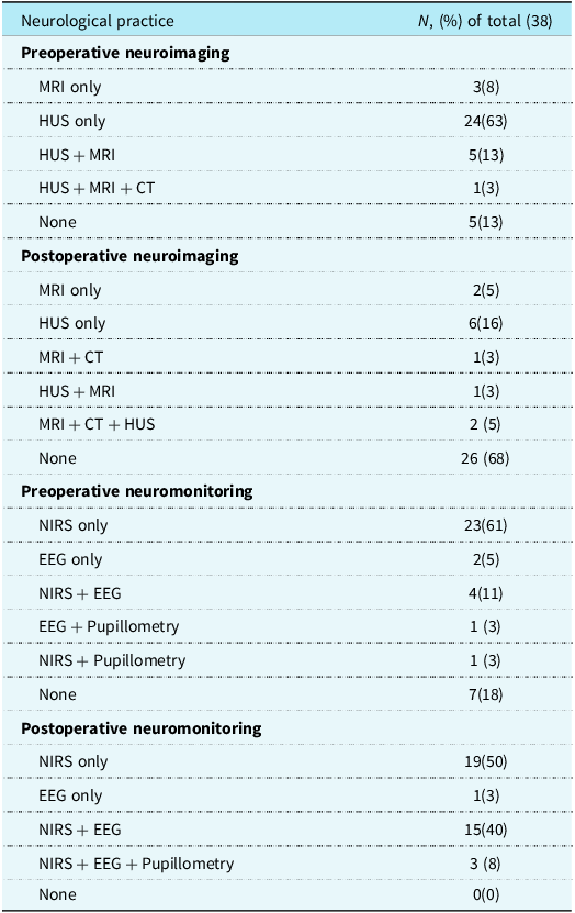
Table 2. Long description
Medical director responses to the question asking which forms of neuroimaging and neuromonitoring are routinely ordered before and after cardiopulmonary bypass.ÊMRI, magnetic resonance imaging; HUS, head ultrasound; CT, computed tomography; NIRS, near-infrared spectroscopy; EEG, electroencephalography.
Medical director responses to the question asking which forms of neuroimaging and neuromonitoring are routinely ordered before and after cardiopulmonary bypass.
MRI = magnetic resonance imaging; HUS = head ultrasound; CT = computed tomography; NIRS = near-infrared spectroscopy; EEG = electroencephalography.
Routine postoperative neuroimaging was less common (32%) and occurred significantly less frequently than preoperative imaging (p < 0.001). Routine postoperative MRI use was similar among centres with and without neurocritical care teams (3/16, 19% vs. 3/22, 14%). Common indications for postoperative MRI outside routine protocols included concern for stroke or intracranial haemorrhage, postoperative cardiac arrest, seizures, and postoperative mechanical circulatory support (Supplementary Table 1). Routine perioperative neuroimaging practices did not differ by centre type, surgical volume, or neurocritical care team presence (Supplementary Tables 2–4).
Neuromonitoring practices
All 38 centres reported using postoperative neuromonitoring, compared to 31 centres (82%) preoperatively (p = 0.02). Utilisation of neuromonitoring did not differ significantly between academic and non-academic centres or between high- and low-volume surgical centres (Supplementary Tables 2 and 3). Centres without a dedicated neurocritical care team were more likely to report routine preoperative neuromonitoring compared with centres with an neurocritical care team (20/22, 91% vs. 10/16, 63%, p = 0.05), although postoperative neuromonitoring was universally adopted in both groups (Supplementary Table 4). Preoperatively, near-infrared spectroscopy was the most used modality, reported by 28/38 centres (74%). Near-infrared spectroscopy was used alone in 23 centres, and 5 combined near-infrared spectroscopy with electroencephalography and pupillometry (Table 2). Postoperatively, near-infrared spectroscopy use was nearly universal (37/38, 97%). Routine electroencephalography use was uncommon preoperatively (18%), but utilisation increased substantially after surgery (50%). The most common postoperative neuromonitoring strategies were near-infrared spectroscopy alone (50%) and near-infrared spectroscopy combined with electroencephalography (40%). Outside of routine protocols, the most common indications for postoperative electroencephalography were concern for seizure activity, postoperative cardiac arrest, and suspected ischaemic stroke or intracranial haemorrhage (Supplementary Table 4). Fifteen centres (40%) also reported protocolised use of postoperative electroencephalography in patients requiring mechanical circulatory support with extracorporeal membrane oxygenation or a ventricular assist device. Routine postoperative electroencephalography use was more frequently reported at centres with a dedicated neurocritical care team than at centres without one (11/16, 69% vs. 8/22, 36%), although this difference did not reach statistical significance (p = 0.1).
Individual perceptions
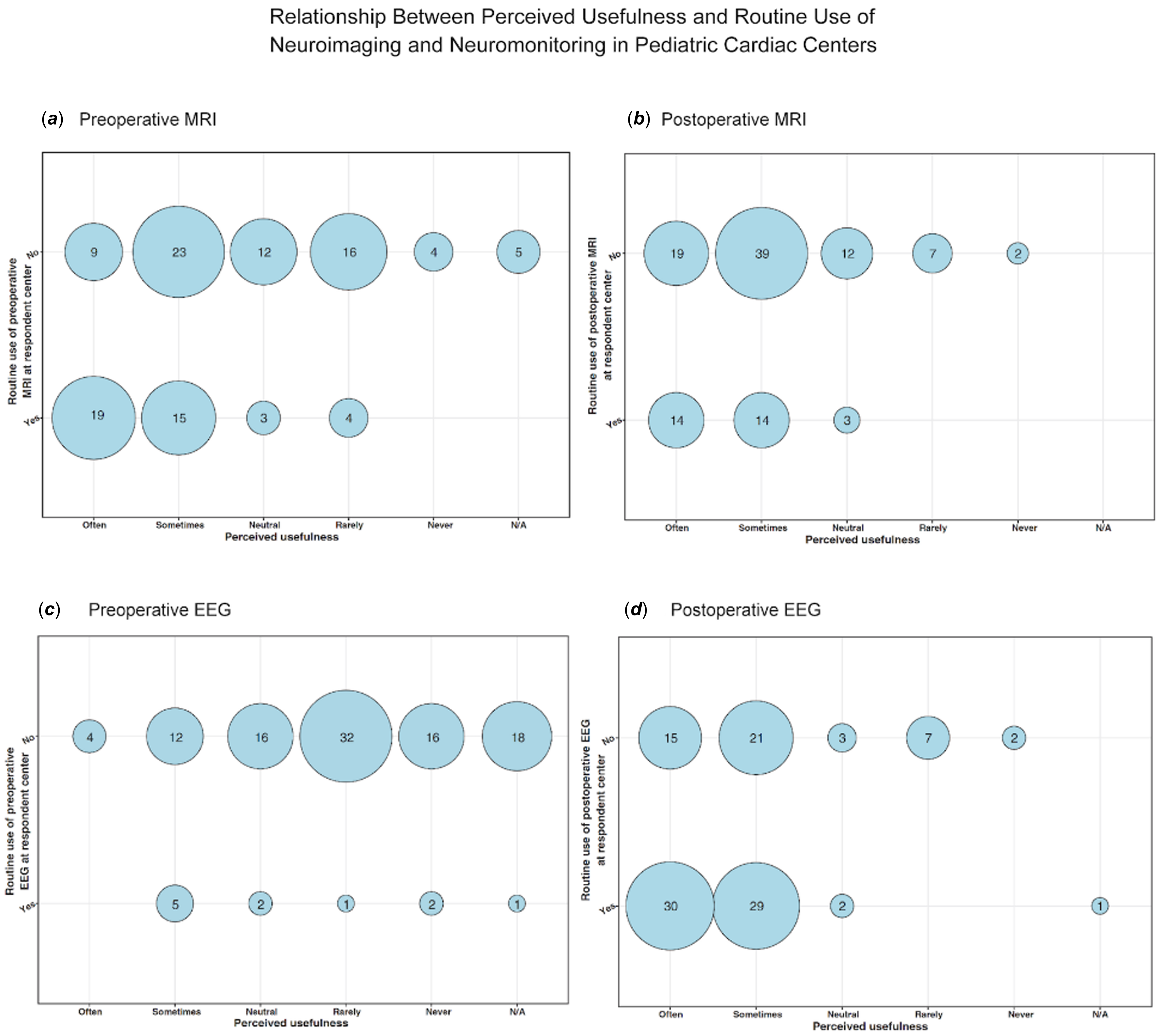
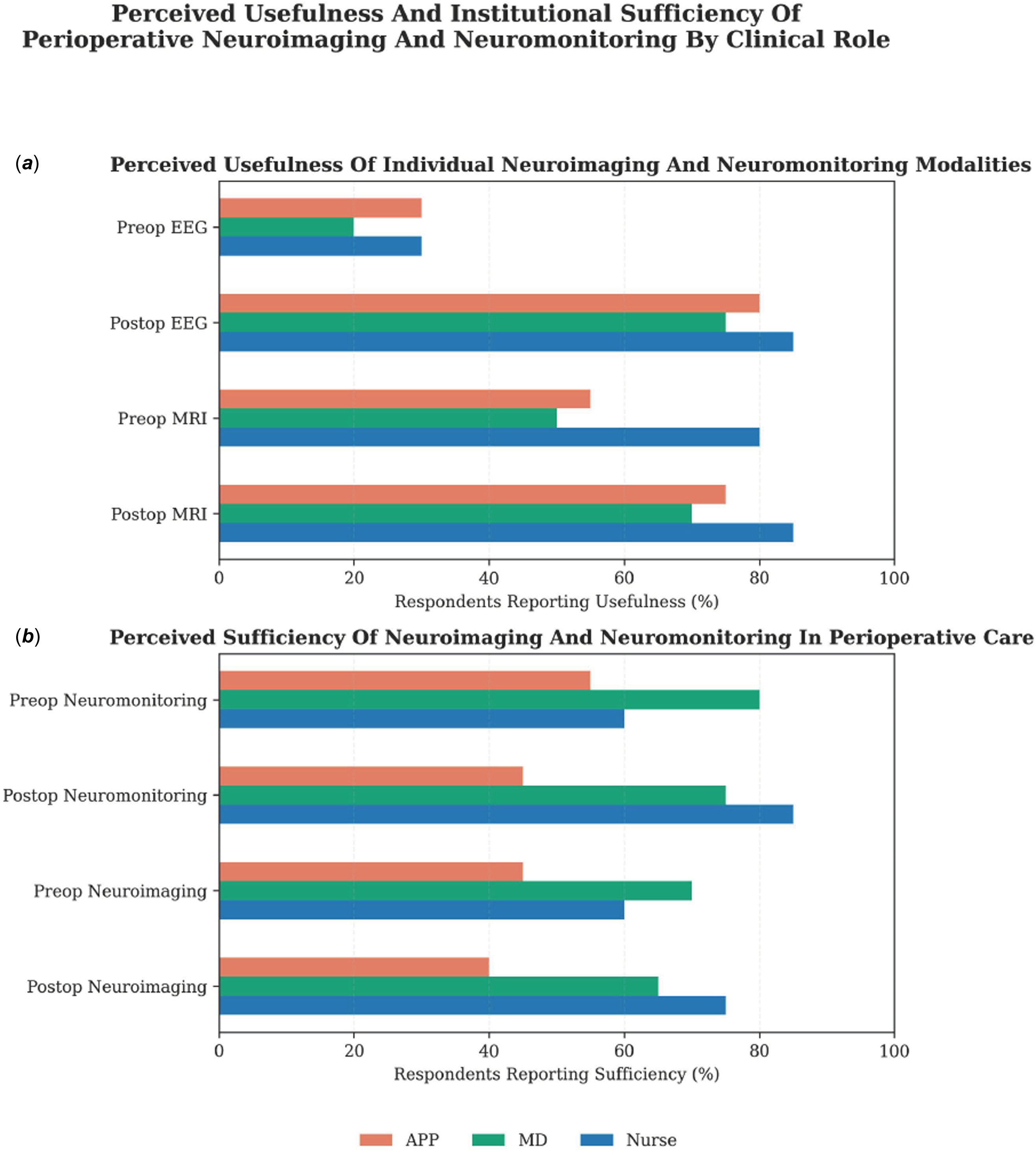
Brain MRI was perceived as clinically useful by 56% (80/142) of respondents preoperatively, compared to 78% (110/140) postoperatively. Respondents from centres with routine preoperative MRI protocols were significantly more likely to perceive the modality as useful compared to those from centres without such a protocol (34/41, 83% vs. 32/69, 46%, p < 0.001; Figure 1( a )). A similar pattern emerged postoperatively, with higher perceived usefulness among respondents from centres performing routine MRI, although this did not reach statistical significance (28/31, 90% vs. 58/79, 73%; p = 0.08; Figure 1( b )). Nurses were more likely than physicians to find both preoperative and postoperative MRI useful (preoperative: OR 6.4 (1.2–34), p = 0.03; postoperative: OR 4.05 (1.21–18.64), p = 0.01; Figure 2( a )). Advanced practice providers had similar perceptions to physicians for both preoperative and postoperative MRI.
Provider perceptions regarding the usefulness of neuroimaging and neuromonitoring practices, stratified by institutional practice patterns. ( a ) Preoperative MRI; ( b ) Postoperative MRI; ( c ) Preoperative EEG; and ( d ) Postoperative EEG. The x-axis demonstrates the distribution of provider responses regarding perceived usefulness, while the y-axis indicates whether the respondent’s institution routinely performs the practice. Bubble size corresponds to the number of respondents.

Figure 1. Long description
ProviderÊperceptionsÊregardingÊthe usefulness of neuroimaging and neuromonitoring practices, stratified by institutional practice patterns. (A) Preoperative MRI; (B) Postoperative MRI; (C) Preoperative EEG; and (D) Postoperative EEG. The x-axisÊdemonstratesÊthe distribution of provider responsesÊregardingÊperceived usefulness, while the y-axisÊindicatesÊwhether theÊrespondentÕsÊinstitution routinely performs the practice. Bubble size corresponds to the number of respondents.
Perceptions of the usefulness and sufficiency of neuromonitoring and neuroimaging, stratified by clinical role. (APP, Advanced Practice Provider) The x-axis shows the percentage of positive responses; the y-axis lists modalities or modality categories. Bars are grouped by provider role. ( a ) Usefulness: positive responses were “often” or “sometimes” useful; negative responses were “not applicable,” “neutral,” “rarely useful,” or “never useful.” Nurses were more likely than physicians to view pre- and postoperative MRI as useful (preop: OR 6.4 [1.2–34], p = 0.03; postop: OR 4.05 [1.21–18.64], p = 0.01). ( b ) Sufficiency: positive responses indicated institutional practices were sufficient. Significant differences were observed for preoperative neuromonitoring (APPs: OR 0.28 [0.10–0.80], p = 0.02; nurses: OR 0.16 [0.03–0.88], p = 0.04). Compared with physicians, APPs were less likely to view pre- and postoperative neuroimaging and postoperative neuromonitoring as sufficient (preop imaging: OR 0.20 [0.05–0.73], p = 0.02; postop imaging: OR 0.33 [0.11–0.94], p = 0.04; postop monitoring: OR 0.33 [0.11–0.94], p = 0.04).
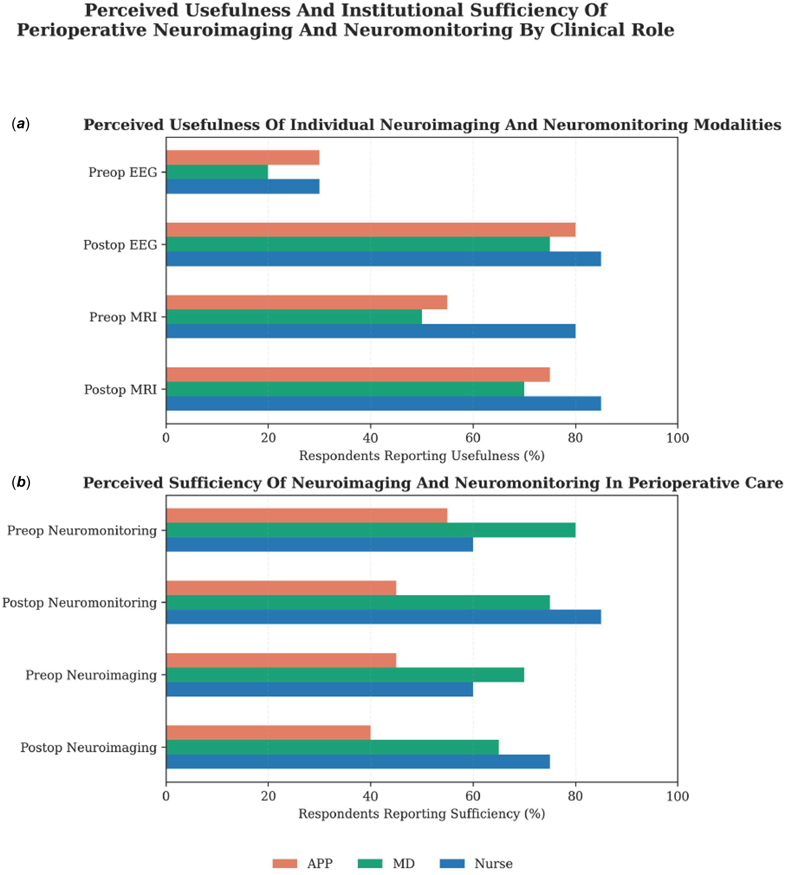
Figure 2. Long description
PerceptionsÊof the usefulness and sufficiency of neuromonitoring and neuroimaging, stratified by clinical role. (APP, Advanced Practice Provider) The x-axis shows the percentage of positive responses; the y-axis lists modalities or modality categories. Bars are grouped by providerÊrole. (A) Usefulness: positive responses were ÒoftenÓ or ÒsometimesÓ useful; negative responses were Ònot applicable,Ó Òneutral,Ó Òrarely useful,Ó or Ònever useful.Ó Nurses were more likely than physicians to view pre- and postoperative MRI asÊuseful (preop: OR 6.4 [1.2Ð34],Êp=0.03; postop: OR 4.05 [1.21Ð18.64],Êp=0.01). (B) Sufficiency: positive responsesÊindicatedÊinstitutional practices were sufficient. Significant differences wereÊobservedÊfor preoperative neuromonitoring (APPs: OR 0.28 [0.10Ð0.80],Êp=0.02; nurses: OR 0.16 [0.03Ð0.88],Êp=0.04). Compared with physicians, APPs were less likely to view pre- and postoperative neuroimaging and postoperative neuromonitoring as sufficient (preop imaging: OR 0.20 [0.05Ð0.73],Êp=0.02; postop imaging: OR 0.33 [0.11Ð0.94],Êp=0.04; postop monitoring: OR 0.33 [0.11Ð0.94],Êp=0.04).Ê
For neuromonitoring, most respondents reported near-infrared spectroscopy as useful both preoperatively (116/142, 86%) and postoperatively (134/141, 95%). In contrast, preoperative electroencephalography was infrequently perceived as useful (26/141, 18%), but its perceived utility increased substantially in the postoperative period (125/141, 89%). Individuals from centres that perform routine preoperative electroencephalography were more likely to perceive it as useful compared with individuals from centres without this practice (p = 0.04; Figure 1( c )). In the postoperative setting, the perceived usefulness of electroencephalography was high regardless of the presence of an institutional protocol (Figure 1( d )), and no differences were observed across provider roles (Figure 2( a )).
Participants rated the sufficiency of neuromonitoring and neuroimaging practices at their institution and perceived barriers to their broader implementation. Most respondents reported that neuromonitoring and neuroimaging practices at their institution were sufficient, both preoperatively (neuromonitoring 103/134, 77%; neuroimaging 93/131, 70%) and postoperatively (neuromonitoring 100/130, 77%; neuroimaging 82/131, 63%; Figure 2( b )). However, advanced practice providers and nurses were significantly less likely than physicians to report sufficient preoperative neuromonitoring (APP: OR 0.28 [0.10–0.80], p = 0.02; nurses: OR 0.16 [0.03–0.88], p = 0.04). Advanced practice providers were less likely than physicians to report that preoperative neuroimaging was sufficient (APP: OR 0.20 [0.05–0.73], p = 0.02). Similarly, advanced practice providers were less likely to report sufficiency of postoperative neuromonitoring (OR 0.20 [0.05–0.73], p = 0.02) and neuroimaging (OR 0.33 [0.11–0.94], p = 0.04) compared to physicians (Figure 2( b )). Among 68 respondents who perceived neurological monitoring and/or imaging at their institution to be insufficient, the most frequently cited barrier was a perceived lack of benefit reported by 56 participants (56/68, 81%). Additional barriers included limited physical resources (28%), insufficiently trained personnel (24%), and inadequate institutional funding (19%).
Discussion
Our multicentre survey provides a comprehensive look at the perioperative neurological care landscape for neonates undergoing cardiac surgery. Despite well-documented evidence that this population experiences substantial rates of perioperative brain injury, we identified significant variability in neuromonitoring and neuroimaging practices across respondent Pediatric Cardiac Intensive Care Society centres and a discordance between perceived utility and clinical adoption. Reference Peyvandi, Xu and Barkovich3,Reference Stegeman, Feldmann and Claessens5,Reference Andropoulos, Hunter and Nelson6,Reference Petit, Rome and Wernovsky17 Several key practical themes emerged: (1) a preference for preoperative over postoperative imaging, with heavy reliance on head ultrasound; (2) neuromonitoring was more consistently implemented postoperatively, with near-infrared spectroscopy as the predominant modality; (3) a lack of correlation between specialised neurocritical care teams and increased diagnostic utilisation; and (4) a perceptual mismatch regarding the necessity and sufficiency of these tools among physicians, advanced practice providers, and nursing staff. Collectively, these findings highlight an implementation gap and the urgent need for multidisciplinary guidelines to standardise neurological care in this vulnerable population.
We found a clear preference for routine neuroimaging before rather than after cardiopulmonary bypass (87% vs. 32%). Brain MRI was protocolised more commonly preoperatively than postoperatively, but HUS remained the predominant modality in both periods, despite its well-documented limited sensitivity for detecting white matter injury and stroke. Reference Rios, Welty and Gunn18 Our findings mirror previous survey studies within the United States and Europe, which report wide variability in preoperative MRI use, performed in 17–72% of centres, and near universal use of screening HUS. Reference Feldmann, Hagmann and de Vries19–Reference Belfi, Vega and Aguar21 Similarly, we found that postoperative MRI was largely reserved for symptomatic neonates or those requiring mechanical circulatory support. Reference Feldmann, Hagmann and de Vries19,Reference Belfi, Vega and Aguar21 This predominantly reactive rather than proactive approach to postoperative imaging is concerning, as brain MRI detects new postoperative injuries in up to 50% of neonates with CHD, many of which are clinically silent. Reference Stegeman, Feldmann and Claessens5,Reference Andropoulos, Hunter and Nelson6,Reference Alablani, Chan and Beishon22 Reliance on head ultrasound or symptom-based imaging triggers, therefore, likely underestimates the true burden of postoperative brain injury. Further, perioperative brain injury has been linked with slowed postoperative brain growth and substantial neurodevelopmental impairment. Reference Marino, Lipkin and Newburger23–Reference Dimitropoulos, McQuillen and Sethi26 Early identification of injury may therefore inform neuroprotective strategies and facilitate early neurodevelopmental referral and family counselling.
Perioperative neuromonitoring was widely utilised among centres in our survey, with near-universal use of near-infrared spectroscopy, consistent with prior studies of paediatric cardiac centres. Reference Feldmann, Hagmann and de Vries19–Reference Belfi, Vega and Aguar21,Reference Hoskote, Tume and Trieschmann27 Near-infrared spectroscopy is widely regarded as a noninvasive, readily available neuromonitoring tool that provides continuous real-time feedback and may facilitate early detection of acute neurologic events. Reference Nagdyman, Fleck and Schubert28,Reference Loomba, Rausa and Sheikholeslami29 While near-infrared spectroscopy serves as a valuable trend-monitoring tool, evidence remains limited regarding its correlation with neuroimaging abnormalities or long-term neurodevelopmental outcomes. Reference Hoskote, Tume and Trieschmann27,Reference Pardo, Carrasco and Wintermark30 Our findings demonstrate a substantial increase in electroencephalography utilisation compared with historical cohorts, with 50% of centres now reporting postoperative electroencephalography use, compared with 17–20% in prior studies. Reference Feldmann, Hagmann and de Vries19,Reference Leon, Levy and Hu20 This shift likely reflects a growing recognition of the high incidence and clinical impact of postoperative seizures and an evolving alignment with recommendations published by the American Clinical Neurophysiology Society. Reference Andropoulos, Mizrahi and Hrachovy9–Reference Clancy, Sharif and Ichord11,Reference Feng, Lin and Zhang31,Reference Wusthoff, Numis and Pressler32 Importantly, our findings regarding clinician perceptions suggest that electroencephalography was highly valued across all provider roles, regardless of whether it was routinely incorporated into institutional practice. This suggests that electroencephalography is broadly perceived as a high-utility tool in the postoperative care of these patients, supporting its inclusion in future consensus guidelines and underscoring the need for formal cost-benefit analyses to inform its optimal implementation.
To our knowledge, this is the first study to quantify the presence of neurocritical care consultation services in paediatric heart centres (42%). Neurocritical care consultants are increasingly recognised as essential stakeholders in the comprehensive care of critically ill children. Reference LaRovere, Murphy and Horak33 This is particularly important in the cardiac ICU, where patients are at high risk for neurological injury due to complex physiology and operative interventions. In our survey, the presence of a neurocritical care team did not increase neuroimaging or neuromonitoring utilisation, and while noteworthy, the trend toward higher electroencephalography use in centres with neurocritical care teams did not reach statistical significance. While our limited sample size may have obscured more significant findings, the data suggests that specialised expertise alone cannot drive clinical adoption without the support of formal institutional protocols. Nonetheless, as neurocritical care teams expand, they are well-positioned to play a central role in developing guidelines, implementing targeted interventions, and integrating neurocritical care into postoperative management.
A key finding was the divergence between the perceived utility of perioperative neurological practices and their actual clinical utilisation, alongside significant perceptual variation across clinical roles. While brain MRI was widely recognised as valuable, this perception was notably stronger among nurses than among physicians or advanced practice providers. This trend may reflect the nursing role’s focus on neurodevelopmental advocacy and the need for prognostic clarity during family counselling. However, this high perceived value did not consistently translate into clinical practice, particularly within postoperative neuroimaging protocols. This disconnect suggests that implementation barriers are not driven solely by clinician beliefs, but rather by a combination of structural, logistical, and evidence-based gaps. Specifically, clinicians may be hesitant to navigate the logistical hurdles of neonatal transport and the structural costs of MRI without definitive data proving these diagnostics directly alter immediate management or improve long-term outcomes.
Furthermore, a significant discrepancy emerged regarding the beliefs about the institutional adequacy of neurological practices. Physicians were more likely than advanced practice providers and nurses to report their centre practices as sufficient. These conflicting perceptions likely stem from differences in training, proximity to bedside care, and clinical workflow. Addressing these gaps will require interdisciplinary dialogue, targeted education, and the establishment of consensus guidelines to align multidisciplinary perceptions with evidence-based practice and standardised care.
While this study provides novel information about the landscape of neurological practices within the paediatric cardiac ICU, several limitations should be considered. Participation in the Pediatric Cardiac Intensive Care Society is voluntary, and the self-selected nature of the sample may have preferentially captured centres with a greater interest in neurological care. The modest individual response rate of 13.8%, the high response rate by physicians relative to other provider roles and the predominance of academic centres in the United States may limit generalisability to smaller or international programmes. In addition, reported institutional practices were not independently verified and may therefore be subject to misclassification bias. Practices were based on self-reports by medical directors and therefore may not reflect actual clinical utilisation of neuroimaging and neuromonitoring at each institution. Future studies should validate reported institutional practiced using objective data from sources such as chart review and registry-based analysis. An additional limitation was that the terms “useful” and “sufficient” were not standardised within the survey, which may have resulted in variable interpretation across respondents and influenced perception-based analyses.
Importantly, the perceptions captured in this study should be considered hypothesis-generating, particularly among advanced practice providers and nursing providers, who were under-represented relative to physicians. Future studies should aim to improve multidisciplinary engagement, incorporate standardised terminology, and evaluate the relationship between neuromonitoring and neuroimaging practices and patient-centred outcomes. Broader recruitment efforts targeting nursing and APP subgroups within the Pediatric Cardiac Intensive Care Society, as well as collaboration with external professional organisations representing non-physician providers, may further enhance participation and improve the representativeness of future studies.
Conclusion
This survey highlights considerable variability in perioperative neuroimaging and neuromonitoring practices surrounding neonatal CHD surgery. The discordance between clinician-perceived utility and institutional implementation highlights the need for multidisciplinary consensus and evidence-based guidelines to standardise care in this population. Future efforts should focus on creating protocol-driven pathways that integrate neurocritical care expertise and bridge the gap between immediate perioperative management and long-term neurodevelopmental outcomes.
Supplementary material
The supplementary material for this article can be found at https://doi.org/10.1017/S1047951126123567.
Acknowledgements
We are grateful to the Pediatric Cardiac Intensive Care Society for their support in survey distribution and engagement. We also thank the individuals who generously shared their time and insights by completing this survey.
Financial support
No funding was received for this study.
Competing interests
The authors declare none.
Ethical standard
This study was approved by the University of Texas Southwestern Institutional Review Board (STU-2024-0494). A waiver of informed consent was granted. The study was conducted in accordance with institutional ethical standards and the Helsinki Declaration of 1975, as revised in 2013.


 Open access

