
Sometimes in science you experience an “aha” moment. It can happen while sitting in a talk, reading a paper, doing an experiment or doing the evening dishes. And when you have this jump in understanding it is hard to contain the thrill and excitement. Of course, you might be wrong – the next morning may reveal all the holes. But scientists cherish these infrequent moments when they make a new scientific connection that could change the way they – and subsequently others – think about a problem. This is precisely what happened in May 1993 when the young group leader Chris Walsh heard Peter Huttenlocher present at a neurology meeting focused on epilepsy in Venice, Italy.
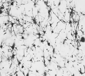
Early on, Peter recognized the importance of having a clinic focused on developmental brain disorders. Many of these patients presented with symptoms that did not fit a specific condition or genetic disorder. Peter was especially interested in patients with tuberous sclerosis and related disorders, and examined brain tissue from families with these disorders using the “Golgi stain” (Figure 13.1). Tuberous sclerosis is a rare genetic condition associated with the formation of “tumors” or “tubers” in the brain. In Venice, Peter presented a talk about a family with a developmental brain disorder who he had seen through his tuberous sclerosis clinic at the University of Chicago. However, the family he described in Italy did not have tuberous sclerosis but rather had a kind of tuberous sclerosis mimic that was particularly interesting. Family members presented with seizures and had nodular subependymal masses of the brain by MRI scan. In this family there were six affected family members, all female, from four different generations. This was consistent with an X-linked dominant inheritance with a lack of survival in affected males. Periventricular heterotopia is the term used to describe the disorder of cortical brain development found in this family, characterized by neural cell masses along the lateral ventricles of the brain.
Chris Walsh was invited to the Venice meeting to present his work on cell lineage analysis in the developing brains of mice. Walsh was particularly interested in cell migration defects that lead to developmental disorders. As Walsh heard the description of this family in detail, he realized that he could “study this family using brain scans to define anatomical abnormalities as a way of defining whether they were sick or not, and combining that with genetics.” In a conversation in December 2021, Chris Walsh said, “This was just a pivotal moment, and my palms were sweating, my heart was racing when he was presenting it, and I just couldn’t wait to talk to him and try to collaborate with him.” Walsh said that this collaboration changed the trajectory of what was to become a highly successful research career. He became a geneticist. Instead of understanding the molecular mechanisms or pathogenesis of a disease first, the approach was to map and identify the genes and “understand mechanisms later.”
The patients in the affected family had periventricular heterotopia, now confirmed as an X-linked dominant cause of “epilepsy” or seizure disorder. The patients had no evidence of developmental delay or other stigmata of disease. For Chris Walsh, this was a turning point in his science. It was the first gene he mapped, and he mapped it based on MRI findings. Walsh knew Peter from his training as a medical student at the University of Chicago. When he approached Peter at the meeting, he asked, “Would you like to collaborate to map the gene in this family?” Chris said that Peter looked “baffled” – and asked, “Map a gene?” This was before it was common to map genetic disorders, and the challenges were many. Walsh said that the four of them – Janellen, Peter, Chris Walsh and Chris’ wife Ming Hui, also a scientist – went out to dinner in Venice and toasted to celebrate the new collaboration. DNA samples were sent and the gene was mapped in a few months, and published in the journal Neuron [Reference Eksioglu, Scheffer and Cardenas1]. It was a serendipitous encounter that changed the trajectory of a career, but as Louis Pasteur famously noted a century earlier, chance favors the prepared mind.
During embryonic development, cortical neurons embark on a long-range directed migration from deep in the brain, near fluid-filled spaces known as the ventricles, to the outer region of the brain in the cerebral cortex. This developmental migration of neurons has been thoroughly characterized in mouse models. Perturbation of this normal developmental navigation can contribute to human developmental disorders like the double cortex syndrome. In many of these syndromes, the neurons still are able to migrate, although aberrantly. In the case of periventricular heterotopia, the neurons fail to perform this developmental migration. Surprisingly, the female patients with this disorder still have normal intelligence despite this defect in neuronal cell migration, and the presentation of a seizure disorder in the adolescent years.
It took the Walsh laboratory over five years to identify the gene responsible for “Huttenlocher’s” classic X-linked bilateral form of periventricular heterotopia [Reference Fox, Lamperti and Eksioglu2]. The gene was in a dense region of the genome and was a challenge to uncover. The responsible gene was identified as the FLNA gene that encodes the cytoskeletal protein filamin A. The cytoskeleton of the cell is a meshwork of membrane-associated proteins that provides structure to the cell. Its most abundant member is the cytoskeletal protein actin that polymerizes to form microfilaments. Dynamic regulation of the actin cytoskeleton is critical for cell movement. While doing the research for this book, as a physician scientist, I was particularly excited by the identification of filamin as the underlying etiology of periventricular heterotopia. My own research focuses on cell migration and how interactions between the matrix environment in tissues, outside of the cell, link to the cytoskeleton inside the cell to control cell migration. In prior work, we and others had found that the strength of these linkages between cell surface adhesion receptors – integrins – and the cytoskeleton play a key role in controlling cell migration.
Filamin is a cytoskeletal protein that links cell surface adhesion receptors to the actin cytoskeleton. This critical connection enables the appropriate amount of adhesion of a cell to the surrounding extracellular environment to allow cells to migrate the right amount – not too much and not too little. Filamin provides a linkage that allows the cell to sense its outside environment, the matrix tissue in which the cells navigate and move. Without these connections between the cell surface receptors and the intracellular cytoskeletal machinery, cell movement is not possible. Due to the point mutations found in patients with periventricular heterotopia, the function of filamin is disabled. The Walsh group’s study was the first to show that filamin plays a critical role in neuronal cell migration. It is a classic story of a basic biomedical science discovery. One physician scientist was sufficiently alert to identify a patient family with an apparently heritable defect, and sufficiently curious to define some of the resulting tissue defects underpinning the disease. A second physician scientist heard them talk at a meeting and was sufficiently insightful to prioritize investigating the matter for multiple years, and sufficiently talented to identify the causal gene and reveal a previously unknown core mechanism involved in brain development.
The finding that filamin plays a role in neuronal cell migration was unexpected. It is an example of how genetic analysis often uncovers new biochemical or cellular functions. Sometimes this type of discovery launches entire new fields by uncovering new functions and signaling networks. It was already known that filamin regulates the cytoskeleton in leukocytes, but this was the first evidence that it regulates neuronal migration. In this case, although the discovery revealed a neuronal mechanism and provided an explanation for a genetic syndrome, it has not yet led to new treatments. This gap between understanding a malady and learning how to intervene and treat a disease is a common “next challenge” in biomedical research.
These types of cases are the ultimate motivation for physician scientists. Can we make discoveries that affect the treatment and outcome for our patients? Chris Walsh is now a global leader whose research group has used genetics to uncover the causes of many other human neurodevelopmental disorders. In a recent interview published in the journal PNAS [Reference Ravindran3], he noted that more recently he has been switching his focus to understand even more about the “basic biology” that regulates human brain development. He is in good company as a physician scientist whose medical training has helped him excel at uncovering important molecular mechanisms – the foundational knowledge – rather than focusing on the development of disease treatments or other applications of that knowledge.

