

Introduction
This paper presents new evidence for some of the foundation burials at the Late Bronze Age and Early Iron Age settlement of Cladh Hallan having been mummified. The female mummy’s knee (lower femur and upper tibia) was broken off prior to burial but long after death when the bones had lost most of their collagen.
Cladh Hallan is one of those remarkable archaeological sites whose investigation provides archaeologists with discoveries which upset our conventional understanding of prehistoric life. In 2005 we reported on human skeletal remains from this settlement as having been formerly mummified (Reference Parker PearsonParker Pearson et al. 2005). The interpretation of the site and its finds would certainly be a good deal simpler if these sub-floor burials were actually components of an earlier cemetery: there would be a straight-forward chronological progression at Cladh Hallan from cemetery to settlement, raising no challenging questions about the inter-relationship between domestic life and ritual; there would be no requirement to consider the time-depth of ancestral history in prehistory or the role of cosmology; and no raised eyebrows at seemingly unlikely practices involving long-term curation of human corpses.
New evidence from post-excavation analysis puts the mummification theory beyond reasonable doubt. It also refutes any potential interpretation that the burials were part of an earlier cemetery unconnected with the houses.
The mummy’s knee
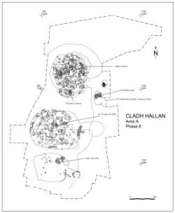
Within the northern roundhouse there were three human burials among the 211 cut features beneath its two lowest floors (Figure 1). Two of these burials lay in the north-east quadrant of the house: an adult male (beneath the primary floor, a composite skeleton consisting of bones from three separate individuals) and an infant (buried when the house was re-built in a later phase). The third skeleton was an adult female lying on her left side and buried beneath the primary floor at the southern edge of the house. Her body was tightly crouched and her grave fill was packed with large stones including a broken fragment of a saddle quern (see Reference Parker PearsonParker Pearson et al. 2005: Figure 6). The western edge of her grave was cut by a pit (2581) containing a dog's articulated vertebral column and the articulated right back foot of a sheep. However, this pit did not impinge on the skeleton or the central grave fill, which remained undisturbed after burial.
A plan of the main area excavated at Cladh Hallan, showing the construction phase (Phase 8) of the roundhouse row.

The female skeleton was, however, incomplete when excavated. The distal end of her left femur was missing, as were her left patella and the proximal ends of her left tibia and fibula. The distal end of her right radius was also absent. Her right arm lay across the tightly flexed right leg. Although the right leg covered the left in the crouched burial, the left knee (consisting of the distal femur, patella and proximal tibia) was missing. In addition, her right hand lay above the break of the left tibia and fibula.
The bones forming the left knee can only have been removed prior to the body’s burial within the pit. There was no sign of disturbance within the grave fill except for the deeper pit (2581) cut into the western edge of the grave away from the body (ibid.: right side of Figure 6). Furthermore, the broken-off and removed leg bones were from that part of the left leg which was hidden below the right knee and right hand. Any intrusion from above would have resulted in the bones of the right leg being disturbed.
Outside the northern roundhouse, to the south of its east-facing doorway, a figure-of-eight shaped pit (2551) was dug and filled during the period of construction and initial use of the roundhouse, dated by its stratigraphic relationship to other features on the site (Phase 8, the same phase as the female skeleton’s burial pit). It lay about 5m east of the female burial. Its primary fill (2748) consisted of red peat ash and other domestic debris and contained the missing distal femur, proximal tibia and distal right radius fragments of the female skeleton. The missing left patella was retrieved in one of the uppermost of the six layers within this pit, about 0.5m above these bones. There was no sign of the proximal fibula fragment. The broken-off left distal femur and proximal tibia were still in anatomical articulation, although at a wider angle of flexion than they had been when part of the corpse. This osteological and stratigraphic evidence demonstrates that the bones of the left knee were broken off a body that was still articulated; that this was done prior to burial; and that the removed bones were buried still in articulation at the time that the house was being constructed and initially used.
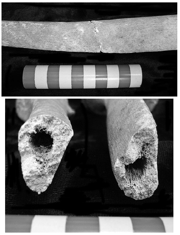
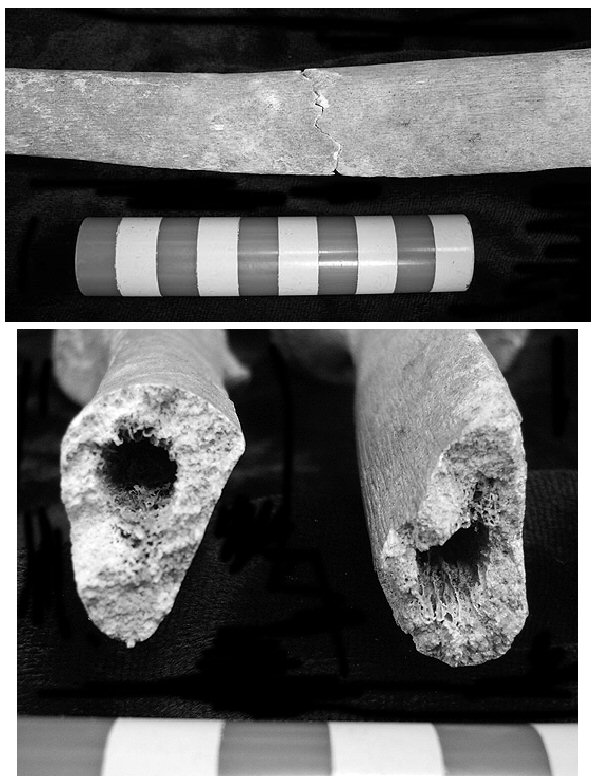
Most significantly, the fractures on the broken-off bones are dry (Figure 2). There are no cut or chop marks on the bone. The cleanly snapped ends of the tibia and radius are characteristic of long bones which have broken after they have lost most of their collagen and are no longer ‘green’ or fresh (Knüsel & Outram 2006: 262). As far as we know, there are no data on the length of time post mortem for bones to reach this state of decay although, even in the most exposed of environments, it is likely to take many months or even years.
The conjoining bones of the ‘female’ burial’s tibia, showing the ‘dry’ fracture.
Two hypotheses must be considered. Could the skeleton have been disinterred from an earlier grave, with the prehistoric excavators somehow preserving full anatomical articulation, at a moment sufficiently long after death that the bones had lost their collagen, were then broken around the knee, and finally reburied still fully articulated, with the knee being placed in a separate pit? We argue that a more acceptable hypothesis is that the body was kept above ground, tightly wrapped and in a protective environment (conditions which preserved not only the bones themselves but also their articulation), with the knee being broken and deposited in the pit around the time that the full skeleton was buried, at the time of the houses’ construction.
The composite male mummy
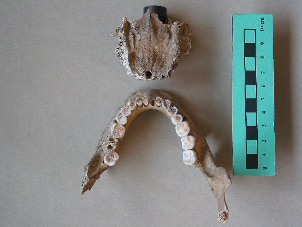
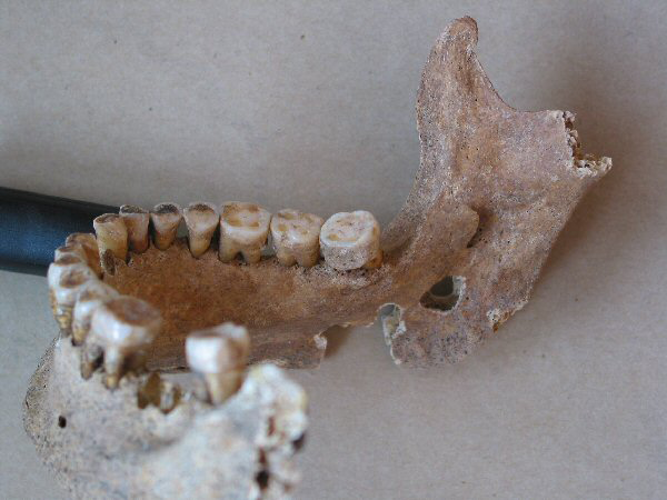
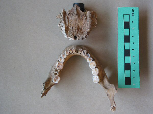
There is no doubt that this skeleton (Figure 3) is composed of bones from three separate individuals. The δ15N stable isotope levels of both the skull and the mandible (10.8‰) are incompatible with those of the tibia (9.9‰). More importantly, the mandible with its full set of teeth is from a different individual to that of the skull whose cheek teeth had been lost many years before death, leading to alveolar resorption on both sides of the upper jaw (Figure 4). The original owner of the mandible possessed a maxilla with functioning upper dentition which is absent in the skull with which it is now associated.
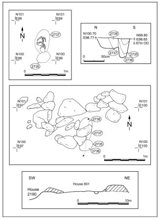
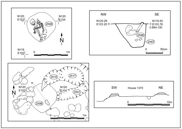
The burial pit of the composite male mummy in plan and section.
Mandible (left) and maxilla (right) from the composite male skeleton, showing the bilateral ante-mortem loss of maxillary cheek teeth with complete alveolar resorption in the molar and premolar region, contrasting with the presence of functioning dentition and maintenance of a normal occlusal plane in the lower jaw. Note that the right central lower incisor was not recovered at excavation, and the left lower second molar has been extracted for isotopic analysis.
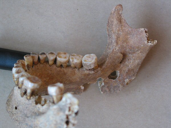
We can be confident of this because the lower molars have substantial build-up of dental calculus on their lingual surfaces (grade 2 to grade 3, following the scoring system of Reference DobneyDobney & Brothwell 1987) but there is a complete absence of calculus on their occlusal surfaces (Figure 5).
Medial view of right premolars and molars in the composite male skeleton, showing 2mm thick in situ calculus deposit on the lingual surface of the third molar. Remnants of dental calculus are also present on the lingual surfaces of the first and second molars..
The 3 year-old child buried beneath the south house
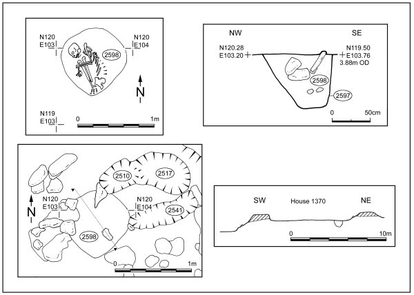
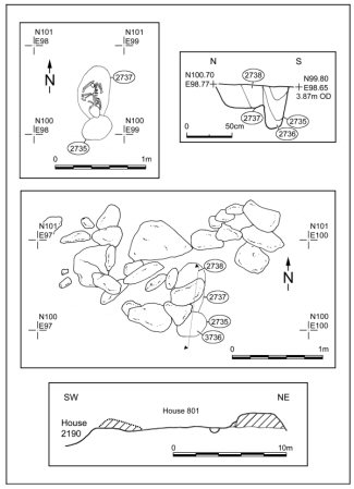
There were many fewer features below the south house than were dug beneath the central and north houses. None the less, the burial pit containing this child’s skeleton was cut on its south side by one of the house’s four roof-bearing postholes (Figure 6). This may just have been coincidental but the juxtaposition of burial and post is noteworthy: it strengthens the case for the burial being a foundation deposit interred immediately before house construction. More importantly, the burial can be demonstrated to be much later than the underlying cremation cemetery (Phase 3; Parker Pearson et al. 2004: 62-3, colour plate 5) which was sealed beneath an earlier house.
The burial pit of the 3 year-old child in plan and section.
The cemetery consists of five cremation burials only, two of which date to 1750-1530 cal BC (95% confidence, 3375±35 BP, SUERC-10717, and 95% confidence, 3270±35 BP, SUERC-10716). Three of these were sealed beneath a U-shaped house (Reference Parker PearsonParker Pearson et al. 2004: Figures 25 & 26) which is dated by carbonised grain from its floor to 1410-1270 cal BC (95% probability; 3040±35 BP; SUERC-10818). This house was stratigraphically earlier than both the roundhouse terrace and the 3 year-old child burial beneath the southern roundhouse (this burial is dated to 1440-1210 cal BC [95% probability; 3070±50 BP; GU-9841] although its unusually high percentage [16% ± 10%] of marine carbon probably makes this determination earlier than it should be).
Since the construction of the row of roundhouses can now be estimated more precisely, thanks to new AMS radiocarbon determinations and further mathematical modelling, to 1310-1150 cal BC (95% probability) and probably 1270-1180 cal BC (68% probability), the child’s skeleton dates to the same period as this event.
The burials beneath the north-east quadrants of the houses
The burials beneath the house floors were marked on the floor surfaces by spreads of stone. Rather than being positioned directly over the graves, as a capping of an earlier grave would be, the majority of stones in each case were located on the north-west side of the grave. Furthermore, they exhibited none of the characteristics of disturbed Early to Middle Bronze Age burial cairns (for undisturbed examples, see Reference BraniganBranigan 2000; Reference Close-brooksClose-Brooks 1995; Reference CrawfordCrawford 1977; Reference MerronyMerrony 2000), or stone-lined short-cist burials (Reference DunwellDunwell et al. 1995; Reference CookCook 2006; Reference SimpsonSimpson et al. 2006: 155-7) or encircling rings such as those found at Northton on Harris, and in Cladh Hallan’s earlier cremation cemetery (Phase 3; Reference SimpsonSimpson et al. 2006: 160-1; Reference Parker PearsonParker Pearson et al. 2004: colour plate 5).
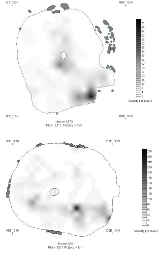
That the burials were an integral feature of the roundhouses is supported by the distinctive use of space within the north-east quadrants. Not only were the stone settings visible to its first inhabitants within the peat-formed floor matrix but they were also in the one part of the house which was kept clean. In each house’s primary floor layer (Phase 9), the distributions of pottery, potting clay, tools of bone, flint, pumice and antler, and micro-debris all avoid this zone (see Figure 7 for the distribution of pottery in the north and middle houses).
The distribution of pottery sherds (>10mm) within the northern and middle roundhouses in Phase 9: a) Floor 2211 of House 1370; b) Floor 1311 of House 401.
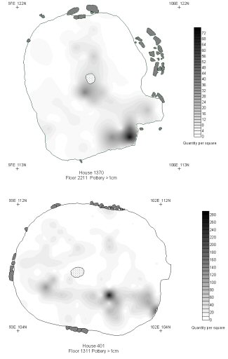
Indeed, the only categories of artefact to be deposited in this quadrant of the central and northern houses were white quartz chippings and copper alloy items. The latter were deliberately left – a bracelet in the northern house and two chisels in the middle house, forming ‘closing’ deposits placed on the floor before it was covered by a new floor layer during refurbishment. Explaining this avoidance of the north-east in ‘practical’ terms simply does not work. This is the sector of the roundhouse where there would have been most light, particularly in the mornings, and yet it was avoided for domestic tasks of craft working and cooking.
Wind direction as the motivating force in the organisation of British Iron Age domestic space has been thoroughly discredited by recent research (for Scottish and Hebridean roundhouses, see Reference Parker PearsonParker Pearson et al. 1996 and Parker Reference Parker PearsonPearson & Sharples 1999: 16-23): it does not adequately explain why the east-facing house is an island-wide phenomenon, nor why the first millennium BC is the only prehistoric period to show such a concern with wind direction, nor why the British Isles only and no other Atlantic coastline suffered so badly that all its inhabitants kept their backs to the wind, nor why most of the exceptions to the rule face westwards, into the teeth of the wind.
Other human remains
Further human bone fragments have been identified during post-excavation analysis. Many of these single human bones (as opposed to teeth) come from contexts associated with the period of roundhouse construction (Phase 8). Of the 21 so far recorded, 11 are from construction layers (within house walls and in under-floor pits). Thus the deposition of articulated remains, mummified and otherwise, was just part of a wider series of deposits of human body parts during the founding and occupation of the roundhouse row.
Evidence for post-mortem treatment of the bodies
We remain uncertain of the post-mortem treatment of the skeletons, but the following features are apparent and give further clues to possible interventions.
Acidity of the burial environment The bones of the male torso display changes to the mineral, identified by X-ray scattering. There has been further work on wide- and small-angle X-ray scattering (WAXS and SAXS; see Reference HillerHiller et al. 2003; Reference Hiller2004; 2006a & b) but a wider range of controls is still required to fully interpret the data. The enhanced size of the bone mineral crystals strongly suggests that the skeletons were exposed to an acid environment for a brief period of time, but the bone was buried in calcareous soil. Although bone dissolution can occur in calcareous soils, in a large scale study Nielsen-Marsh et al. (in press) observed a strong correlation between dissolution and pH only in burials in which pH was less than 6.0 (Figure 6 loc. cit.). The alkaline pH in the composite male’s grave fill was 7.2 (taken on a distilled water extract using a Hanna pHep 3 pH meter). We speculate that a reduction in pH during soft-tissue decay was responsible for the process – driven by anaerobic fermentation - which led to the formation of ‘microbial spongiform porosity’ (Reference Turner-walkerTurner-Walker et al. 2002). But this process was apparently arrested, as evidenced by the intense but localised distribution of the histological change.
If the bodies were exposed to acid, it could be proposed that they were chanced-upon bog bodies reclaimed for proper burial, but this seems unlikely, given the composite nature of the male adult. Furthermore exposure a highly acid peat environment appears to have been sufficiently brief to cause to only partial mineral alteration.
Soft tissue preservation and the possibility of evisceration In our earlier paper (Reference Parker PearsonParker Pearson et al. 2005), we put forward the idea of evisceration to explain the observation that microbial spongiform porosity (MSP) was intense but localised, leaving much of the bone histologically unaltered. The decay porosity is likely to be indicative of arrested putrefaction given new results about the role of gut-bacteria in a large survey (261 bones from 41 sites) of human and animal bones (Jans et al. 2004; Jans 2005). Although our control sample was dog bone, which is not the ideal control for human inhumation, it is interesting to note that both dog samples studied by Reference JansJans (2005) from an Iron Age refuse tip (Visongo: 118 loc. cit) and a Viking Age-medieval cemetery (Hag: 111 loc. cit.) displayed MSP similar to that observed in human bone. Conversely, Reference JansJans et al. (2004) rarely observed MSP in butchered animal bone.
We admit that our ideas regarding the biostratinomy of these samples are speculative; the precise cause is less relevant than the central observation, namely a pattern of intense but localised decay. This pattern of decay suggests an active process, suddenly curtailed.
Conclusion
The breaking-off of bones from the composite ‘female’ mummy, when the bones were already dry (but prior to burial) is difficult to interpret in terms other than that the skeleton was held together by preserved soft tissues long after her death.
The distribution of her broken bones, together with other evidence from the burial contexts, also refutes the counter-hypothesis that the sub-floor skeletons at Cladh Hallan are components of an otherwise unrevealed earlier inhumation cemetery whose regular positioning in the houses might be coincidental.
Acknowledgements
Figures 1, 3 and 6 were drawn by Irene de Luis whilst Figure 7 was drawn by Harry Manley. In addition to those acknowledged in the original article, we also thank Rob Craigie for carrying out the pH analysis. At Bradford University, we thank Dr Chris Knüsel and Dr Holger Schutkowski for useful comments. At Durham University, we thank Professor Charlotte Roberts for her expert advice and opinion on the human remains. The new osteological work on the skeletons has been carried out by Christie Cox.