


Main text
Membrane proteins, or more precisely, membrane-embedded proteins and protein complexes, are responsible for many crucial functions of cells. For example, G-protein-coupled receptors (GPCRs) and ligand-gated, voltage-gated, and mechanosensitive ion channels allow information from outside the cell to affect the activity of the cell; electron transport chain complexes, together with the ATP synthase, power countless biochemical processes by enabling synthesis of ATP; and transporters and channels ensure that useful molecules are imported into cells while toxic substances and waste products are removed. Understanding these functions requires high-resolution knowledge of the structures of the proteins that carry out the processes.
For decades, X-ray crystallography was the predominant way of determining protein structures. During this time, the structural study of membrane proteins lagged behind the structural study of soluble proteins and protein complexes. There were multiple reasons for this deficit. First, membrane proteins tend to be harder to express recombinantly than soluble proteins at the high levels required for X-ray crystallography. Second, a reagent, usually a detergent, that can extract a membrane protein from its lipid bilayer prior to crystallization while preserving its structure, needs to be available. Finally, even when a suitable detergent for protein extraction exists, the detergent may impede crystallization: mild detergents that enable protein extraction without denaturation tend to form large micelles that can interfere with crystallization, while detergents with small micelles that facilitate inter-molecular crystal contacts tend to be more destabilizing (Bamber et al., Reference Bamber2006). This conundrum can sometimes be resolved through the introduction of stabilizing mutations in the protein (Serrano-Vega et al., Reference Serrano-Vega2008) or by adding additional protein mass to facilitate crystallization (Rasmussen et al., Reference Rasmussen2007; Rosenbaum et al., Reference Rosenbaum2007). However, structure determination for membrane proteins became much easier and more successful when single-particle electron cryomicroscopy (cryo-EM) became a mature method (Kühlbrandt, Reference Kühlbrandt2014; Smith and Rubinstein, Reference Smith and Rubinstein2014). Cryo-EM does not require intermolecular crystal contacts and is more tolerant of conformational and compositional variability in samples than crystallography. Nonetheless, overexpression, detergent extraction, and purification of membrane proteins are often sub-problems that one must solve before getting to the more interesting problem of determining a membrane protein’s structure by cryo-EM.
Recombinant protein expression can be avoided by studying endogenous proteins
When aiming to determine the structure of a protein, the experimentalist must decide between attempting to isolate the endogenous protein from cells or tissues or attempting to overexpress the protein recombinantly. Recombinant overexpression has the potential to provide a large yield of the protein and allows straightforward incorporation of affinity tags into the protein sequence. However, an immediate disadvantage of recombinant overexpression is that it is not necessarily trivial to do: many proteins are difficult to overexpress or do not fold natively on overexpression. The first advantage of structure determination with endogenous membrane proteins is that the investigator does not need to determine conditions for overexpression. However, an important disadvantage of working with endogenous proteins is that many are not found in high abundance in cells and tissues. This low abundance would not be a problem except for the fact that single-particle cryo-EM, although usually requiring less protein than crystallization, is remarkably inefficient. For the 104 to 106 molecular images that are typically used in three-dimensional (3D) reconstruction, orders of magnitude more protein particles must be isolated, applied to the specimen support grid, and then usually blotted away and discarded to prepare a suitable specimen. Further, protein purification strategies for endogenous proteins are often more complicated and customized than for recombinant proteins. To isolate an endogenous protein, the investigator will often develop a bespoke purification strategy with conventional chromatography, introduce sequences encoding tags into the chromosomal DNA of the cells if the protein is being isolated from cultured cells (Murphy et al., Reference Murphy2018; Zhao et al., Reference Zhao2022), or use a high-affinity antibody or binding protein for purification if one is available (Abbas et al., Reference Abbas2020; Schenck et al., Reference Schenck2024). The recent development of computational methods to design binding proteins may dramatically simplify the isolation of endogenous proteins (Cao et al., Reference Cao2022), but endogenous protein purification remains a formidable challenge for many proteins of interest.
A second major advantage of structure determination with endogenous membrane proteins is that unexpected subunits or interactions can often be discovered. Small membrane proteins are frequently too hydrophobic to stain well on an SDS-PAGE gel. If they are hydrophobic, they also often lack polar lysine and arginine residues where trypsin cleaves, decreasing the chance of detecting them by standard mass spectrometry of tryptic fragments. Consequently, the first evidence that a small membrane protein interacts with a large membrane protein complex is frequently the protein being found in a structure of the complex after it was obtained from a native source. For example, cryo-EM of endogenous yeast V-ATPase led to the discovery that the protein encoded by hypothetical open reading frame YPR170W-B (Mazhab-Jafari et al., Reference Mazhab-Jafari2016), subsequently named subunit f, and the protein Voa1p (Roh et al., Reference Roh2018), are part of the enzyme. Cryo-EM of the endogenous mammalian V-ATPase showed that the protein RNAseK, which was found in the structure, corresponds to subunit f in mammals (Abbas et al., Reference Abbas2020). Cryo-EM also revealed the full subunit composition of the mitochondrial respiratory Complex I (Vinothkumar et al., Reference Vinothkumar2014). When the endogenous Plasmodium falciparum sodium efflux pump PfATP4 was isolated from parasite-infected human red blood cells, its structure revealed an apicomplexan-specific binding partner that the authors named PfABP (Haile et al., Reference Haile2025). Similarly, new and unexpected subunits were discovered when studying mycobacterial Complex I (Liang et al., Reference Liang2023), the mycobacterial Complex III2IV2 supercomplex (Gong et al., Reference Gong2018; Wiseman et al., Reference Wiseman2018), and the Pseudomonas aeruginosa bcc-cbb 3 supercomplex (Di Trani et al., Reference Di Trani2023), to name just a few examples. In fact, it seems new subunit interactions are detected regularly when endogenous membrane protein complexes are subjected to structural analysis by cryo-EM for the first time. Therefore, not having to attempt overexpression of a protein complex when the full subunit composition of the complex is not certain is an extremely strong motivation to work with endogenous proteins.
Cryo-EM of endogenous human proteins
Human proteins are important drug targets. However, unless a protein can be purified from human blood, placenta, or cultured cells, it can be difficult to obtain starting material for isolating the endogenous protein. Consequently, cryo-EM of human proteins is usually performed with recombinant human proteins. CRISPR-Cas gene editing approaches allow the introduction of affinity tags for purification and cryo-EM of endogenous protein complexes from cultured human cells (Zhao et al., Reference Zhao2022). Nonetheless, costs associated with culturing mammalian cells still limit these experiments to relatively abundant protein complexes. Methods that improve the ‘efficiency’ of cryo-EM may help address this limitation. Conventional cryo-EM specimen preparation for a 1 MDa protein complex typically requires ~2 μL of a protein at ~3 mg/mL (3 μM) concentration to be applied to the EM grid. Three-dimensional reconstructions are often calculated using 104–106 particle images from these 3.6 × 10¹² complexes. This ratio corresponds to an efficiency of one complex used out of every 3.6 × 10⁶ to 3.6 × 10⁸ complexes applied to the grid. Improving this efficiency by even two orders of magnitude, so that one particle image is obtained for every 104–106 complexes applied to the grid, could drastically decrease the required yield from endogenous protein preparations. Purification procedures that previously needed a gram of tissue or cultured cells could be performed from tens of milligrams of material. This amount of material could come from a small volume of cultured cells or even a tumor biopsy if studying variant proteins expressed in transformed cells. Gains in cryo-EM specimen preparation efficiency may be obtained from developing appropriate affinity grids (Glaeser, Reference Glaeser2021), an old idea with many variations, or specimen preparation devices that make use of nanoliters of sample instead of microliters (Jain et al., Reference Jain2012; Ravelli et al., Reference Ravelli2020).
Cryo-EM of purified proteins lends itself to reductionist experiments
For many structural biologists, the ideal structure determination method would enable the study of all proteins in situ within the context of an intact, living cell. An ideal workflow would be to freeze a cell, cut it into thin sections with an instrument like a focused ion beam scanning electron microscope (FIB SEM), perform electron tomography on the resulting thin sections, and calculate 3D tomograms. From there, the molecule of interest would have to be identified, and images of independent realizations of the molecule would be averaged to calculate a 3D map at sufficient resolution to construct an atomic model. Already, this technique is providing near-atomic resolution structures of ribosomes in a variety of cells (Tegunov et al., Reference Tegunov2021; Xue et al., Reference Xue2022). The technique is also close to high resolution for complexes like adenosine triphosphate (ATP) synthase in mitochondria (Dietrich et al., Reference Dietrich2024). Further, new approaches are showing that proteins in cells can be identified in 2D images without tomographic reconstruction (Lucas et al., Reference Lucas2021). Nonetheless, there are numerous practical problems in extending these in situ methods to all proteins of interest. First, many proteins are not so abundant within cells that one can confidently expect to find a copy in a single FIB SEM section. Second, unlike the large and high-contrast RNA-protein ribosome, numerous proteins are not discernible above the protein background in a crowded tomogram of unstained frozen-hydrated material. These are practical problems that improved hardware, software, and molecular labelling approaches may eventually address. However, beyond these concerns, there are situations where the study of purified proteins lends itself to reductionist experiments that cannot easily be accomplished within cells. Elegant examples from early cryo-EM reveal the power of this approach, which allowed investigation of the activation of the nicotinic acetylcholine receptor by spraying the specimen with acetylcholine immediately before freezing (Berriman and Unwin, Reference Berriman and Unwin1994; Unwin and Fujiyoshi, Reference Unwin and Fujiyoshi2012). Similarly, studies of conformational changes in bacteriorhodopsin performed by irradiating the specimen with specific wavelengths of light prior to freezing revealed short-lived conformational changes linked to the photocycle of the protein (Subramaniam et al., Reference Subramaniam1993). More recent experiments show the structural consequences of glutamate binding to ionotropic glutamate receptors (Gangwar et al., Reference Gangwar2024) and visualized early intermediates in bacterial RNA polymerase promoter melting (Saecker et al., Reference Saecker2024). While some of these experiments may have in-cell equivalents, studying the systems outside of cells allows for a level of control and removal of confounding factors that will not be possible in the foreseeable future with in situ methods. As a result, the future of in vitro structural biology remains bright.
The abundance of some proteins facilitates studying them in their native membrane
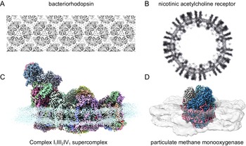
Some proteins, including bacteriorhodopsin and the nicotinic acetylcholine receptor mentioned above, are naturally abundant in a specific membrane. Isolating the appropriate membrane with minimal biochemical perturbation allows for the structural analysis of the protein by electron microscopy. Bacteriorhodopsin is abundant and forms well-ordered 2D arrays in the membranes of the archaeon Halobacterium salinarum. These arrays are suitable for structural analysis by electron diffraction and imaging with the electron microscope (Henderson and Unwin, Reference Henderson and Unwin1975; Henderson et al., Reference Henderson1990) (Figure 1A). Similarly, the nicotinic acetylcholine receptor is packed into the postsynaptic membranes of the electric organ of Torpedo marmorata, with the isolated membrane readily forming lipid tubules that contain helical arrays of the protein. These helical arrays can be imaged and analyzed with helical reconstruction methods (Brisson and Unwin, Reference Brisson and Unwin1984) (Figure 1B). Imaging these proteins within the membrane gives confidence that the proteins are in a native conformation and ensures that they form native protein–lipid interactions (Grigorieff et al., Reference Grigorieff1995; Miyazawa et al., Reference Miyazawa2003; Unwin, Reference Unwin2022).
Cryo-EM of abundant protein complexes in native membranes. (A) A projection map of bacteriorhodopsin in the purple membrane of H. salinarum (from Grigorieff et al., Reference Grigorieff1995). (B) Cross-section through a 3D map of a tubular crystal of the nicotinic acetylcholine receptor from the postsynaptic membranes of the electric organ of T. marmorata (from Unwin, Reference Unwin2013). (C) A 3D reconstruction of a respiratory supercomplex from the mitochondrial membrane of Sus scrofa (from Zheng et al., Reference Zheng2024). (D) A 3D reconstruction of the particulate methane monooxygenase from the plasma membrane of M. capsulatus (Bath) (from Tucci and Rosenzweig, Reference Tucci and Rosenzweig2025).

Other examples exist where specific proteins are abundant in a membrane, but their orientations in the membrane are less constrained than in 2D crystals or helical arrays. These samples require the use of single-particle methods to determine the pose of individual protein molecules in images prior to using the images to calculate 3D maps. For example, the mitochondrial inner membrane from eukaryotes is crowded with the proteins that carry out oxidative phosphorylation: the electron transport chain complexes (respiratory Complexes I through IV) and the ATP synthase. This phenomenon has allowed for high-resolution single-particle cryo-EM analysis to be performed by freezing mitochondrial membrane preparations on EM grids and imaging the specimen without tilting the microscope stage (Zheng et al., Reference Zheng2024) (Figure 1C). The analysis allowed direct visualization of respiratory supercomplexes, or respirasomes, that include Complexes I, III, and IV. Other beautiful examples of abundant membrane proteins allowing the application of single-particle methods to native membranes include studies of methane monooxygenase from Methylococcus capsulatus and ammonia monooxygenase from Nitrosomonas europaea (Tucci and Rosenzweig, Reference Tucci and Rosenzweig2025). The active forms of the proteins were reconstructed to high-resolution from images of the membranes without specimen tilting, revealing important protein–lipid interactions and α helices not seen after extraction of the protein from the lipid bilayer with detergent (Figure 1D).
Imaging of membrane proteins in lipid vesicles allows functional studies
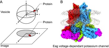
In the early 2000s, Fred Sigworth recognized the value of studying membrane proteins in lipid vesicles (Jiang et al., Reference Jiang2001). A vesicle-embedded membrane protein has fewer degrees of freedom in its pose than a membrane protein in a detergent micelle, amphipol, or lipid nanodisc (Figure 2A). Further, a membrane protein embedded in a well-sealed vesicle could be subjected to a transmembrane ion or voltage gradient that cannot be achieved in bulk solution, allowing study of the effects of transmembrane forces on the conformation of the protein of interest. Numerous methods exist to reconstitute membrane proteins into proteoliposomes (Rigaud and Lévy, Reference Rigaud and Lévy2003). These approaches usually involve mixing a detergent-solubilized membrane protein with detergent-solubilized lipids or detergent-saturated pre-formed lipid vesicles and allowing the mixture to equilibrate. Detergents are then removed by dialysis, adsorption onto polystyrene beads (Bio-Beads), or dilution, allowing protein reconstitution. These procedures can be difficult to perform successfully for fragile membrane protein complexes; the methods often require extensive customization, and the yield of successfully reconstituted proteoliposomes is typically low. However, working with reconstituted proteoliposomes, Sigworth and colleagues developed procedures to swell vesicles to be spherical, concentrate them on cryo-EM grids (Wang and Sigworth, Reference Wang and Sigworth2010), and computationally remove the contribution of the vesicle to the image (Liu and Sigworth, Reference Liu and Sigworth2014). The study of membrane proteins reconstituted into proteoliposomes has revealed force-induced conformational changes in the mechanosensitive PIEZO1 channel (Lin et al., Reference Lin2019), voltage-sensor movements in the Eag Kv channel under an applied electric field (Mandala and MacKinnon, Reference Mandala and MacKinnon2022), and shown how the transmembrane electric field regulates the PIP2-binding site to gate the KCNQ1 channel (Mandala and MacKinnon, Reference Mandala and MacKinnon2023) (Figure 2B). Cryo-EM of mitochondrial Complex I reconstituted in proteoliposomes has shown how ischemia regulates the activity of the complex (Grba et al., Reference Grba2024). Cryo-EM of a bacterial V/A-type ATP synthase reconstituted in proteoliposomes with a light-powered proton pump has even allowed analysis of the ATP synthase during proton translocation-driven ATP synthesis (Nakano et al., Reference Nakano2025). Because the yield of reconstituted protein is typically low, purification of overexpressed recombinant proteins or extremely abundant proteins like Complex I or a V/A-type ATP synthase is usually needed to obtain a proteoliposome sample that is suitable for cryo-EM. Further, any disruption of the protein that occurs on detergent extraction from its native membrane would still occur prior to reconstitution into a non-native proteoliposome. An interesting and powerful variant of this technique is the use of cell-derived lipid vesicles following over-expression of the protein of interest (Tao et al., Reference Tao2023). This approach can preserve some protein–lipid and protein–protein interactions. However, the proteins are still not studied in their native membrane, potentially removing other interactions.
Cryo-EM of membrane proteins in vesicles. (A) As described by Sigworth, the 3D poses of vesicle-embedded membrane proteins are constrained by vesicle geometry (based on, and showing the angle convention from, Jiang et al., Reference Jiang2001). While the in-membrane rotation, ψ, can take on any value, the elevation, θ, and the in-image rotation, φ, are constrained by where the image of the particle was found relative to the location of the vesicle. (B) A 3D reconstruction of overexpressed and purified Eag Kv channel reconstituted in a lipid vesicle (from Mandala and MacKinnon, Reference Mandala and MacKinnon2022).

Imaging of membrane proteins in native lipid vesicles preserves fragile interactions
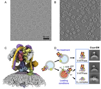
Another system where a specific protein is abundant in a particular membrane is the proton-pumping V-type ATPase (V-ATPase) in mammalian brain synaptic vesicle membranes (reviewed in Vasanthakumar and Rubinstein, Reference Vasanthakumar and Rubinstein2020). In these ~40 nm diameter organelles, the ~20 nm long V-ATPase generates a transmembrane proton motive force, powering the loading of the vesicle with neurotransmitter. The enzyme comprises an ATP-hydrolyzing soluble catalytic V1 region and a proton-translocating membrane-embedded VO region. Single-particle cryo-EM analysis of V-ATPases in synaptic vesicles was carried out in two studies published nearly simultaneously (Coupland et al., Reference Coupland2024; Wang et al., Reference Wang2024). In one study, synaptic vesicles were isolated using the high-affinity interaction between a Legionella pneumophila effector protein and the V1 region of the complex (Coupland et al., Reference Coupland2024) (Figure 3A–C). In the other, a nanobody developed to bind the vGlut1 transporter present in some synaptic vesicles was used to isolate vesicles (Wang et al., Reference Wang2024). Both efforts revealed that, in the context of a native synaptic vesicle membrane, V-ATPase stoichiometrically binds the synaptic vesicle protein synaptophysin, an interaction that is lost when the enzyme is extracted from the membrane with detergent (Abbas et al., Reference Abbas2020). In both studies, single-particle analysis was able to achieve higher resolution than subtomogram averaging (Kravčenko et al., Reference Kravčenko2024; Wang et al., Reference Wang2024). However, differences in the two single-particle studies provide valuable methodological lessons for this type of experiment. When purified with the bacterial effector protein, which binds only to intact V-ATPase, the purification appears to select for vesicles that contain many V-ATPase complexes (~1 M intact V-ATPase particle images from ~18 k micrographs), probably owing to avidity effects. In contrast, when synaptic vesicles were isolated with the vGlut1 nanobody, there were fewer vesicles that contained intact V-ATPase (~300 k intact V-ATPase particle images from ~22 k micrographs). Analysis of vesicles isolated with the nanobody revealed that a substantial portion of the V-ATPase complexes are in the dissociated state (~700 k VO complex images in 22 k micrographs), while the VO complex was not detectable in vesicles isolated with the bacterial effector. The sample with more intact V-ATPase complexes facilitated high-resolution structural analysis and in vitro experiments to look at substrate-induced dissociation of V1 and VO (Figure 3D), which is an important part of synaptic vesicle function (Bodzęta et al., Reference Bodzęta2017). Together, these observations suggest that purifying vesicles via the target of the structural studies facilitates high-resolution structure determination, while purification via a different protein in the vesicle provides a more realistic snapshot of the components of the vesicle.
3D reconstruction of V-ATPase in native synaptic vesicle membranes (A) An example micrograph of purified rat brain synaptic vesicles on a graphene-oxide coated cryo-EM grid. (B) A slice through an example tomogram of the same specimen as in part A. (C) A 3D reconstruction of V-ATPase in synaptic vesicles). (D) Structural experiment showing that treating synaptic vesicles with a buffer that induces loading results in dissociation of some of the soluble catalytic V1 complex from the membrane-embedded VO complex. (All panels are adapted from Coupland et al. Reference Coupland2024).

Native membrane vesicles can be generated and isolated for structural analysis
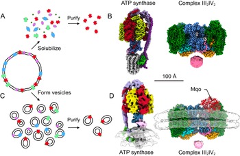
From the above text, it may seem that it is possible to study endogenous membrane proteins in their native lipid bilayer only when they are abundant in a specific membrane. However, it is also possible to enrich membrane proteins in native lipid vesicles without extraction from the membrane (Di Trani et al., Reference Di Trani2025). In this experiment, native membranes are homogenized to form vesicles, vesicles containing the protein of interest are enriched, and the resulting preparation is imaged (Figure 4). In principle, this approach can be applied to any membrane protein, even if it is not particularly abundant in its native membrane. To date, published examples from the approach have come from Mycobacterium smegmatis cytosolic membranes, yielding a low-resolution map of ATP synthase, and a high-resolution map of the respiratory Complex III2IV2 supercomplex (Figure 4D). The latter map revealed an additional bound protein, identified as the malate:quinone (Mqo) oxidoreductase enzyme from the mycobacterial Krebs cycle, which was absent in structures of the detergent-solubilized enzyme (Wiseman et al., Reference Wiseman2018; Yanofsky et al., Reference Yanofsky2021). While the interaction of Mqo with the Complex III2IV2 supercomplex could conceivably be studied at high resolution within the cell by in situ electron tomography, the small membrane vesicles used for single-particle analysis also enabled stopped-flow spectroscopic measurements (Di Trani et al., Reference Di Trani2025). These measurements showed that the interaction of Mqo with the respiratory Complex III2IV2 supercomplex allows rapid electron transfer from malate to the supercomplex. This phenomenon would not have been detected without studying the Complex III2IV2 supercomplex in this simplified membrane system, supporting the utility of cryo-EM of endogenous membrane proteins in small lipid vesicles derived from their native lipid bilayer.
Generation of membrane vesicles for structure determination of proteins in their native lipid bilayer. (A) Endogenous membrane proteins are typically extracted from the membrane with detergents before purification, which can lead to loss of protein–protein interactions. (B) Example structures of proteins from the M. smegmatis plasma membrane extracted with detergent: ATP synthase (left, from Guo et al., Reference Guo2021) and the Complex III2IV2 respiratory supercomplex (right, from Yanofsky et al., Reference Yanofsky2021). The disconnected pink density corresponds to the tethered superoxide dismutase subunit of the complex. (C) Membrane vesicles can be formed from the plasma membrane before purification of the protein of interest, which may preserve native protein–protein interaction. (D) Example structures of proteins from the M. smegmatis plasma membrane determined from vesicles and vesicle fragments: The ATP synthase (left, from Di Trani et al., Reference Di Trani2025) and the Complex III2IV2 respiratory supercomplex (right, from Di Trani et al., Reference Di Trani2025). When detergents are avoided during protein purification, the interaction between the respiratory supercomplex and malate:quinone oxidoreductase (Mqo, red) is preserved.

New tools are needed for cryo-EM of membrane proteins in lipid vesicles
Imaging endogenous membrane proteins in vesicles derived from their native membrane has proven a useful approach for high-resolution structural analysis. The method allows downstream functional studies and preserves interactions that are lost in detergent. However, when detergent extraction does not remove protein–protein interactions and functional experiments do not benefit from the presence of the lipid bilayer, structure determination in detergents is usually easier, faster, and achieves higher resolution. Challenges with cryo-EM of membrane proteins in native membrane vesicles originate from four disadvantages relative to more conventional approaches: (1) low image quality, (2) low yields of vesicles, (3) difficulty with selecting particles of interest, and (4) a lack of computational tools for their analysis.
First, contrast in images of vesicles tends to be low because the vesicles require a thick layer of ice. Without flattening the vesicles on the EM grid or thinning the specimen, perhaps with a focused ion beam, this thickness will remain an inherent issue with vesicle specimens compared to isolated proteins and protein complexes. Thick cryo-EM specimens will benefit from continued improvement in direct electron detector and energy filter technology, as well as higher voltage electron microscopes and perhaps chromatic aberration-corrected microscopes (Wu et al., Reference Wu2025).
Second, the yield of vesicles enriched for the protein of interest can be surprisingly low. From experience, affinity matrices tend to recover less of a protein when it is embedded in a vesicle than when it is extracted from the membrane with detergent. Further, the centrifuged concentrator devices typically used to concentrate samples of purified proteins lose a significant fraction of vesicle samples. The low yield and low concentration of vesicles often necessitate the use of graphene oxide-coated cryo-EM grids, which decrease image quality even further by contributing noise to images. Cryo-EM of native membrane vesicles would benefit from improved biochemical tools for affinity isolation of vesicle-embedded membrane proteins, improved techniques for concentrating vesicle samples, and improved specimen grids that can enrich vesicles on the grid surface without adding noise to images.
Third, unlike synthetic membrane vesicles reconstituted with a single protein, native membranes are often crowded with many different proteins, making it hard to find the protein of interest in the vesicle image. Further, native membrane vesicle preparations are often contaminated with soluble complexes, such as ribosomes, which stick to membranes and complicate analysis. As a result, particle selection is one of the major challenges in this type of study. The structures described above are relatively large, which facilitates particle selection, and better ways of finding particles in micrographs are needed. At present, the program Topaz is one of the best tools for identifying particles in images of native vesicles (Bepler et al., Reference Bepler2019). Another useful tool is the program Vesicle Picker (Karimi et al., Reference Karimi2024), which uses the Segment Anything model (Kirillov et al., Reference Kirillov2023) to find vesicles in images, and then transfers all possible particle locations to CryoSPARC (Punjani et al., Reference Punjani2017) to identify complexes of interest by 2D classification. Further advances in particle selection, probably using machine learning methods, will facilitate efficient retrieval of particle images from micrographs of native membrane vesicles.
Finally, during image analysis, the signal from the lipid bilayer can dominate image alignment and classification, leading to classes that show similar membranes and not similar particles. Methods for subtracting the contribution of membranes to images have improved structure determination in some cases (Zheng et al., Reference Zheng2024). However, it is not clear that manipulating the raw particle image through lipid bilayer subtraction, which will modify the noise characteristics of the image, is the optimal way of accounting for the membrane during structure determination. Including Sigworth’s simple constraints on the 3D pose of particle images could be extremely valuable for image analysis of proteins in vesicles. Appropriate masking of different regions in the map to reduce the contribution of the membrane to image alignment could also improve map calculation dramatically. However, adding these constraints and procedures to the highly developed computational workflows in software packages like CryoSPARC and Relion (Scheres, Reference Scheres2012; Punjani et al., Reference Punjani2017) is not trivial.
Despite the challenges listed above, the steadily increasing number of examples of 3D structures determined for endogenous membrane proteins in their native lipid bilayer supports the utility of this method. It may not show a completely native environment for the protein of interest, but it allows for an essential aspect of the scientific endeavor: performing reductionist experiments to interrogate the relationship between structure and function.
Acknowledgments
I thank members of my research group and our collaborators for experiments and conversations that shaped the ideas described in this review. I thank Niko Grigorieff, Rod MacKinnon, Amy Rosenzweig, Nigel Unwin, and Kai Zhang for providing high-resolution images from their manuscripts.
Financial support
I was supported by the Canada Research Chairs program. Development of methods for cryo-EM of membrane proteins in vesicles was supported by a Natural Sciences and Engineering Research Council Discovery Grant.
Competing interests
I declare no competing interests.

 Open access

