





Introduction
Coarctation of the aorta refers to a localised narrowing of the thoracic aorta, typically located distal to the origin of the left subclavian artery at the site of the ductus arteriosus. It accounts for 6 to 8% of all CHDs and is one of the rare causes of secondary hypertension in children and young adults, as well as an uncommon cause of cerebral haemorrhage and cerebellar infarction. Coarctation of the aorta is not merely a simple stenosis issue but a complex vascular pathology with multiple potential risk factors and long-term complications. Reference Kim, Andrade and Cook1,Reference Prisant, Mawulawde and Kapoor2 Coarctation of the aorta patients can be diagnosed through the detection of brachial and femoral artery pulsations and blood pressure measurements in the limbs. However, reports indicate that coarctation of the aorta diagnosis is often missed, and many asymptomatic individuals may not be identified until adulthood, when related complications arise. Reference Vriend, Lam and Mulder3 We report a case of a young patient with atrial fibrillation, renal infarction, and acute cerebral infarction, which are consequences of untreated hypertension due to coarctation of the aorta. The purpose of this report is to emphasise the importance of early diagnosis and management of coarctation of the aorta as a cause of secondary hypertension in children and young adults.
Case presentation
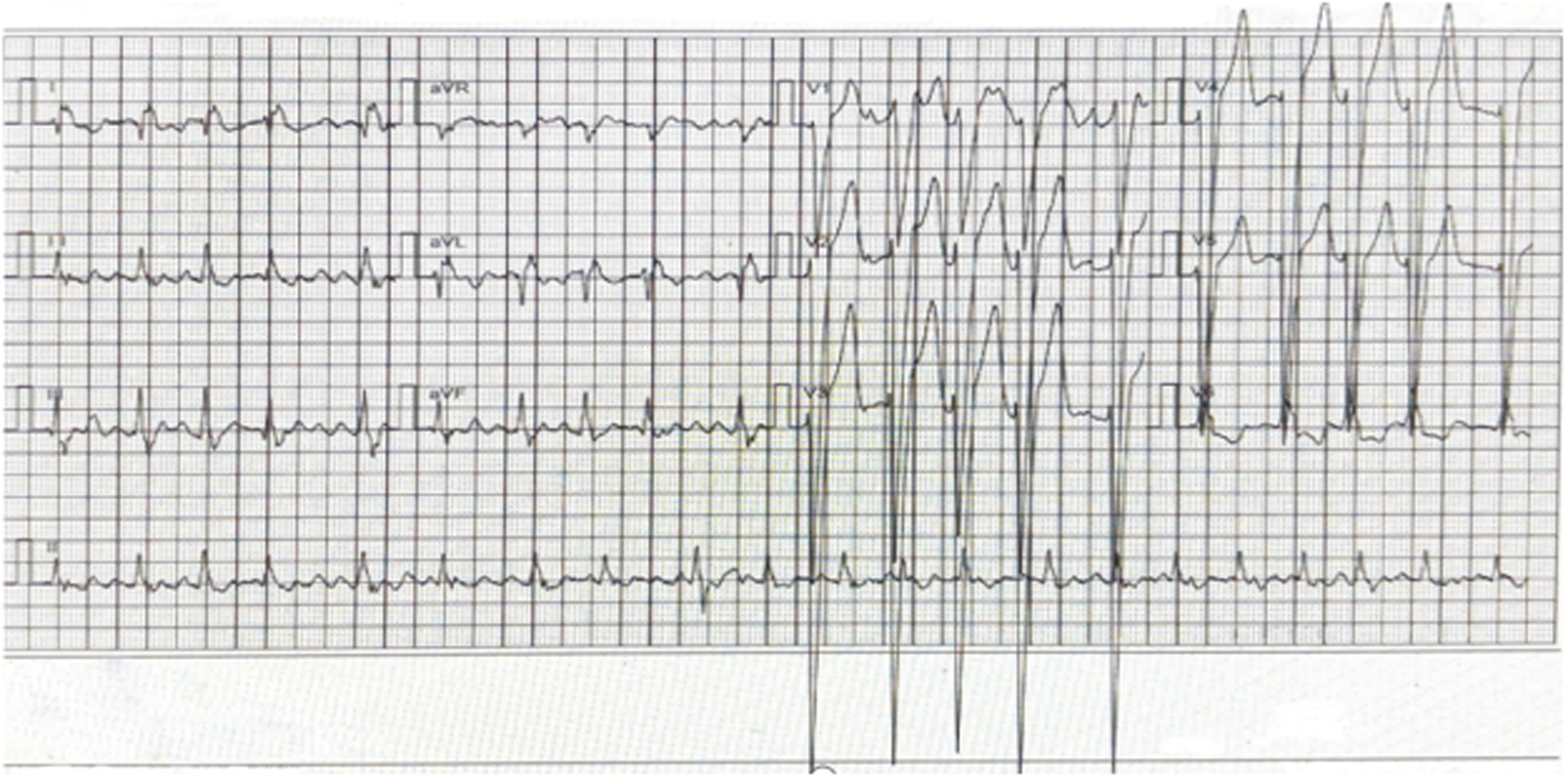
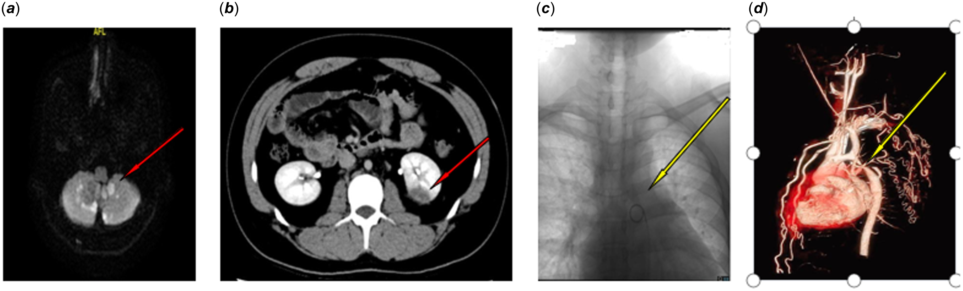
The patient is a 19-year-old male who was admitted due to “dizziness, fatigue, and palpitations for 12 hours.” There was no history of heart disease or positive family history. On admission, blood pressure was 190/108 mmHg, pulse rate was 95 beats per minute, heart rate was 115 beats per minute with an irregular rhythm, the first heart sound was variable in intensity, and no third or fourth heart sounds were heard. A rough, high-pitched systolic murmur was audible at the cardiac apex, with no murmurs in other valve areas. No significant abnormalities were found on the rest of the physical examination. The electrocardiogram indicated: 1) rapid atrial fibrillation with intraventricular conduction defect; 2) poor R-wave progression in leads V1–V5; 3) complete left bundle branch block; and 4) ST-T changes (T-wave peaking in leads V2–V5)(see Figure 1). Echocardiography showed enlargement of the left and right atria, abnormal left ventricular wall motion, mitral regurgitation (moderate), tricuspid regurgitation (mild); reduced left ventricular systolic function; chest X-ray suggested cardiomegaly. Cerebral magnetic resonance angiography suggested bilateral cerebellar acute infarction (see Figure 2A); pituitary MRI showed no significant abnormalities. Abdominal CT with contrast enhancement showed an abnormal density lesion in the lower pole of the left kidney, suggestive of renal infarction (see Figure 2B). Upon admission, urapidil was administered intravenously to control blood pressure, and the doses of amlodipine besylate tablet and perindopril tert-butylamine tablets were increased to 10 mg/day and 4 mg/day, respectively, to lower blood pressure, and metoprolol 50 mg/day was used to control the ventricular rate. After the above treatment, the patient’s blood pressure stabilised. On day 19 of admission, cerebral angiography was performed due to bilateral cerebellar acute infarction, and a 5F vertebral artery catheter met resistance near the descending aorta, close to the aortic arch, and could not pass through, suggesting aortic stenosis (Figure 2C). Postoperative thoracic aortic CTA showed a stenosis at the junction of the aortic arch and descending aorta, with a diameter of approximately 3 mm and a stenotic segment length of about 5 mm, with the distal descending aorta slightly slender. Multiple tortuous collateral small vessels were visible in the aorta, left subclavian artery, left common carotid artery, and right brachiocephalic trunk, running along the thoracic wall and mediastinum (Figure 2D). Bilateral upper and lower limb blood pressure measurements were taken, with BP 132/83 mmHg and BP 105/64 mmHg in the upper limbs and BP 117/75 mmHg and BP 108/65 mmHg in the lower limbs, confirming the diagnosis of aortic stenosis.
Electrocardiogram of a 19-year-old male with hypertension upon admission.1) rapid atrial fibrillation with intraventricular conduction defect; 2) poor R-wave progression in leads V1–V5; 3) complete left bundle branch block; and 4) ST-T wave changes (T-wave peaking in leads V2–V5).

( a ) diffusion magnetic resonance imaging of the brain shows acute infarction in both cerebellar hemispheres. ( b ) contrast-enhanced computed tomography reveals abnormal density lesions in the upper abdomen, localized to the inferior pole of the left kidney, indicative of renal infarction. ( c ) during cerebral angiography, the advancement of a 5F vertebral artery catheter, navigated by a guidewire, was met with substantial resistance at the level of the aortic arch within the descending aorta, impeding catheter progression and suggesting the presence of aortic stenosis. ( d ) the thoracic aorta CTA reveals a stenosis at the junction of the aortic arch and descending aorta, with a lumen diameter of approximately 3 mm and a stenotic segment length of about 5 mm. The distal portion of the descending aorta appears attenuated. Multiple tortuous collateral vessels are visible in the thoracic wall and mediastinum, originating from the aorta, left subclavian artery, left common carotid artery, and right brachiocephalic trunk.

Discussion
Coarctation of the aorta is a narrowing of the thoracic aorta at the isthmus. Literature reports that 90% of cases occur at the aortic isthmus, typically presenting as localised stenosis, related to embryonic development. The incidence rate is about 7–14% of all CHDs. Coarctation of the aorta is a lifelong disease closely associated with long-term hypertension. Reference Chiu, Tsai and Ho4,Reference Salciccioli and Zachariah5 Coarctation of the aorta significantly increases the prevalence of hypertension in adolescents, related to subsequent risks of early onset and mortality. Reference Kenny and Hijazi6 The aortic stenosis lesion is in a special location with severe consequences, and long-term survival is very low, with high rates of disability and mortality; the high mortality rate is due to cardiovascular complications such as heart failure, aortic dissection, and rupture of cerebral aneurysms. Reference Lara-Rojas, Bernal-Lopez and Lopez-Carmona7
The cause of hypertension in coarctation of the aorta is that during coarctation of the aorta, the heart compensatorily strengthens its contraction, but the blood it ejects cannot pass through the stenosis smoothly, causing a large amount of blood to accumulate in the proximal aorta and its branches with limited capacity, thus increasing the blood pressure in the arteries above the upper limbs but not in the lower limbs. In some cases with long-term survival and atypical clinical manifestations, over time, the body may develop highly effective compensatory mechanisms, forming collateral circulation to bypass the constriction. Reference Law, Collier and Sharma8 The characteristic of coarctation of the aorta is chronic left ventricular pressure overload, affecting the left ventricle and left atrium, leading to left ventricle hypertrophy, left atriumremodelling, fibrosis, and dysfunction, as well as the development of atrial fibrillation. Reference Labombarda, Bonopéra and Maragnes9 In this case, the patient developed atrial fibrillation on the basis of undetected long-term hypertension, and atrial fibrillation itself can cause embolic events. The chronic behaviour of aortic coarctation also causes ischaemia in important abdominal organs, and the combined effect of the three led to renal infarction and cerebellar infarction.
This case is that of a 19-year-old young patient with hypertension. On admission, the physical examination missed the measurement of blood pressure in the limbs, and the lower limb blood pressure was significantly lower than the upper limbs, with the dorsalis pedis artery pulsation lower than the radial artery. The measurement of blood pressure in the limbs aims to establish or verify the diagnosis of hypertension, establish current blood pressure, screen for causes of secondary hypertension, and complete the overall cardiovascular risk assessment. At least once, blood pressure should be measured in both arms and the difference in systolic blood pressure between the two arms should be greater than 20 mmHg and/or the difference in diastolic blood pressure should be greater than 10 mmHg. If confirmed, further research into vascular abnormalities should be conducted . Reference Law, Collier and Sharma8,Reference Reis, Gomes and Teixeira10
A comprehensive medical history and physical examination are crucial for identifying the key clinical features of coarctation of the aorta. The abnormal differences in blood pressure between the upper and lower limbs found during the physical examination can also lead to a presumptive diagnosis, playing a key role in accurately diagnosing the causes of hypertension. The information obtained will eliminate unnecessary, frequent, and expensive laboratory and imaging tests, especially for the discovery of potential secondary causes.
Data availability statement
No data was used for the research described in the article.
Acknowledgements
Not applicable.
Author contributions
Suchen Tang: Writing—review & editing. Ronghua Huang: Writing—original draft. Yi Zhou: Visualisation, software, and validation. Zhengjiang Liu: Provided essential resources for the project, reviewed and edited the manuscript, and administered the project.
Financial support
This study was supported by Batch of High-level Talent Scientific Research Projects of the Affiliated Hospital of Youjiang Medical University for Nationalities in 2022(R202210305), Key Laboratory of Research on Clinical Molecular Diagnosis for High Incidence Diseases in Western Guangxi of Guangxi Higher Education Institutions.
Competing interests
The author(s) declared no potential conflicts of interest with respect to the research, authorship, and/or publication of this article.

 Open access

