

Introduction
Various sulfur-bearing groups are common extra-framework components in tectosilicates (tectoaluminosilicates) belonging to the sodalite, cancrinite and scapolite groups. In other silicate minerals sulfur is a rather rare component. Usually it occurs there as species-defining SO42– groups (e.g. in ternesite, queitite, tuscanite, latiumite, kegelite, innelite, bobmeyerite and roeblingite: Back, Reference Back2022) or as a minor impurity of sulfate groups (in numerous minerals, including delhayelite, members of the günterblassite, britholite and eudialyte groups, etc.). Among S-bearing anions, SO42– is the most common major or impurity species in feldspathoids and some related minerals. In particular, the sulfate anion is a species-defining component in the majority of members of the sodalite group (haüyne, nosean, lazurite, vladimirivanovite and slyudyankaite), two-layer cancrinite-group minerals (vishnevite, davyne, montesommaite and pitiglianoite) and numerous multilayer members of the cancrinite group as well as some minerals related to feldspathoids (wenkite and silvialite).
Less commonly, sulfur is present in silicate minerals as the S2– anion. Examples of such minerals are members of the helvine–genthelvite–danalite solid-solution system, sodalite-related beryllosilicates with the general formula (Mn,Zn,Fe2+)8(Be6Si6O24)S2 (Hassan and Grundy, Reference Hassan and Grundy1985; Bulakh et al., Reference Bulakh, Pekov, Shcherbakov, Vigasina, Karpov and Chukanov2025), as well as some S-bearing varieties of eudialyte-group minerals (Rastsvetaeva et al., Reference Rastsvetaeva, Chukanov, Pekov, Varlamov and Aksenov2020, Reference Rastsvetaeva, Chukanov, Pekov, Varlamov and Kazheva2022). The formula Na6Ca2(Al6Si6O24)S2, with the same additional anion, was attributed initially to lazurite, another cubic mineral with a framework of the sodalite topological type (Hassan et al., Reference Hassan, Peterson and Grundy1985). However, it was subsequently shown (Ostroumov et al., Reference Ostroumov, Fritsch, Faulques and Chauvet2002) that the blue colour of lazurite is associated with the presence of the radical anion S3•– (here and below ‘•–’ means an unpaired electron).
Feldspathoids of the sodalite and cancrinite groups stand apart, not only among natural silicates, but also among minerals in general, demonstrating an outstanding diversity of forms of sulfur, including species-defining minerals. Examples of which are: lazurite, Na7Ca(Al6Si6O24)(SO4)S3•–·H2O (Sapozhnikov et al., Reference Sapozhnikov, Tauson, Lipko, Shendrik, Levitskii, Suvorova, Chukanov and Vigasina2021, Reference Sapozhnikov, Chukanov, Shendrik, Vigasina, Tauson, Lipko, Belakovskiy, Levitskii, Suvorova and Ivanova2022); sapozhnikovite, Na8(Al6Si6O12)(HS–)2 (Chukanov et al., Reference Chukanov, Zubkova, Pekov, Shendrik, Varlamov, Vigasina, Belakovskiy, Britvin, Yapaskurt and Pushcharovsky2022b); vladimirivanovite, (Na+6.0–6.4Ca2+1.5–1.7)(Al6Si6O24)(SO42–,S3•–,S4)1.7–1.9(CO2)0–0.1·nH2O (with S3•– or S4 occurring in the mineral as the components causing commensurate structure modulations) (Bolotina et al., Reference Bolotina, Chukanov, Sapozhnikov, Zubkova, Pekov, Varlamov, Vigasina, Bulakh, Yapaskurt and Ksenofontov2024); slyudyankaite, Na28Ca4(Si24Al24O96)(SO4)6(S6)1/3(CO2)·2H2O (Sapozhnikov et al., Reference Sapozhnikov, Bolotina, Chukanov, Shendrik, Kaneva, Vigasina, Ivanova, Tauson and Lipko2023); steudelite, (Na3□)[(K,Na)17Ca7]Ca4(Al24Si24O96)(SO3)6F6·4H2O (Chukanov et al., Reference Chukanov, Zubkova, Varlamov, Pekov, Belakovskiy, Britvin, Van, Ermolaeva, Vozchikova and Pushcharovsky2022a, where □ = vacancy); bystrite, Na7Ca(Al6Si6O24)S52–Cl–; and sulfhydrylbystrite, Na7Ca(Al6Si6O24)S52–(HS)– (Chukanov et al., Reference Chukanov, Sapozhnikov, Kaneva, Varlamov and Vigasina2023c).
Isomorphic substitutions of extra-framework S-bearing species in feldspathoids belonging to the sodalite and cancrinite groups as well as members of the scapolite (Shendrik et al., Reference Shendrik, Chukanov, Bogdanov, Myasnikova, Pankrushina, Zolotarev, Babkina, Popova, Vigasina, Aksenov, Ilyin and Pekov2024) and vesuvianite (Panikorovskii and Chukanov, Reference Panikorovskii and Chukanov2025) groups have been studied intensively since 2020, mainly by our team. The application of a multimethodic approach based on infrared (IR), Raman and photoluminescence spectroscopy, luminescence excitation spectroscopy, electron spin resonance (ESR), absorption spectroscopy in near infrared, visible and ultraviolet (NIR-Vis-UV) regions, photoelectron spectroscopy (XPS), single-crystal and powder X-ray diffraction as well as electron microprobe and wet chemical analyses has demonstrated a wide diversity of S-bearing species in these minerals and related synthetic compounds (Chukanov et al., Reference Chukanov, Vigasina, Zubkova, Pekov, Schäfer, Kasatkin, Yapaskurt and Pushcharovsky2020a, Reference Chukanov, Sapozhnikov, Shendrik, Vigasina and Steudel2020b, Reference Chukanov, Aksenov and Rastsvetaeva2021a, Reference Chukanov, Zubkova, Pekov, Giester and Pushcharovsky2021b, Reference Chukanov, Zubkova, Varlamov, Pekov, Belakovskiy, Britvin, Van, Ermolaeva, Vozchikova and Pushcharovsky2022a, Reference Chukanov, Zubkova, Pekov, Shendrik, Varlamov, Vigasina, Belakovskiy, Britvin, Yapaskurt and Pushcharovsky2022b, Reference Chukanov, Shendrik, Vigasina, Pekov, Sapozhnikov, Shcherbakov and Varlamov2022c, Reference Chukanov, Vigasina, Shendrik, Varlamov, Pekov and Zubkova2022d, Reference Chukanov, Shchipalkina, Shendrik, Vigasina, Tauson, Lipko, Varlamov, Shcherbakov, Sapozhnikov, Kasatkin, Zubkova and Pekov2022e, Reference Chukanov, Zubkova, Schäfer, Pekov, Shendrik, Vigasina, Belakovskiy, Britvin, Yapaskurt and Pushcharovsky2022f, Reference Chukanov, Aksenov and Pekov2023a, Reference Chukanov, Sapozhnikov, Shendrik, Zubkova, Vigasina, Potekhina, Ksenofontov and Pekov2023b, Reference Chukanov, Sapozhnikov, Kaneva, Varlamov and Vigasina2023c, Reference Chukanov, Bolotina, Shendrik, Sapozhnikov, Zubkova, Pekov, Vigasina, Sandalov and Ksenofontov2024a, Reference Chukanov, Zubkova, Pekov, Ksenofontov and Pushcharovsky2024b, Reference Chukanov, Zubkova, Shendrik, Sapozhnikov, Pekov, Vigasina, Chervonnaya, Varlamov, Bolotina, Ksenofontov and Pushcharovsky2025; Chukanov and Aksenov, Reference Chukanov and Aksenov2024; Radomskaya et al., Reference Radomskaya, Kaneva, Shendrik, Suvorova and Vladykin2021; Sapozhnikov et al., Reference Sapozhnikov, Tauson, Lipko, Shendrik, Levitskii, Suvorova, Chukanov and Vigasina2021, Reference Sapozhnikov, Chukanov, Shendrik, Vigasina, Tauson, Lipko, Belakovskiy, Levitskii, Suvorova and Ivanova2022, Reference Sapozhnikov, Bolotina, Chukanov, Shendrik, Kaneva, Vigasina, Ivanova, Tauson and Lipko2023, Reference Sapozhnikov, Tauson, Lipko, Danilov and Chukanov2025; Bolotina et al., Reference Bolotina, Chukanov and Schäfer2022, Reference Bolotina, Sapozhnikov, Chukanov and Vigasina2023, Reference Blumentritt, Notari and Caplan2024; Shchipalkina et al., Reference Shchipalkina, Vereshchagin, Chukanov, Gorelova, Pekov and Bocharov2023; Zubkova et al., Reference Zubkova, Chukanov, Varlamov, Vigasina, Pekov, Ksenofontov and Pushcharovsky2023; Shendrik et al., Reference Shendrik, Chukanov, Bogdanov, Myasnikova, Pankrushina, Zolotarev, Babkina, Popova, Vigasina, Aksenov, Ilyin and Pekov2024, Reference Shendrik, Chukanov, Panikorovskii, Vigasina and Pekov2025; Fedyaeva et al., Reference Fedyaeva, Lepeshkin, Chukanov and Oganov2024; Bulakh et al., Reference Bulakh, Pekov, Shcherbakov, Vigasina, Karpov and Chukanov2025; Pekov et al., Reference Pekov, Chukanov, Shcherbakov, Vigasina, Shendrik, Sandalov, Vyatkin and Turchkova2025; Potekhina et al., Reference Potekhina, Shendrik, Chukanov, Krzhizhanovskaya, Britvin, Yapaskurt, Shcherbakov and Pekov2026). In particular, different extra-framework S- and C-bearing anions (SO42–, SO32–, S2–, S52–, HS–, CO32–, C2O42– and HCO4–), radical anions (S2•–, S3•–, cis- and trans-S4•– and SO4•–) and neutral molecules (CO2, COS, H2S, cis- and trans-S4 and S6) have been identified in feldspathoids and products of their thermal and radiation-induced transformations under reducing and oxidizing conditions (in natural processes and under laboratory conditions).
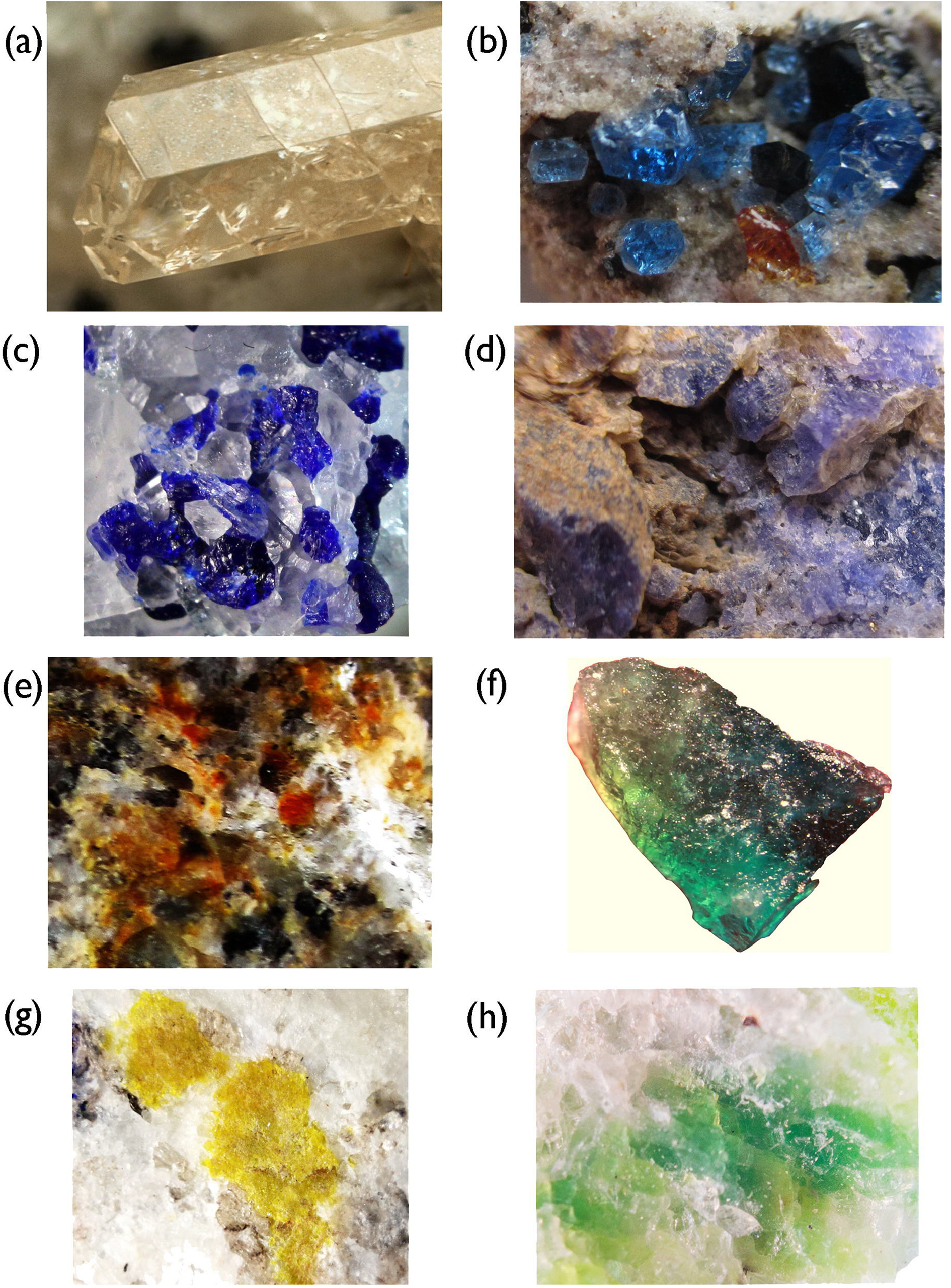
It is noteworthy that all polysulfide groups are chromophores (see Fig. 1) and show a high Raman activity (Clark and Cobbold, Reference Clark and Cobbold1978; Clark et al., Reference Clark, Dines and Kurmoo1983; Eckert and Steudel, Reference Steudel and Steudel2003; Kowalak et al., Reference Kowalak, Jankowska, Zeidler and Wiećkowski2007; Wong, Reference Wong and Steudel2013; Steudel and Chivers, Reference Steudel and Chivers2019; Rejmak, Reference Rejmak2020).
Feldspathoids containing polysulfide colour centres: (a) twin of S2•–-bearing bolotinaite (holotype sample); (b) crystals of S3•–-bearing haüyne on sanidinite (both from Eifel, Germany); (c) intermediate member of the haüyne–lazurite series from Ladgvardara, Tajikistan; (d) cis-S4- and S3•–-bearing haüyne from the Malobystrinskoye lazurite deposit, Baikal Lake area, Siberia, Russia; (e) orange sulfhydrylbystrite containing species-defining S52– anion (yellow chromophore) and cis-S4 impurity (red chromophore) from the Malobystrinskoye deposit; (f) holotype sample of slyudyankaite containing species-defining S6 molecule (yellow chromophore) as well as S4 and S3•– impurities under an incandescent lamp; (g) S4-free bystrite from the Malobystrinskoye deposit; and (h) balliranoite containing S52–, trans-S4 and minor S3•–. The field of view widths are (a) 0.5 mm, (b) 8 mm, (c) 6 mm, (d) 15 mm, (e) 7 mm, (f) 0.7 mm, (g) 5 mm and (h) 3 mm.

Figure 1 Long description
Eight close-up photographs of mineral specimens are arranged in a grid, each labeled with a lowercase letter from a through h. Photograph a shows a pale, translucent crystal with a blocky, elongated twin form set against a plain background. The crystal surfaces are smooth with visible internal fractures. Photograph b shows several small, rounded, deep blue crystals scattered across a pale gray rocky matrix. The crystals appear compact and irregular in shape. Photograph c shows a cluster of deep blue angular crystal fragments mixed with pale translucent pieces, all resting on a light gray matrix surface. Photograph d shows a rough rocky surface with patches of blue-gray mineral material distributed across a pale brown and gray host rock. The blue areas appear granular and uneven. Photograph e shows a rocky surface covered with a mixture of orange, brown, black and pale patches. The surface texture is coarse and irregular with no distinct crystal forms visible. Photograph f shows a single, flat, irregular specimen with a dark surface on one side and a bright green surface on the other, photographed against a plain pale background. Photograph g shows a cluster of yellow mineral material sitting on a pale white and gray rocky surface. The yellow material appears granular and loosely aggregated. Photograph h shows a pale rocky surface with patches of yellow-green mineral material distributed across it. The surface texture is fine-grained and uneven.
This work is a review which summarizes the most important, from our viewpoint, results and conclusions reported in the above-cited works and related data published elsewhere.
Spectroscopic properties of S-bearing groups occurring in silicate minerals
Anions
The SO42– anion is the most common sulfur-bearing species in feldspathoids and scapolites. A regular SO4 tetrahedron has four fundamental vibrational modes: A 1(ν1) (a non-degenerate totally symmetric stretching mode, which is IR inactive); E(ν2) (a doubly degenerate bending mode); F 2(ν3) (a triply degenerate asymmetric stretching mode); and F 2(ν4) (a triply degenerate bending mode). In the case of distorted SO42– groups, the degeneracy is removed and the corresponding E and F bands are split, whereas the A1(ν1) IR band intensity becomes non-zero. In the Raman spectra, the A 1(ν1) band is the strongest. The wavenumbers of the A 1(ν1), E(ν2), F 2(ν3) and F 2(ν4) bands in the Raman spectra of feldspathoids are in the ranges of 975–990, 436–447, 1115–1160 and 613–630 cm–1, respectively (Chukanov et al., Reference Chukanov, Zubkova, Varlamov, Pekov, Belakovskiy, Britvin, Van, Ermolaeva, Vozchikova and Pushcharovsky2022a, Reference Chukanov, Shendrik, Vigasina, Pekov, Sapozhnikov, Shcherbakov and Varlamov2022c, Reference Chukanov, Vigasina, Shendrik, Varlamov, Pekov and Zubkova2022d, Reference Chukanov, Shchipalkina, Shendrik, Vigasina, Tauson, Lipko, Varlamov, Shcherbakov, Sapozhnikov, Kasatkin, Zubkova and Pekov2022e; Pekov et al., Reference Pekov, Chukanov, Shcherbakov, Vigasina, Shendrik, Sandalov, Vyatkin and Turchkova2025; Shendrik et al., Reference Shendrik, Chukanov, Panikorovskii, Vigasina and Pekov2025). In the IR spectra of SO42–-bearing minerals belonging to the cancrinite and sodalite groups, only the F 2(ν3), and F 2(ν4) bands are observed (at 1135–1145 and 613–625 cm–1, respectively) whereas the A 1(ν1) and E(ν2) bands are very weak and overlap with bands of the aluminosilicate framework.
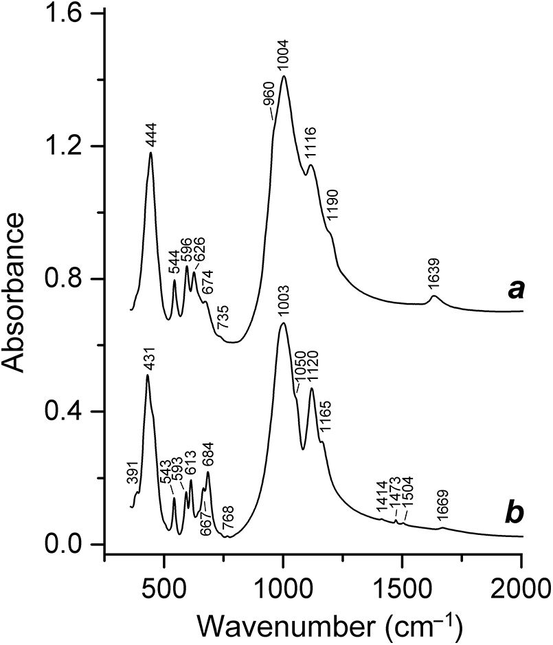
Among silicate minerals, only some multilayer members of the cancrinite group contain sulfite anionic groups, SO32–. These minerals are: afghanite (Chukanov et al., Reference Chukanov, Zubkova, Shendrik, Sapozhnikov, Pekov, Vigasina, Chervonnaya, Varlamov, Bolotina, Ksenofontov and Pushcharovsky2025); its hydrous F- and SO3-dominant analogue steudelite (Chukanov et al., Reference Chukanov, Zubkova, Varlamov, Pekov, Belakovskiy, Britvin, Van, Ermolaeva, Vozchikova and Pushcharovsky2022a); a hydrous Cl-deficient analogue alloriite (Rastsvetaeva et al., Reference Rastsvetaeva, Ivanova, Chukanov and Verin2007); the sulfite-dominant analogue of alloriite (Chukanov et al., Reference Chukanov, Zubkova, Pekov, Giester and Pushcharovsky2021b); the sulfite analogue of marinellite (Zubkova et al., Reference Zubkova, Chukanov, Varlamov, Vigasina, Pekov, Ksenofontov and Pushcharovsky2023); and the SO32–-bearing variety of tounkite (Rozenberg et al., Reference Rozenberg, Sapozhnikov, Rastsvetaeva, Bolotina and Kashaev2004). A sodalite cage (in minerals of the cancrinite and sodalite groups) as well as larger Losod, liottite and giuseppettite cages (in multilayer members of the cancrinite group) can host up to 1, 2, 3 and 4 SO32– and/or SO42– groups, respectively (Chukanov et al., Reference Chukanov, Aksenov and Rastsvetaeva2021a). However, SO32– occurs in the liottite cage of the aluminosilicate framework in all known cases. The assignment of the bands of SO32– group in the IR and Raman spectra of the listed minerals is ambiguous because of their overlapping with stronger bands of the framework and SO42– groups, respectively. In IR spectra, indirect signs of partial replacement of sulfate groups by sulfite groups are the reduced intensity of the SO42– bands (compared to purely sulfate varieties and analogues of the corresponding minerals) and a shoulder at ∼960 cm–1 (Fig. 2).
Powder infrared absorption spectra of (a) steudelite, (Na,□)4(K,Na,Ca,□)18Ca4(Al24Si24O96)(SO3,SO4)6(F,Cl)6(H2O)4, and (b) afghanite from the Ladgvardara lazurite deposit, Tajikistan.
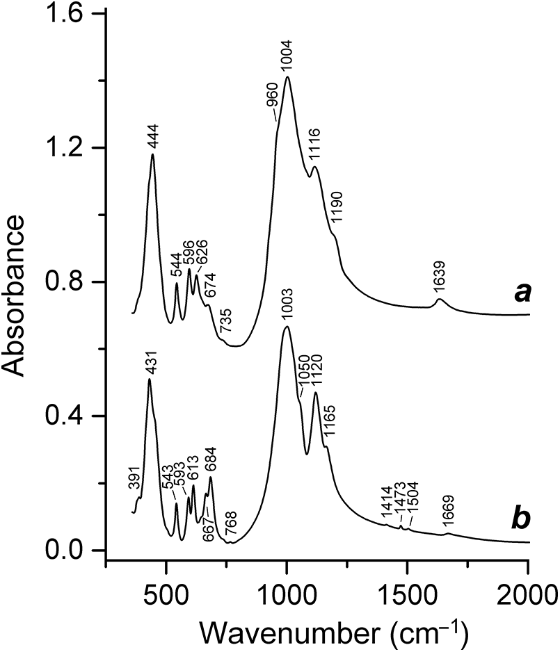
Figure 2 Long description
Two stacked line graphs labeled a and b. The x-axis is labeled Wavenumber (cm superscript minus 1), ranging from 400 to 2000. The labeled ticks are 500, 1000, 1500 and 2000. The y-axis is labeled Absorbance, ranging from 0.0 to 1.6. The labeled ticks are 0.0, 0.4, 0.8, 1.2 and 1.6.
In the Raman spectra of the sulfite minerals hannebachite, CaSO3·H2O, and orschallite, Ca3(SO3)2(SO4)·12H2O, doublets of SO32– stretching vibrations (969+1005 and 971+1005 cm–1, respectively) are observed (Frost and Keefe, Reference Frost and Keeffe2009). Similar Raman bands of cancrinite-group minerals are observed as shoulders of the stronger band of SO42– with a maximum at 991–992 cm–1 (Zubkova et al., Reference Zubkova, Chukanov, Varlamov, Vigasina, Pekov, Ksenofontov and Pushcharovsky2023; Chukanov et al., Reference Chukanov, Zubkova, Shendrik, Sapozhnikov, Pekov, Vigasina, Chervonnaya, Varlamov, Bolotina, Ksenofontov and Pushcharovsky2025).
The HS– anion is a species-defining component in the rock-forming sodalite-group mineral sapozhnikovite, Na8(Al6Si6O24)(HS–)2 (Chukanov et al., Reference Chukanov, Zubkova, Pekov, Shendrik, Varlamov, Vigasina, Belakovskiy, Britvin, Yapaskurt and Pushcharovsky2022b), and the cancrinite-group mineral, sulfhydrylbystrite, Na5K2Ca(Al6Si6O24)S52–HS– (Chukanov et al., Reference Chukanov, Sapozhnikov, Kaneva, Varlamov and Vigasina2023c) and is a common impurity in numerous varieties of other feldspathoids that crystallized under reducing conditions, including sodalite, haüyne, slyudyankaite, kyanoxalite, balliranoite, bystrite and tounkite (Chukanov et al., Reference Chukanov, Vigasina, Shendrik, Varlamov, Pekov and Zubkova2022d, Reference Chukanov, Sapozhnikov, Kaneva, Varlamov and Vigasina2023c, Reference Chukanov, Bolotina, Shendrik, Sapozhnikov, Zubkova, Pekov, Vigasina, Sandalov and Ksenofontov2024a, Reference Chukanov, Zubkova, Shendrik, Sapozhnikov, Pekov, Vigasina, Chervonnaya, Varlamov, Bolotina, Ksenofontov and Pushcharovsky2025; Sapozhnikov et al., Reference Sapozhnikov, Bolotina, Chukanov, Shendrik, Kaneva, Vigasina, Ivanova, Tauson and Lipko2023; Pekov et al., Reference Pekov, Chukanov, Shcherbakov, Vigasina, Shendrik, Sandalov, Vyatkin and Turchkova2025). Sodalite and sapozhnikovite form a continuous isomorphous series [with the Cl:HS ratio variation (in mol.%) from Cl100(HS)0 to Cl12(HS)88] (Pekov et al., Reference Pekov, Chukanov, Shcherbakov, Vigasina, Shendrik, Sandalov, Vyatkin and Turchkova2025) whereas all known samples of minerals belonging to the bystrite–sulfhydrylbystrite solid-solution series are close in composition to the end-members of this series.
Wavenumbers of the HS– stretching mode are in the range of 2550–2565 cm–1. Extinction coefficients of the IR absorption band of S–H stretching vibrations are very low (Sheppard, Reference Sheppard1949; Bragin et al., Reference Bragin, Diem, Guthals and Chang1977). For this reason, Raman spectroscopy is usually used to confirm the presence of HS– in minerals. Distinct narrow Raman peaks of HS– are observed at 2553 cm–1 for sapozhnikovite (Chukanov et al., Reference Chukanov, Zubkova, Pekov, Shendrik, Varlamov, Vigasina, Belakovskiy, Britvin, Yapaskurt and Pushcharovsky2022b; Pekov et al., Reference Pekov, Chukanov, Shcherbakov, Vigasina, Shendrik, Sandalov, Vyatkin and Turchkova2025), 2556–2562 cm–1 for the minerals of the bystrite–sulfhydrylbystrite solid-solution series and 2564 cm–1 for sulfide-bearing balliranoite (Chukanov et al., Reference Chukanov, Sapozhnikov, Shendrik, Zubkova, Vigasina, Potekhina, Ksenofontov and Pekov2023b). The bands at 2575 cm–1 in the Raman spectrum of slyudyankaite (Sapozhnikov et al., Reference Sapozhnikov, Bolotina, Chukanov, Shendrik, Kaneva, Vigasina, Ivanova, Tauson and Lipko2023) and 2577 cm–1 in the Raman spectrum of ‘haüyne-45Å’ (Chukanov et al., Reference Chukanov, Zubkova, Shendrik, Sapozhnikov, Pekov, Vigasina, Chervonnaya, Varlamov, Bolotina, Ksenofontov and Pushcharovsky2025) may correspond to stretching vibrations of the H2S molecule rather than HS– (Graneri et al., Reference Graneri, Spagnoli, Wild and McKinley2024).
Based on Raman active vibrational frequencies calculated by Tossell (Reference Tossell2012), Farsang et al. (Reference Farsang, Caracas, Adachi, Schnyder and Zajacz2023) assigned the bands at 223, 443 and 481–485 cm–1 in the Raman spectra of lapis lazuli, blue haüyne and synthetic ultramarine blue to the S42– anion. However, this assignment is questionable because the bands at 223 and 481–485 cm–1 can be assigned to the neutral S4 molecule which is a common component in sodalite-group minerals and the band at 443 cm–1 can correspond to the SO42– anionic group [the E(ν2) mode] (Pekov et al., Reference Pekov, Chukanov, Shcherbakov, Vigasina, Shendrik, Sandalov, Vyatkin and Turchkova2025; Shendrik et al., Reference Shendrik, Chukanov, Panikorovskii, Vigasina and Pekov2025).
The S52– anion is a species-defining component in the cancrinite-group minerals bystrite and sulfhydrylbystrite (Sapozhnikov et al., Reference Sapozhnikov, Ivanov, Piskunova, Kashaev, Terentieva and Pobedimskaya1991, Reference Sapozhnikov, Kaneva, Suvorova, Levitsky and Ivanova2017; Kaneva et al., Reference Kaneva, Sapozhnikov and Suvorova2017; Chukanov et al., Reference Chukanov, Sapozhnikov, Kaneva, Varlamov and Vigasina2023c). These minerals, as well as an insufficiently studied bystrite-related mineral with the idealized formula Na5K2Ca(Al6Si6O24)S52–Cl–, are the only mineral species containing S52– as a dominant S-bearing extra-framework component.
The yellow colour of bystrite is caused by S52– anions having a chain configuration and occurring in Losod cages of the aluminosilicate framework with the ABAC stacking sequence. Trans- or cis-conformers of S52– alternate in the structure of bystrite (Kaneva et al., Reference Kaneva, Sapozhnikov and Suvorova2017). The presence of admixed cis-S4 (the planar non-cyclic C2v isomer), that is a strong red chromophore (Rejmak, Reference Rejmak2020), in orange sulfhydrylbystrite (the analogue of bystrite with HS– > Cl–) is in agreement with its colour, different from the yellow colour of holotype bystrite (Chukanov et al., Reference Chukanov, Sapozhnikov, Kaneva, Varlamov and Vigasina2023c).
A minor amount of S52– anions was detected in green tounkite, a cancrinite-group mineral with the CACACBCBCACB stacking sequence (Chukanov et al., Reference Chukanov, Bolotina, Shendrik, Sapozhnikov, Zubkova, Pekov, Vigasina, Sandalov and Ksenofontov2024a). The green colour of this tounkite variety is due to the presence of the S52– anion (yellow chromophore) and S3•– radical anion (blue chromophore).
The absorption band of the S52– anion in the green commensurately modulated haüyne analogue was observed at 455 nm, along with the bands of the S2•– and S3•– radical anions at 390 and 590 nm, respectively (Chukanov et al., Reference Chukanov, Zubkova, Shendrik, Sapozhnikov, Pekov, Vigasina, Chervonnaya, Varlamov, Bolotina, Ksenofontov and Pushcharovsky2025).
The geometry of cis-S52– in the solid state (in the compound Na2S5) is characterized by the bond lengths d 12 = 2.061 Å and d 23 = 2.066 Å, bond angles φ1 = 107.31° and φ1 = 108.11°, and torsion angle τ = 88.61° which differs from corresponding values for cis-S52– in the gas phase calculated with the PCM78 correction (2.079 Å, 2.137 Å, 116.6°, 116.5° and 107°, respectively) (Steudel and Steudel, Reference Steudel and Steudel2013). The helical non-flat iso-conformer of S52– is slightly less stable than cis-S52–. The geometrical parameters of iso-S52– in the polarizable medium are: d 12 = 2.084 Å, d 23 = 2.127 Å, φ1 = 113.7°, φ1 = 116.5°, and τ = 91.5° (Steudel and Chivers, Reference Steudel and Chivers2019). S52– is thermodynamically favoured over polysulfide anions with other chain lengths. In the aqueous solution with the phosphate buffer, the S52– anion partly transforms to a series of Sn 2– anions (n = 2–8) among which S62– and S42– are most abundant and S22– occurs in trace amounts (Kamyshny et al., Reference Kamyshny, Goifman, Gun, Rizkov and Lev2004).
Based on high-level ab initio calculations, the wavenumbers of fundamental S–S stretching vibrations predicted for S52− coordinated by Li+ are 471, 463 and 416 cm−1 (Steudel and Chivers, Reference Steudel and Chivers2019). Similar bands are observed in the IR spectra of bystrite and sulfhydrylbystrite in the ranges of 413–422 and 454–466 cm−1 (Chukanov et al., Reference Chukanov, Sapozhnikov, Kaneva, Varlamov and Vigasina2023c). No bands in these ranges are in the IR spectrum of carbobystrite, a carbonate cancrinite-group mineral with the bystrite-type framework. The IR spectrum of bottle-green S52−-bearing tounkite contains a strong peak at 448 cm−1; in the IR spectrum of blue tounkite this band is observed as a shoulder on the background of a band of bending vibrations of the aluminosilicate framework (Chukanov et al., Reference Chukanov, Bolotina, Shendrik, Sapozhnikov, Zubkova, Pekov, Vigasina, Sandalov and Ksenofontov2024a).
The Raman bands in the range of 410–510 cm−1 correspond to stretching vibrations of different S52– conformers, and bands in the ranges of 170–190 and 250–270 cm−1 are due to S–S–S bending vibrations of S52– (Steudel and Chivers, Reference Steudel and Chivers2019; Chukanov et al., Reference Chukanov, Sapozhnikov, Kaneva, Varlamov and Vigasina2023c, Reference Chukanov, Bolotina, Shendrik, Sapozhnikov, Zubkova, Pekov, Vigasina, Sandalov and Ksenofontov2024a; Pekov et al., Reference Pekov, Chukanov, Shcherbakov, Vigasina, Shendrik, Sandalov, Vyatkin and Turchkova2025; Bogdanov et al., Reference Bogdanov, Chukanov, Shendrik and Pekov2025). The peak at 505 cm−1 in the Raman spectrum of bystrite and the peak at 508 cm−1 in the Raman spectrum of sulfhydrylbystrite were assigned to S–S stretching vibrations of the S52– anion (Chukanov et al., Reference Chukanov, Sapozhnikov, Kaneva, Varlamov and Vigasina2023c). In the Raman spectrum of green S52−-bearing tounkite, bands of S52− are observed at 174, 267 and 452 cm−1 (Chukanov et al., Reference Chukanov, Bolotina, Shendrik, Sapozhnikov, Zubkova, Pekov, Vigasina, Sandalov and Ksenofontov2024a).
Radical anions
The S2•– radical anion is a yellow chromophore (Steudel and Chivers, Reference Steudel and Chivers2019; Chukanov et al., Reference Chukanov, Zubkova, Schäfer, Pekov, Shendrik, Vigasina, Belakovskiy, Britvin, Yapaskurt and Pushcharovsky2022f). This species is a typical impurity component in sodalite-group minerals. In particular, the yellow colour of bolotinaite, ideally (Na6K□)(Al6Si6O24)F·4H2O, is caused by S2•– radical anions (Chukanov et al., Reference Chukanov, Zubkova, Schäfer, Pekov, Shendrik, Vigasina, Belakovskiy, Britvin, Yapaskurt and Pushcharovsky2022f), but in most cases the yellow colour is masked by other chromophores.
According to various estimates made using quantum-chemical calculations (Steudel, Reference Steudel1975; Stevens et al., Reference Stevens, Vrielinck, Callens, Pauwels and Waroquier2002; Wong and Steudel, Reference Steudel and Steudel2003; Chivers and Oakley, Reference Chivers and Oakley2023, Steudel and Steudel, Reference Steudel and Steudel2013; Steudel and Chivers, Reference Steudel and Chivers2019), the isolated S2•– radical anion is characterized by an S–S bond length in the range of 2.00–2.08 Å and a wavenumber of S–S stretching vibrations from 528 to 579 cm–1. In the visible region of the absorption spectrum, the S2•– group appears as an absorption band with a maximum at ∼400 nm (Chivers and Elder, Reference Chivers and Elder2013; Steudel and Chivers, Reference Steudel and Chivers2019). A similar band in the absorption spectrum of the S2•–-rich commensurately modulated haüyne analogue (‘haüyne-45Å’) is observed at 390 nm (Chukanov et al., Reference Chukanov, Zubkova, Shendrik, Sapozhnikov, Pekov, Vigasina, Chervonnaya, Varlamov, Bolotina, Ksenofontov and Pushcharovsky2025).
The luminescence spectrum of the S2•– radical anion has a maximum at ∼665 nm and a vibrational structure determined by the stretching vibrations of S2•– (Kirk et al., Reference Kirk, Schulman and Rosenstock1965). The position of the absorption band associated with the transition from the ground b2u state to the excited b2g state depends on the environment of the S2•– radical anion (Shendrik et al., Reference Shendrik, Chukanov, Bogdanov, Myasnikova, Pankrushina, Zolotarev, Babkina, Popova, Vigasina, Aksenov, Ilyin and Pekov2024).
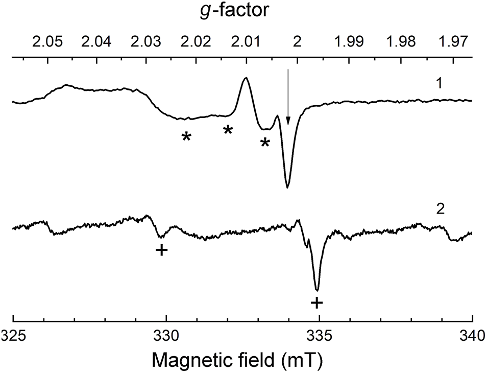
The ESR spectra of S2•– in crystals of halides of alkaline and rare-earth metals are characterised by the g-tensor components: g 11 = 1.6–1.99, g 22 = 2.002–2.019 and g 33 = 2.2–2.6 (Schneider et al., Reference Schneider, Dischler and Räuber1966; Vannotti and Morton, Reference Vannotti and Morton1967; de Siebenthal and Bill, Reference de Siebenthal and Bill1979). The ESR spectra of alkali halide crystals calculated using DFT methods (Stevens et al., Reference Stevens, Vrielinck, Callens, Pauwels and Waroquier2002) are in good agreement with the experimental data. In particular, theoretical calculations were performed for the configuration in which S2•– occupies two neighbouring vacancies, and it was shown that in this case an ESR signal with g 11 = 0.948; g 22 = 0.95 and g 33 = 3.43 should be observed. A similar signal was observed in the ESR spectra of some alkali halide crystals (Schneider et al., Reference Schneider, Dischler and Räuber1966). The experimentally measured g-tensor components of S2•– in sodalite-group minerals are in the ranges g 11 = 1.82–1.995 (the latter value is for tugtupite), g 22 = 2.00–2.02 and g 33 = 2.20–2.34 (Chukanov et al., Reference Chukanov, Zubkova, Pekov, Shendrik, Varlamov, Vigasina, Belakovskiy, Britvin, Yapaskurt and Pushcharovsky2022b, Reference Chukanov, Shchipalkina, Shendrik, Vigasina, Tauson, Lipko, Varlamov, Shcherbakov, Sapozhnikov, Kasatkin, Zubkova and Pekov2022e, Reference Chukanov, Zubkova, Shendrik, Sapozhnikov, Pekov, Vigasina, Chervonnaya, Varlamov, Bolotina, Ksenofontov and Pushcharovsky2025). The isotropic g-factor of S2•– in sapozhnikovite and most other lazurite-related sodalite-group minerals is close to 2.002 (Fig. 3). These data show that g factors of the S2•– radical anion strongly depend on its environment, but in all cases an extremely strong splitting of the ESR signal is observed, which is a specific feature of the S2•– ESR spectra.
ESR spectra of (1) S2•–- and S3•–-bearing sapozhnikovite from the Lovozero massif (Kola Peninsula) and (2) lilac S4•–-bearing haüyne from the Malobystrinskoye deposit (this work). The arrow, asterisks and crosses indicate positions of the signals associated with S2•–, S3•– and S4•–, respectively.
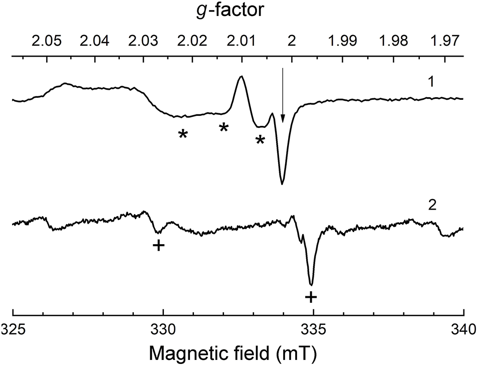
Figure 3 Long description
G-factor A two-trace line plot with two stacked spectra labeled 1 and 2. The x-axis is labeled Magnetic field (mT). The axis shows labeled ticks at 325, 330, 335 and 340. The top axis is labeled g-factor. The labeled values run from 2.05, 2.04, 2.03, 2.02, 2.01, 2.00, 1.99, 1.98, to 1.97.
The main S-bearing paramagnetic species in ‘haüyne-45Å’ is S2•–, for which the ESR spectrum has g components of 2.00 and 2.34 (Chukanov et al., Reference Chukanov, Zubkova, Shendrik, Sapozhnikov, Pekov, Vigasina, Chervonnaya, Varlamov, Bolotina, Ksenofontov and Pushcharovsky2025). The latter value indicates the occurrence of S2•– in a rather strong crystal field.
The stretching mode of the S2•– radical anion is inactive in the IR spectrum because the dipole moment of the S–S bond is close to zero. The stretching and combination-mode (with the participation of librational vibrations) bands of S2•– are observed in the Raman spectra in the ranges of 530–550 and 600–620 cm–1, respectively. Similar bands of the S2•–-containing oxalate member of the cancrinite group, kyanoxalite, have wavenumbers of 564 and 598 cm–1, respectively. In the Raman spectra of S2•–-bearing feldspathoids, a characteristic luminescence with an vibrational structure having a period ranging from 520 to 550 cm–1 is observed (Radomskaya et al., Reference Radomskaya, Kaneva, Shendrik, Suvorova and Vladykin2021; Chukanov et al., Reference Chukanov, Sapozhnikov, Shendrik, Vigasina and Steudel2020b, Reference Chukanov, Zubkova, Pekov, Shendrik, Varlamov, Vigasina, Belakovskiy, Britvin, Yapaskurt and Pushcharovsky2022b, Reference Chukanov, Sapozhnikov, Shendrik, Zubkova, Vigasina, Potekhina, Ksenofontov and Pekov2023b; Shendrik et al., Reference Shendrik, Chukanov, Panikorovskii, Vigasina and Pekov2025).
The formation of S2•– radical anions in tugtupite upon excitation with a light having a wavelength of ∼400 nm was confirmed by the presence of luminescence with a S2•– vibrational structure (Shendrik et al., Reference Shendrik, Chukanov, Panikorovskii, Vigasina and Pekov2025). Tugtupite, when irradiated with short-wave UV light acquires a purple colour and the appearance of an absorption band at ∼520 nm as well as a strong ESR signal with g 1 = 2.210; g 2 = 2.010 and g 3 = 1.995 related to the S2•– radical anions. The purple colour of irradiated tugtupite may arise as a result of partial dimerization of S2•– followed by subtraction of electron and formation of S4•–. This process is reversible: the purple colour disappears after irradiation by light with the wavelength in the range of 500–600 nm.
Using the DFT method (Cavignac et al., Reference Cavignac, Latouche and Jobic2022), it was shown that the luminescence maximum of the S2•– radical anion, located in a strong crystal field, shifts to the short-wavelength region of the spectrum. The luminescence of the S2•– defect in alkali halide crystals has a maximum in the wavelength range of 580–640 nm (Kirk et al., Reference Kirk, Schulman and Rosenstock1965; Rolfe, Reference Rolfe1968). A similar photoluminescence was observed for synthetic sodalite (Sidike et al., Reference Sidike, Sawuti, Wang, Zhu, Kobayashi, Kusachi and Yamashita2007). The yellow–orange photoluminescence observed upon irradiation of S-bearing sodalite (hackmanite) with long-wavelength UV light (Sidike et al., Reference Sidike, Sawuti, Wang, Zhu, Kobayashi, Kusachi and Yamashita2007; Gaft et al., Reference Gaft, Panczer, Nagli and Yeates2009; Kaiheriman et al., Reference Kaiheriman, Maimaitinaisier, Rehiman and Sidike2014) has been attributed to the S2•– radical anion (Taylor et al., Reference Taylor, Marshall, Forrester and McLaughlan1970; Warner, Reference Warner2011; Kaiheriman et al., Reference Kaiheriman, Maimaitinaisier, Rehiman and Sidike2014; Finch et al., Reference Finch, Friis and Maghrabi2016) or the S22− anion (Kirk, Reference Kirk1955; Ballentyne and Bye, Reference Ballentyne and Bye1970; Warner, Reference Warner2011), although the latter assignment is unlikely because of the low stability of S22− (Berghof et al., Reference Berghof, Sommerfeld and Cederbaum1998; Steudel and Chivers, Reference Steudel and Chivers2019). The cathodoluminescence intensity of sodalite paradoxically decreases with increasing sulfur content. A possible cause of this phenomenon is that some sulfide groups are luminescence quenchers (Zahoransky et al., Reference Zahoransky, Friis and Marks2016).
According to XANES data (Goettlicher et al., Reference Goettlicher, Kotelnikov, Suk, Kovalski, Vitova and Steininger2013), hackmanite contains sulfide sulfur. As was shown recently, it occurs in hackmanite mainly in the form of HS– (Pekov et al., Reference Pekov, Chukanov, Shcherbakov, Vigasina, Shendrik, Sandalov, Vyatkin and Turchkova2025). The mechanism of photochromism (tenebrescence) of hackmanite and tugtupite (i.e. their colour change under the influence of light) has been discussed in many publications (Hassib et al., Reference Hassib, Beckman and Annersten1977; Jensen and Petersen, Reference Jensen and Petersen1982; Pizani et al., Reference Pizani, Terrile, Farach and Poole1985; Warner and Hutzen, Reference Warner and Hutzen2012; Norrbo et al., Reference Norrbo, Gluchowski, Hyppänen, Laihinen, Laukkanen, Mäkelä, Mamedov, Santos, Sinkkonen, Tuomisto, Viinikanoja and Lastusaari2016; Curutchet and le Bahers, Reference Curutchet and le Bahers2017; Agamah et al., Reference Agamah, Vuori, Colinet, Norrbo, de Carvalho, Nakamura, Lindblom, van Goethem, Emmermann, Saarinen, Laihinen, Laakkonen, Linden, Konu, Vrielinck, van der Heggen, Smet, le Bahers and Lastusaari2020; Colinet et al., Reference Colinet, Gheeraert, Curutchet and le Bahers2020; Blumentritt and Fritsch, Reference Blumentritt and Fritsch2021; Colinet et al., Reference Colinet, Byron, Vuori, Lehtio, Laukkanen, van Goethem, Lastusaari and le Bahers2022; Song et al., Reference Song, Guo, Liu, Rao and Liao2023, Reference Song, Guo, Wang and Liao2024; Blumentritt et al., Reference Blumentritt, Notari and Caplan2024; Yang et al., Reference Yang, Guo and Liao2024). Most authors associate this phenomenon with the transfer of an electron from a sulfide anion or a bisulfide radical anion to a chlorine or oxygen vacancy under the influence of UV irradiation, which leads to the formation of F-centres with an absorption band near 550 nm.
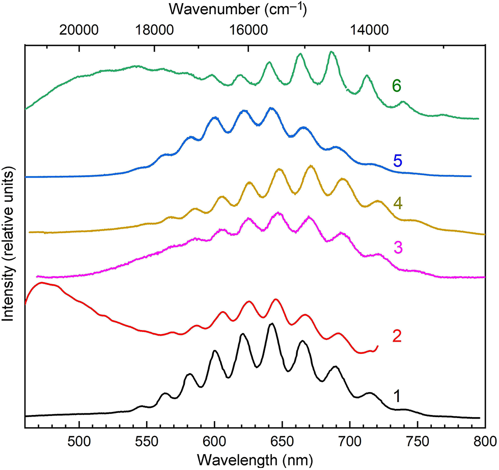
In the structures of cancrinite and other minerals with the same topological type of framework, there is a wide channel passing through twelve-membered rings (Hassan et al., Reference Hassan, Antao and Parise2006; Chukanov et al., Reference Chukanov, Aksenov and Rastsvetaeva2021a). The yellow or yellow–green (in the presence of the S3•– impurity) colouration of some samples of such minerals, including sulfide-bearing balliranoite from the Tultuy lazurite deposit, Baikal Lake region, Siberia, Russia is associated with the presence of the S2•– radical anion, which is confirmed by the characteristic luminescence spectrum with a maximum at 645 nm and vibrational structure with a period of ∼530 cm–1 upon excitation with a laser with a wavelength of 405 nm at 77 K (Chukanov et al., Reference Chukanov, Sapozhnikov, Shendrik, Zubkova, Vigasina, Potekhina, Ksenofontov and Pekov2023b). The vibration frequency of S2•– in sapozhnikovite determined from the distance between vibrational satellites is equal to 536 cm–1 (Chukanov et al., Reference Chukanov, Zubkova, Pekov, Shendrik, Varlamov, Vigasina, Belakovskiy, Britvin, Yapaskurt and Pushcharovsky2022b). Similar luminescence spectra with maxima between 600 and 650 nm and distinct vibrational structure of S2•– (with a period of 530 to 580 cm–1) were observed for various minerals belonging to the cancrinite group (kyanoxalite, marinellite, afghanite and biachellaite), sodalite group (tugtupite, hackmanite, members of the sodalite–sapozhnikovite series, haüyne and nosean) as well as scapolites (Chukanov et al., Reference Chukanov, Sapozhnikov, Shendrik, Vigasina and Steudel2020b, Reference Chukanov, Zubkova, Pekov, Shendrik, Varlamov, Vigasina, Belakovskiy, Britvin, Yapaskurt and Pushcharovsky2022b, Reference Chukanov, Shendrik, Vigasina, Pekov, Sapozhnikov, Shcherbakov and Varlamov2022c, Reference Chukanov, Vigasina, Shendrik, Varlamov, Pekov and Zubkova2022d, Reference Chukanov, Shchipalkina, Shendrik, Vigasina, Tauson, Lipko, Varlamov, Shcherbakov, Sapozhnikov, Kasatkin, Zubkova and Pekov2022e, Reference Chukanov, Zubkova, Schäfer, Pekov, Shendrik, Vigasina, Belakovskiy, Britvin, Yapaskurt and Pushcharovsky2022f, Reference Chukanov, Sapozhnikov, Shendrik, Zubkova, Vigasina, Potekhina, Ksenofontov and Pekov2023b, Reference Chukanov, Bolotina, Shendrik, Sapozhnikov, Zubkova, Pekov, Vigasina, Sandalov and Ksenofontov2024a; see Fig. 4).
Vibrational structure of S2•– in the luminescence spectra of S2•–-containing feldspathoids: (1) kyanoxalite from the Lovozero massif, Kola Peninsula (Chukanov et al., Reference Chukanov, Vigasina, Shendrik, Varlamov, Pekov and Zubkova2022d); (2) hackmanite from the Inagli massif, Aldan Shield (Radomskaya et al., Reference Radomskaya, Kaneva, Shendrik, Suvorova and Vladykin2021); (3) nosean from the Eifel sanidinite, Germany (Chukanov et al., Reference Chukanov, Sapozhnikov, Shendrik, Vigasina and Steudel2020b); (4) haüyne from the Malobystrinskoye deposit, Baikal Lake region (Chukanov et al., Reference Chukanov, Sapozhnikov, Shendrik, Vigasina and Steudel2020b); (5) sapozhnikovite; and (6) sodalite from Mount Flora, Lovozero massif (Chukanov et al., Reference Chukanov, Zubkova, Pekov, Shendrik, Varlamov, Vigasina, Belakovskiy, Britvin, Yapaskurt and Pushcharovsky2022b), obtained upon excitation with radiation with a wavelength of 405 nm at 77 K.
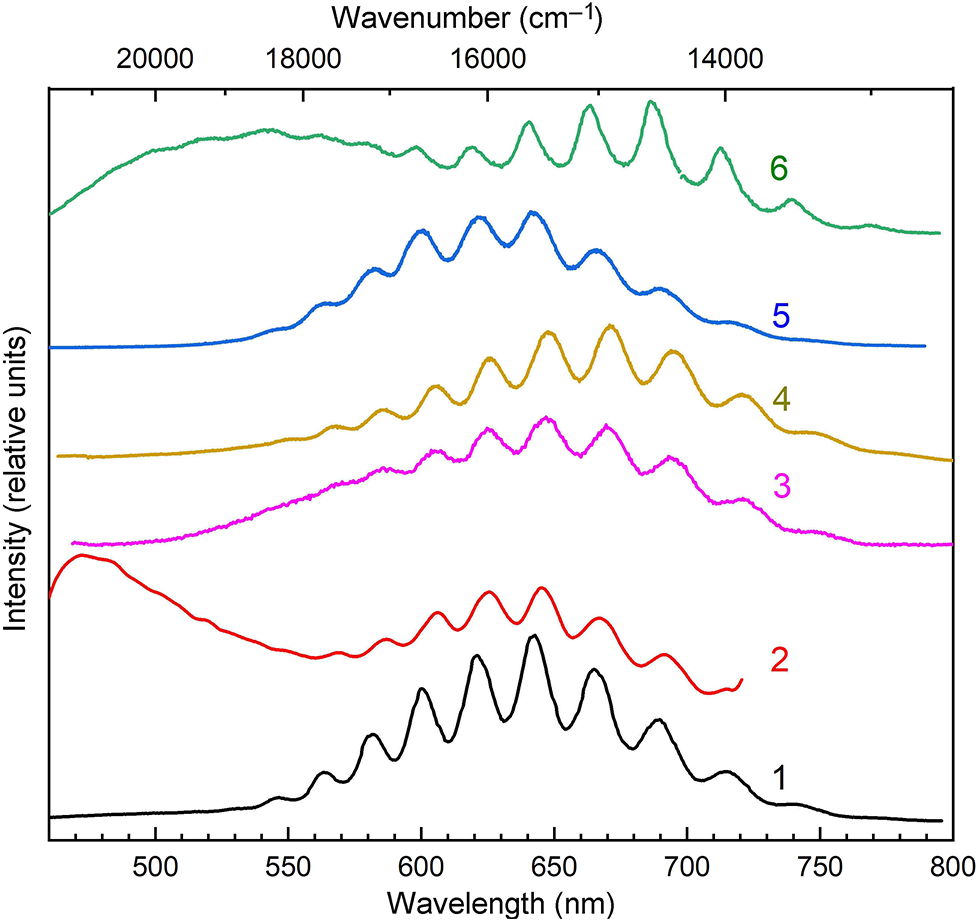
Figure 4 Long description
The graph displays six spectra labeled 1 to 6, showing intensity in relative units versus wavelength in nanometers. The horizontal axis is labeled Wavelength (nm) ranging from 500 to 800, with tick marks at 500, 550, 600, 650, 700, 750 and 800. The vertical axis is labeled Intensity (relative units) without numeric tick marks. Each spectrum is visually distinguished by color: 1 is black, 2 is red, 3 is magenta, 4 is gold, 5 is blue and 6 is green.
The calculated Raman shift for the stretching band of S2•– in scapolites is in the range of 618–630 cm–1 and depends on the cation environment of the trisulfide radical anion, but wavenumbers of experimentally observed Raman bands of S-bearing scapolites are in the range of 602–607 cm–1 (Shendrik et al., Reference Shendrik, Chukanov, Bogdanov, Myasnikova, Pankrushina, Zolotarev, Babkina, Popova, Vigasina, Aksenov, Ilyin and Pekov2024). The occurrence of S2•– at the sites coordinated by three cations and a vacancy, which would be energetically more favourable than S2•– coordinated by four cations, may be a cause of this discrepancy (it should be noted that empirical formulae of most S-bearing scapolites calculated on 12 Si + Al + Fe apfu have a slight deficit of extra-framework metal cations). Photoluminescence with vibrational structure having a period of ∼ 600 cm–1 is observed for S2•–-bearing scapolites (Shendrik et al., Reference Shendrik, Chukanov, Bogdanov, Myasnikova, Pankrushina, Zolotarev, Babkina, Popova, Vigasina, Aksenov, Ilyin and Pekov2024).
The luminescence excitation spectrum of sulfide-bearing balliranoite shows bands at 255, 265 and 375 nm associated with S2•−.
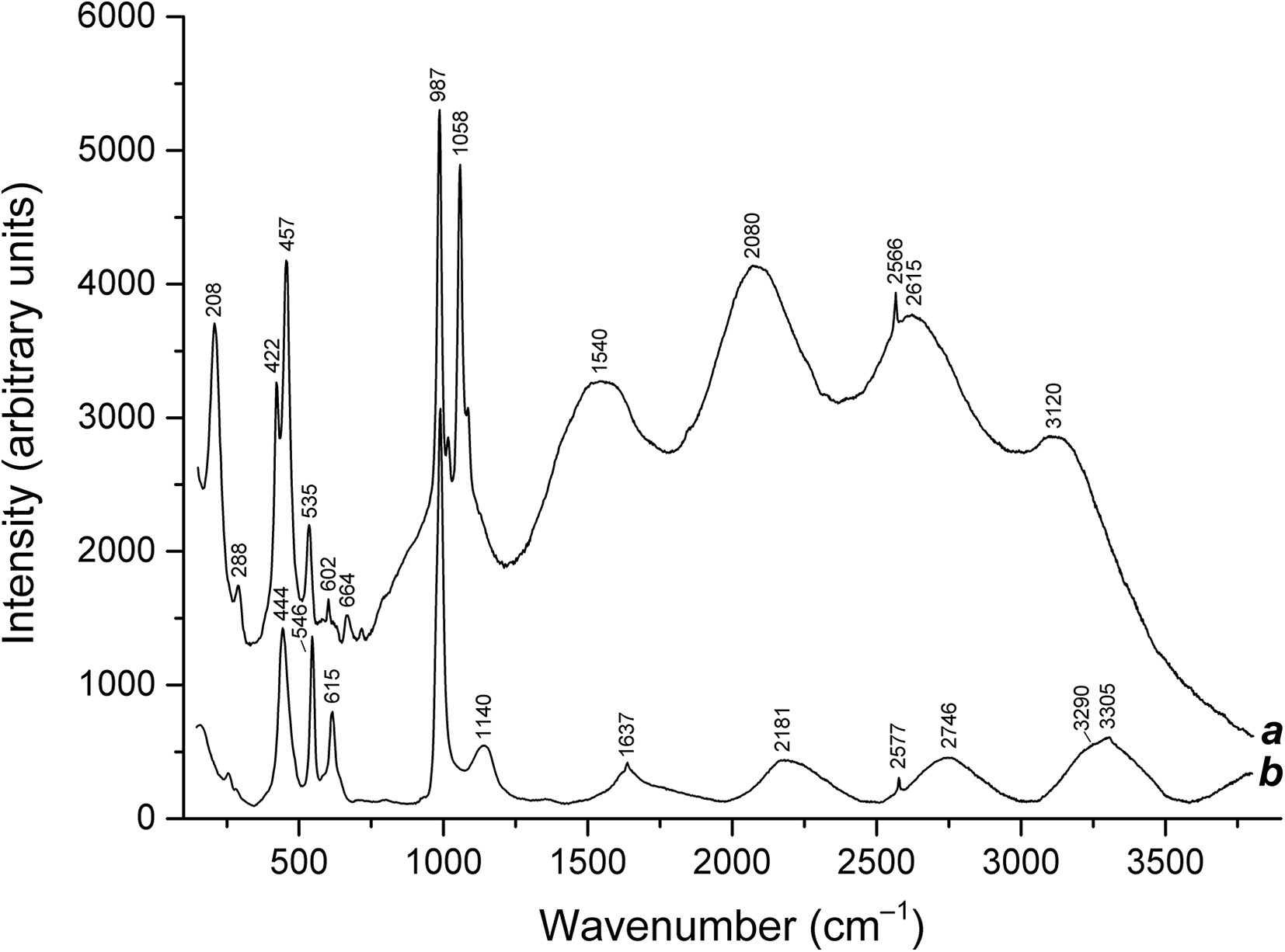
Luminescence with vibrational structure of S2•− in the range of 1200–3500 cm–1 is observed in the Raman spectra of some minerals belonging to the cancrinite and sodalite groups as well as products of their thermal conversions. In particular, after calcination of the sulfide-bearing balliranoite in air at 600°C, the luminescence is enhanced due to the decomposition of impurity ions S52– (with the formation of S2•–) and is observed in the Raman spectrum (Fig. 5).
Raman spectra of (a) the product of calcination in air at 600°C of S52−-containing balliranoite from the Tultuy lazurite deposit, Baikal Lake region (Chukanov et al., Reference Chukanov, Sapozhnikov, Shendrik, Zubkova, Vigasina, Potekhina, Ksenofontov and Pekov2023b) and (b) S2•–-, S3•–-, S52–-, H2S- and HS–-containing green haüyne with a commensurately modulated crystal structure and a fivefold increased cubic unit cell parameter from the Malobystrinskoye lazurite deposit, Baikal Lake region (Chukanov et al., Reference Chukanov, Zubkova, Shendrik, Sapozhnikov, Pekov, Vigasina, Chervonnaya, Varlamov, Bolotina, Ksenofontov and Pushcharovsky2025).
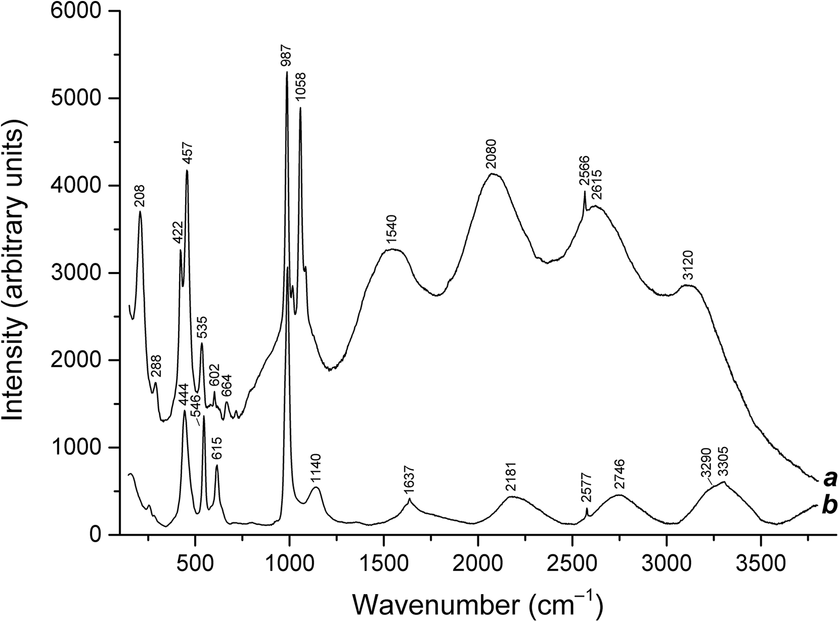
Figure 5 Long description
Two overlaid line plots labeled a and b. The x-axis is labeled Wavenumber (cm minus 1), with tick labels at 500, 1000, 1500, 2000, 2500, 3000 and 3500. The y-axis is labeled Intensity (arbitrary units), ranging from 0 to 6000 with tick labels at 0, 1000, 2000, 3000, 4000, 5000 and 6000.
Most likely, in cancrinite-group minerals with the simplest two-layer structure, the impurity radical anion S2•– is located in these channels and enters the coordination sphere of Na+ with a Na–S distance of ∼2.75 Å (Shendrik et al., Reference Shendrik, Chukanov, Panikorovskii, Vigasina and Pekov2025).
When afghanite samples are excited by a 405 nm laser at a temperature of 77 K, a broad luminescence band caused by S2•− is observed with a maximum at ∼16,600 cm–1 (Chukanov et al., Reference Chukanov, Vigasina, Shendrik, Varlamov, Pekov and Zubkova2022d). This band has a vibrational structure with repetitions at the distance of 545 cm–1 from each other, which corresponds to the S2•− stretching mode.
In the photoelectron spectra of S2•–-bearing afghanite and vesuvianite, unresolved doublet related to the disulfide radical anion appears in the range of 166–170 eV (Rao et al., Reference Rao, Qingfeng and Libing2024; Shendrik et al., Reference Shendrik, Chukanov, Panikorovskii, Vigasina and Pekov2025; Panikorovskii and Chukanov, Reference Panikorovskii and Chukanov2025).
The isolated S3•– radical anion has a configuration characterized by equivalent S–S bonds of 1.99 Å in length and an S–S–S angle of ∼115° (Chivers and Elder, Reference Chivers and Elder2013). However, in the cages of tectosilicates S3•– is distorted. For example, according to ab initio quantum-chemical calculations under periodic boundary conditions, this group in the structures of Ca-rich scapolites is distorted, with nonequivalent S–S bonds of 1.95 and 1.97 Å and a S–S–S angle of 109.0° (for the Ca4 environment), and S–S bonds of 1.96 and 1.97 Å and a S–S–S angle of 108.4° (for the Ca3Na environment) (Shendrik et al., Reference Shendrik, Chukanov, Bogdanov, Myasnikova, Pankrushina, Zolotarev, Babkina, Popova, Vigasina, Aksenov, Ilyin and Pekov2024).
S3•– is a very strong blue chromophore. For example, the presence of trisulfide radical anions in amounts of the order of 0.01 groups per formula unit is sufficient for the appearance of the blue colour in haüyne (Sapozhnikov et al., Reference Sapozhnikov, Bolotina, Chukanov, Shendrik, Kaneva, Vigasina, Ivanova, Tauson and Lipko2023).
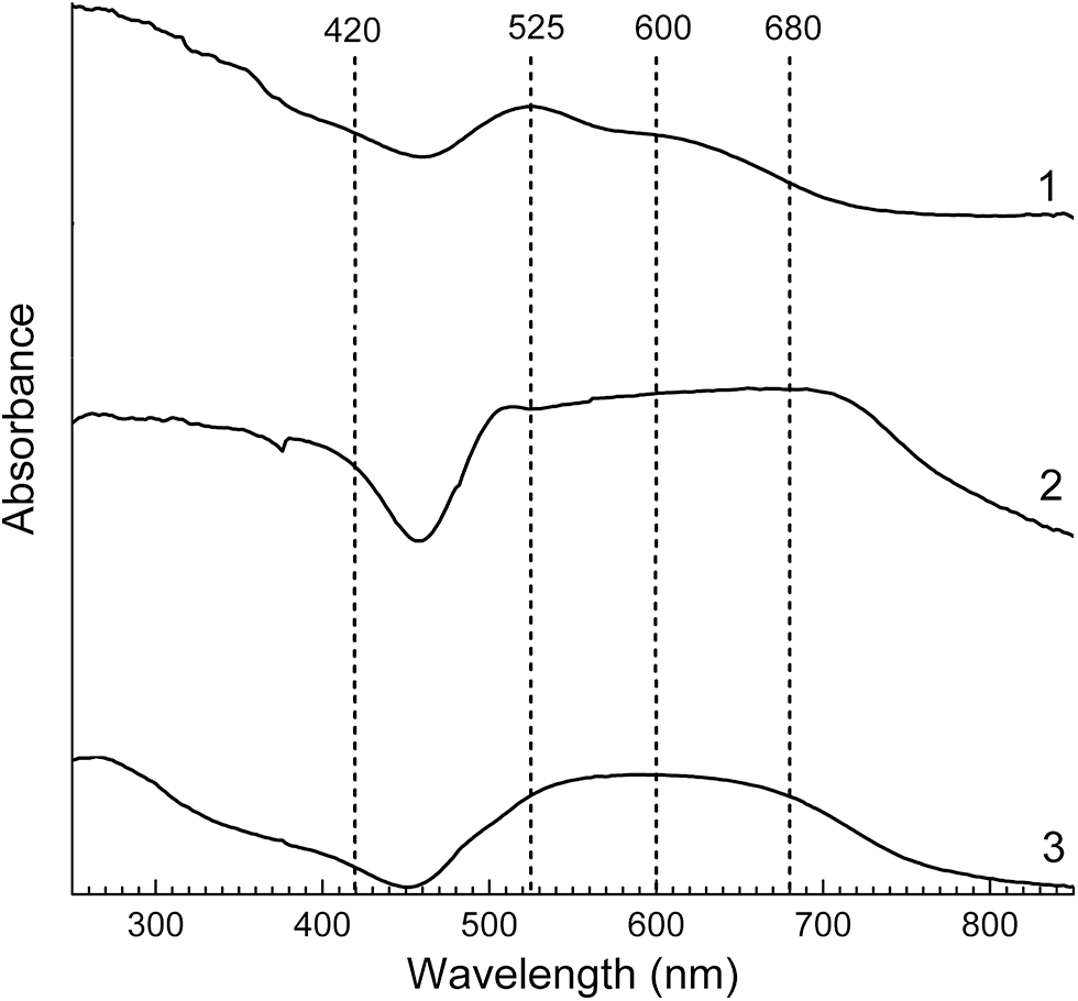
In solution, the trisulfide radical anion shows a solvent-dependent absorption band in the range of 590–620 nm (Chivers and Elder, Reference Chivers and Elder2013; Steudel and Chivers, Reference Steudel and Chivers2019). The lowest wavelength of 590 nm for the maximum of this band was reported for a green commensurately modulated haüyne analogue (Chukanov et al., Reference Chukanov, Zubkova, Shendrik, Sapozhnikov, Pekov, Vigasina, Chervonnaya, Varlamov, Bolotina, Ksenofontov and Pushcharovsky2025). The blue colouration of lazurite, other S3•–-containing sodalite group minerals (lazurite, haüyne, vladimirivanovite and slyudyankaite), and blue and green varieties of cancrinite-group minerals (kyanoxalite, balliranoite and afghanite) is due to the presence of a band in the absorption spectrum in the range of 590–620 nm having a very high molar absorptivity of ∼4500 L mol–1 cm–1 (Chivers and Elder, Reference Chivers and Elder2013; Steudel and Chivers, Reference Steudel and Chivers2019; Chukanov et al., Reference Chukanov, Zubkova, Pekov, Giester and Pushcharovsky2021b, Reference Chukanov, Vigasina, Shendrik, Varlamov, Pekov and Zubkova2022d, Reference Chukanov, Sapozhnikov, Shendrik, Zubkova, Vigasina, Potekhina, Ksenofontov and Pekov2023b; Sapozhnikov et al., Reference Sapozhnikov, Tauson, Lipko, Shendrik, Levitskii, Suvorova, Chukanov and Vigasina2021, Reference Sapozhnikov, Chukanov, Shendrik, Vigasina, Tauson, Lipko, Belakovskiy, Levitskii, Suvorova and Ivanova2022; Ostroumov et al., Reference Ostroumov, Fritsch, Faulques and Chauvet2002). This band is observed in the NIR-Vis-UV absorption spectra of sodalite-group minerals belonging to the haüyne–lazurite solid-solution series (Fig. 6).
NIR-Vis-UV absorption spectra of sodalite-group minerals from the Malo-Bystrinskoe lazurite deposit, Baikal Lake region: (1) lilac S4-bearing haüyne with minor S2•– and S3•– impurities; (2) blue S4-, S4•–- and S3•–-bearing haüyne; and (3) holotype sample of lazurite with species-defining S3•– groups. The wavelengths of 420, 525, 600 and 680 nm nearly correspond to S2•–, cis-S4, S3•– and S4•–, respectively.

Figure 6 Long description
Three line spectra on a single set of axes. The y-axis label is Absorbance. The x-axis label is Wavelength (nm). The x-axis shows tick labels at 300, 400, 500, 600, 700 and 800. Four vertical dashed reference lines are drawn and labeled at the top as 420, 525, 600 and 680. Three curves are labeled at the right edge as 1, 2 and 3.
The bluish violet colour of the sulfite analogue of marinellite is unusual for cancrinite-group minerals (Chukanov et al., Reference Chukanov, Shchipalkina, Shendrik, Vigasina, Tauson, Lipko, Varlamov, Shcherbakov, Sapozhnikov, Kasatkin, Zubkova and Pekov2022e). The components of the ESR spectrum of this mineral with g 1 = 2.220 and g 2 = 2.007 show the presence of a small amount of distorted S3•– radical anions (with a lowered S–S–S angle), which are the most probable chromophore. The maximum of the S3•– absorption band of marinellite is shifted towards a lower wavelength (to 580 nm) compared the typical spectra of most other S3•–-bearing feldspathoids.
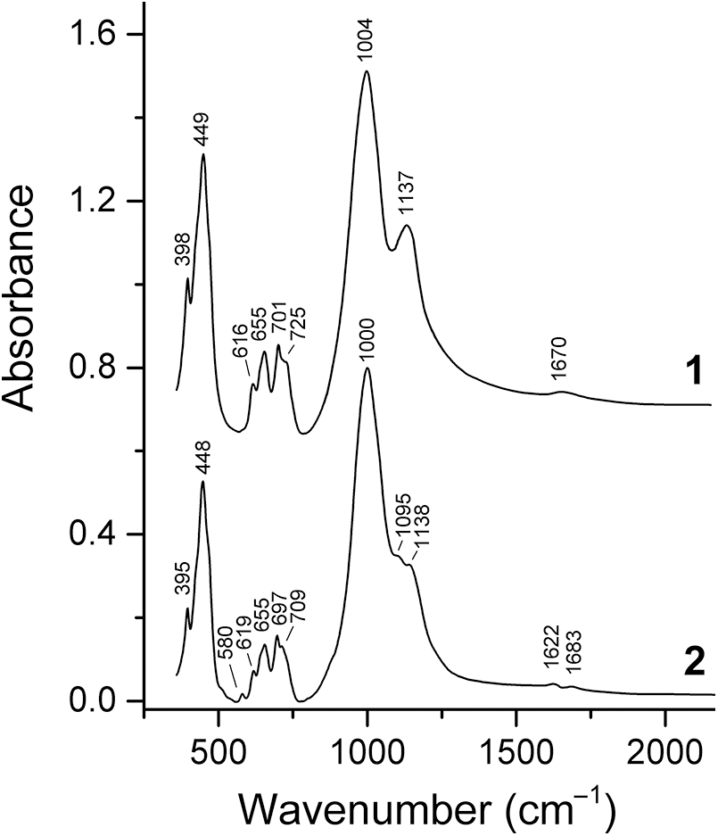
The S–S bond in S3•– is characterized by a low dipole moment. As a result, IR absorption bands of this radical anion are weak and usually are not observed in the IR spectra of feldspathoids. The only exception is lazurite, ideally Na7Ca(Al6Si6O24)(SO4)S3•–·H2O, where a weak band of antisymmetric S–S stretching vibrations is observed at 580 cm–1 (Sapozhnikov et al., Reference Sapozhnikov, Tauson, Lipko, Shendrik, Levitskii, Suvorova, Chukanov and Vigasina2021, Fig. 7).
In contrast, the Raman bands of S3•– are strong and can be used as a reliable tool for the identification of this species. The fundamentals of the trisulfide radical anion in feldspathoids are observed in the ranges of 254–265 (bending mode ν2, a strong band), 542–550 (symmetric stretching mode ν1, a very strong band) and 578–587 cm–1 (antisymmetric stretching mode ν3, a medium-strength band) (Pekov et al., Reference Pekov, Chukanov, Shcherbakov, Vigasina, Shendrik, Sandalov, Vyatkin and Turchkova2025; Shendrik et al., Reference Shendrik, Chukanov, Panikorovskii, Vigasina and Pekov2025). These bands are accompanied by the following bands of overtones and combination modes whose intensities are anomalously high because of a strong anharmonicity: ν1 + ν2 (802–814 cm–1); 2×ν1 (1084–1098 cm–1); 2×ν1 + ν2 (1348–1363 cm–1); 3×ν1 (1632–1642 cm–1); 3×ν2 + ν1 (1891–1908 cm–1, weak); 4×ν1 (2168–2188 cm–1); 4×ν2 + ν1 (2420–2450 cm–1, weak); 5×ν1 (2712–2730 cm–1, weak); and 6×ν1 (3242–3257 cm–1, weak).
The oscillator strength calculated for the ‘blue’ transition in S3•– is ∼0.07 (Fabian et al., Reference Fabian, Komiha, Linguerri and Rosmus2006). In the absorption spectra of lazurite and related blue sodalite-group minerals, the maximum of this band is observed at ∼16,700 cm–1 (Chukanov et al., Reference Chukanov, Sapozhnikov, Shendrik, Vigasina and Steudel2020b; Sapozhnikov et al., Reference Sapozhnikov, Tauson, Lipko, Shendrik, Levitskii, Suvorova, Chukanov and Vigasina2021). In the visible region of the absorption spectra of blue and green afghanites, a broad band with a maximum in the region of 16,800 to 17,100 cm–1 was detected (Chukanov et al., Reference Chukanov, Vigasina, Shendrik, Varlamov, Pekov and Zubkova2022d). This band is related to the blue chromophore S3•–.
The concentration of S3•– can be estimated by the Smakula formula (Smakula, Reference Smakula1930), N·f ≈ 1.29·1017[n/(n 2 + 2)2]a nW where N is the concentration of defects (cm–3), f is the oscillator strength, n is the refractive index at the wavelength if the absorption band, a n is the absorption coefficient at the band maximum, and W is the band width in eW. For afghanite from Sar-e Sang this concentration is ∼1019 cm–3, and for afghanite from Ladgvardara it is one and a half times less.
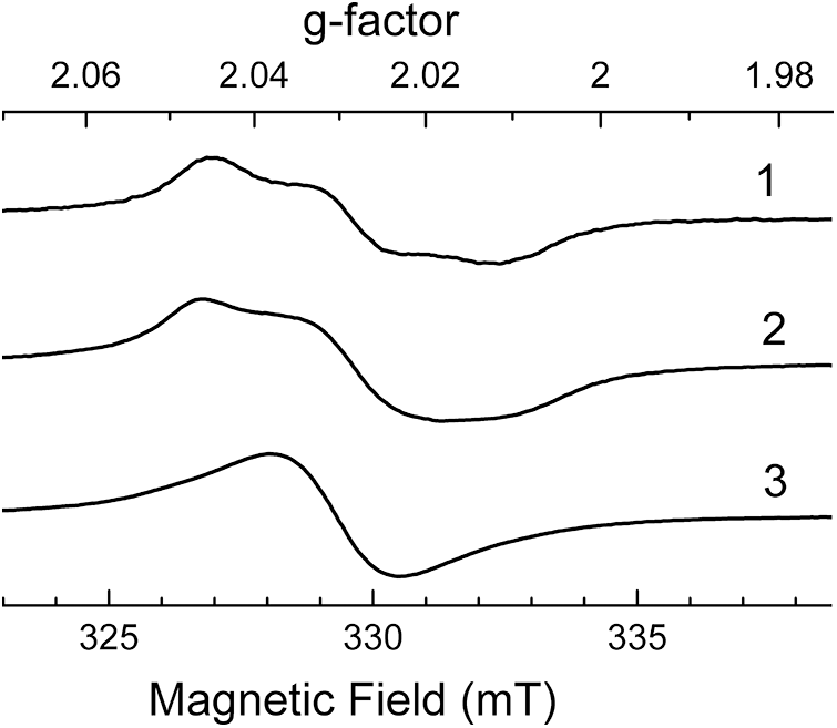
In the ESR spectrum of the blue ultramarine pigment, a synthetic analogue of lazurite, three bands (with g x = 2.0016, g y = 2.0505 and g z = 2.0355) are observed at low temperatures; above 300 K these bands collapse into a single signal at g = 2.028 (Goslar et al., Reference Goslar, Lijewski, Hoffmann, Jankowska and Kowalak2009). In the ESR spectra of S3•–-bearing haüyne samples components of the g-tensor are partly resolved, even at room temperature (curves 1 and 2 in Fig. 8), but in the ESR spectrum of lazurite, the S3•–-dominant sodalite-group mineral, a broad single signal at g = 2.028 is observed (curve 3 in Fig. 8).
IR absorption spectra of: (1) bright blue haüyne with the empirical formula (Na6.45Ca1.36K0.01)Σ7.96(Al5.94Si6.06O24)(SO42–)1.56(S4)0.09(S3•–)0.03Cl0.09(CO2)0.02·nH2O (Chukanov et al., Reference Chukanov, Sapozhnikov, Shendrik, Vigasina and Steudel2020b) and (2) dark blue holotype lazurite sample with the empirical formula (Na6.97Ca0.88K0.10)Σ7.96[(Al5.96Si6.04)Σ12O24](SO42–)1.09(S3•–)0.55S2–0.05Cl0.04·0.72H2O (Sapozhnikov et al., Reference Sapozhnikov, Tauson, Lipko, Shendrik, Levitskii, Suvorova, Chukanov and Vigasina2021).

Figure 7 Long description
Two stacked line graphs labeled 1 and 2. The x-axis is labeled Wavenumber (cm superscript minus 1), ranging from 0 to 2000 with labeled ticks at 500, 1000, 1500 and 2000. The y-axis is labeled Absorbance, ranging from 0.0 to 1.6 with labeled ticks at 0.0, 0.4, 0.8, 1.2 and 1.6.
ESR spectra of haüyne samples with low contents of admixed S3•– groups (1 and 2) and lazurite containing 0.55 S3•– groups per formula unit (3). In the latter case, the ESR triplet of S3•– is unresolved because of some minor signal splitting.

Figure 8 Long description
The plot displays ESR spectra of haüyne samples with low contents of admixed S groups (curves 1 and 2) and lazurite containing 0.55 S groups per formula unit (curve 3). The top axis is labeled g-factor, ranging from 2.06 to 1.98, dimensionless. The bottom axis is labeled Magnetic Field in millitesla, ranging from 325 to 335 millitesla.
The ESR spectrum of the orthorhombic S3•–- and S4-bearing sodalite-group mineral vladimirivanovite shows a strong signal with g 1 = 2.049, g 2 = 2.038 and g 3 = 2.011, associated with S3•–, as well as a band with g = 2 caused by the admixed S4•– radical anion (Chukanov et al., Reference Chukanov, Shchipalkina, Shendrik, Vigasina, Tauson, Lipko, Varlamov, Shcherbakov, Sapozhnikov, Kasatkin, Zubkova and Pekov2022e).
The ESR signals with g x = 2.002, g y = 2.060 and g z = 2.038 for green afghanite from Sar-e Sang, Afghanistan and g x = 2.002, g y = 2.057 and g z = 2.035 for blue afghanite from the Ladgvardara gem lazurite deposit, SW Pamirs, Tajikistan refer to the S3•– radical anion. According to these data, in the Ladgvardara afghanite, this species has a configuration close to a free particle (g x = 2.002, g y = 2.061, g z = 2.039: Hoffmann et al., Reference Hoffmann, Goslar, Lijewski, Olejniczak, Jankowska, Zeidler, Koperska and Kowalak2012), while the S3•– radical anion in the Sar-e-Sang afghanite is similar in its spatial structure to that in synthetic cancrinites. The intensity of the ESR signal of the S3•– centres in afghanites obtained at room temperature is three orders of magnitude lower than the analogous signal of lazurite (Sapozhnikov et al., Reference Sapozhnikov, Tauson, Lipko, Shendrik, Levitskii, Suvorova, Chukanov and Vigasina2021).
After irradiation of a sulfide-bearing variety of the cancrinite-group mineral balliranoite with X-rays, its blue colour becomes more intense as a result of the transformation of S2•–, cis- and trans-S4, S52–, and HS– groups into S3•–, and an ESR signal for S3•– with components at g 1 = 2.053, g 2 = 2.040 and g 3 = 2.001 appears (Chukanov et al., Reference Chukanov, Sapozhnikov, Shendrik, Zubkova, Vigasina, Potekhina, Ksenofontov and Pekov2023b).
The XPS signal of S3•– in lazurite is a doublet with the components located at 163.4 and 164.6 eV (Sapozhnikov et al., Reference Sapozhnikov, Tauson, Lipko, Shendrik, Levitskii, Suvorova, Chukanov and Vigasina2021).
The linear (i.e. non-cyclic) radical anion S4•– can exist in various conformational states, for which the lengths of the terminal and middle bonds and the S–S–S bond angle are approximately equal to 2.0 Å, 2.1 Å and 108° (for the trans conformer); 2.0 Å, 2.2 Å and 110° (for the cis conformer); and 2.1 Å, 2.1 Å and 112–116° (for the gauche conformer), respectively (Steudel and Chivers, Reference Steudel and Chivers2019). Radiation absorption of S4•– in a mixed organic solvent in the visible range has a maximum at 490 nm (Bogdándi et al. Reference Bogdándi, Ida, Sutton, Bianco, Ditrói, Koster, Henthorn, Minnion, Toscano, van der Vliet, Pluth, Feelisch, Fukuto, Akaike and Nagy2019). A similar weak band with a maximum at 505 nm was observed on the background of a strong and broad band for S3•– in the absorption spectrum of vladimirivanovite containing a S4•– impurity (both radical anions were identified by means of ESR and Raman spectroscopy) (Chukanov et al., Reference Chukanov, Shchipalkina, Shendrik, Vigasina, Tauson, Lipko, Varlamov, Shcherbakov, Sapozhnikov, Kasatkin, Zubkova and Pekov2022e).
According to Rejmak (Reference Rejmak2020), wavenumbers of Raman active bands of the isolated S4•– radical anion calculated using different quantum-chemical methods are in the ranges 277–286, 284–296, 583–587 and 609–610 cm–1 (for cis-S4•–), and 180–183, 414–416 and 574 cm–1 (for trans-S4•–). Calculated wavenumbers of harmonic phonons with notable S4 contributions in S4•–-bearing sodalites are in the ranges 328–332, 399–406, 592, 624, 642 and 651 cm–1 for cis-S4•–, and 439–444, 532–583 and 628–691 cm–1 for trans-S4•– (Rejmak, Reference Rejmak2020). However, in the Raman spectra of natural S4•–-bearing feldspathoids, a band at the wavenumber of 298 cm–1, intermediate between the calculated wavenumbers of the bands of bending vibrations of isolated cis-S4•– and cis-S4•– in the sodalite cage, is observed (Shendrik et al., Reference Shendrik, Chukanov, Panikorovskii, Vigasina and Pekov2025). Different calculation models for cis-S4•– in the sodalite cage predict wavelengths of strong absorption bands in the range from 650 to 790 nm (Rejmak, Reference Rejmak2020).
Calculated values of the isotropic g-factor in the ESR spectra of isolated cis-S4•– and cis-S4•– in the sodalite cage are equal to 2.037 and 2.032–2.033, respectively; corresponding values for trans-S4•– are equal to 2.038 and 2.030 (Rejmak, Reference Rejmak2020). The g min value for cis-S4•– in a crystal of lilac haüyne from the Malobystrinskoye lazurite deposit is equal to 1.995 (see Fig. 3).
The presence of Sn •– radical anions with n > 4 in feldspathoids is questionable. However, in the ESR spectrum of nosean from the In den Dellen (Zieglowski) pumice quarry, Eifel region, Germany, a weak signal with g = 2.018, related to a sulfide radical anion, is observed (Chukanov et al., Reference Chukanov, Vigasina, Shendrik, Varlamov, Pekov and Zubkova2022d). A similar signal was assigned to the S6•– radical anion (Steudel, Reference Steudel and Steudel2003). A broad peak at 37,600 cm–1 in the NIR-Vis-UV spectrum of this sample is close to the band at 37,700 cm–1 assigned to S6•– (Steudel et al., Reference Steudel, Jensen, Göbel and Hugo1988).
Ostroumov et al. (Reference Ostroumov, Fritsch, Faulques and Chauvet2002) considered the SO4•– radical anion as a possible colour centre in blue lazurite. In particular, Raman bands at 445–450, 617–620, 640–650, 988–990 and 1090–1095 cm–1 were assigned to this species. However, this assignment is unlikely because the listed wavenumbers coincide with the wavenumbers of fundamental modes of the distorted SO42– anion which is a species-defining component in lazurite. However, ESR signals with g-factors of 2.030, 2.018, 2.011, and 2.007 observed for afghanite samples irradiated by UV radiation may be due to the SO3•– or SO4•– radical anions (Schneider et al., Reference Schneider, Dischler and Räuber1966; Ostroumov et al., Reference Ostroumov, Fritsch, Faulques and Chauvet2002).
Neutral molecules
The isolated S4 neutral molecule can exist as six isomers, linear cis-conformer with the C 2v symmetry being the most stable form. The transformation cis-S4 (C 2v) → trans-S4 (C 2h) requires energy of 41.3 kJ/mol (Eckert and Steudel, Reference Steudel and Steudel2003). Intermediate (gauche) conformation of linear S4 can be realized only in a matrix (e.g. in a sodalite cage). Other (cyclic and branched) isomers of S4 are less stable.
The absorption band of lilac S4-bearing haüyne with a maximum at 525 nm (Chukanov et al., Reference Chukanov, Sapozhnikov, Shendrik, Vigasina and Steudel2020b) was assigned to cis-S4, in accordance with results of quantum-chemical calculations (Wong and Steudel, Reference Steudel and Steudel2003; Rejmak, Reference Rejmak2020). However, this band may correspond to the S4•– radical anion. According to P. Rejmak (Rejmak, Reference Rejmak2020), the absorption maximum of trans-S4 is at 585 nm, which is close to the position of the band at 590 nm in the absorption spectrum of a lilac S4-bearing commensurately modulated cubic haüyne analogue with the modulation parameter of 0.2 and unit-cell parameter of 45.3629(3) Å (Chukanov et al., Reference Chukanov, Zubkova, Shendrik, Sapozhnikov, Pekov, Vigasina, Chervonnaya, Varlamov, Bolotina, Ksenofontov and Pushcharovsky2025). A broad peak in the NIR-Vis-UV absorption spectrum of sulfide-bearing balliranoite has an asymmetric shape and can be decomposed into two Gaussians with maxima at ∼540 and ∼600 nm assigned to neutral S4 molecules having cis- and trans-conformations (Chukanov et al., Reference Chukanov, Sapozhnikov, Shendrik, Zubkova, Vigasina, Potekhina, Ksenofontov and Pekov2023b). Previously, similar bands were observed for lilac S4-containing haüyne (Chukanov et al., Reference Chukanov, Sapozhnikov, Kaneva, Varlamov and Vigasina2023c, see Fig. 8).
The UV-Vis spectrum calculated at the TDDFT level for a cluster model representing a gauche-S4 molecule embedded in a sodalite lattice contains a strong band at ∼700 nm and a weaker band at 505 nm (Rejmak, Reference Rejmak2020). These bands are present in the absorption spectra of some S4-bearing haüyne samples (see e.g. curve 2 in Fig. 8).
Wavenumbers, ν, of vibrational modes of the isolated S4 molecule in the C 2v state (cis-S4, a red chromophore), calculated at the B3LYP/6-31G(2df) level (Eckert and Steudel, Reference Steudel and Steudel2003; Steudel et al., Reference Steudel, Steudel and Wong2003), are (cm–1, relative Raman intensities, I, are given in parentheses): 104(33), 207(35), 330(35), 373(100), 649(13), and 674(76). The band at 649 cm–1 is the strongest one in the IR spectrum, but it could not be identified reliably in the spectra of S4-bearing feldspathoids because of its overlapping with stronger bands of the aluminosilicate framework. Bands at 327–328, 362–363, 394–396, and 667–668 cm−1 in the Raman spectrum of the red–orange sulfhydrylbystrite and orange–yellow bystrite variety are close to the wavenumbers of 330, 373, and 674 cm−1 calculated for cis-S4. These bands are not observed in the Raman spectrum of yellow bystrite.
For the isolated S4 molecule in the C 2h state (trans-conformer), calculated ν(I) values are 93(0), 124(0), 225(55), 471(51), 637(0) and 649(100) – the band at 637 cm–1 being most strong in the IR spectrum. According to Rejmak (Reference Rejmak2020), wavenumbers of Raman active bands of S4 calculated using different quantum-chemical methods are in the ranges 319–334, 326–339, 655–659 and 684–688 cm–1 (for cis-S4), and 209–227, 438–452 and 647 cm–1 (for trans-S4).
Calculated wavenumbers of harmonic phonons with notable S4 contributions in S4-bearing sodalites are in the ranges 335–357, 631–641 and 649–662 cm–1 for cis-S4, and 471, 553–577 and 589–606 cm–1 for gauche-S4 (Rejmak, Reference Rejmak2020). In the Raman spectrum of cis-S4-bearing slyudyankaite, bands of S4 are observed as a peak at 646 and a shoulder at 350 cm–1 (Sapozhnikov et al., Reference Sapozhnikov, Bolotina, Chukanov, Shendrik, Kaneva, Vigasina, Ivanova, Tauson and Lipko2023).
Raman bands of S4 in pink to lilac haüyne from the Malo-Bystrinskoye lazurite deposit, Baikal Lake region are observed at 327, 443 and 684 cm–1, which may indicate the presence of both cis- and trans-conformers of S4 (Sapozhnikov et al., Reference Sapozhnikov, Tauson, Lipko, Danilov and Chukanov2025). The intensities of these bands correlate with the intensity of the colour of the mineral. In the Raman spectrum of natural lilac S4-bearing commensurately modulated cubic haüyne analogue, peaks at 444 and 546 cm–1 and a shoulder at 590 cm–1 are observed, which may correspond to the S4 molecule having trans- or gauche-conformation (Chukanov et al., Reference Chukanov, Zubkova, Shendrik, Sapozhnikov, Pekov, Vigasina, Chervonnaya, Varlamov, Bolotina, Ksenofontov and Pushcharovsky2025). The peak at 546 cm–1 may be a superposition of a symmetric stretching ν1 band of the S3•– radical anion, occurring in the mineral in trace amounts, and a stretching band of S4.
The cyclic hexasulfur (S6) molecule, known as a component of liquid sulfur and studied in the solid state and in the solutions, is a yellow or orange–yellow chromophore (Steudel et al., Reference Steudel, Holdt and Young1986; Eckert and Steudel, Reference Steudel and Steudel2003). The UV absorption spectrum of S6 in a liquid phase has a maximum below 220 nm and shoulders at ∼230, 254 and ∼300 nm with the extinction coefficient at 254 nm equal to 2400 (Steudel et al., Reference Steudel, Jensen, Göbel and Hugo1988). A similar spectrum of the holotype sample of slyudyankaite (a S6-bearing monoclinic sodalite-group mineral) is observed in the range of 210–440 nm (Sapozhnikov et al., Reference Sapozhnikov, Bolotina, Chukanov, Shendrik, Kaneva, Vigasina, Ivanova, Tauson and Lipko2023).
Fundamental vibrational modes of S6 predicted by ab initio calculations have the wavenumbers of 478, 460, 440, 354, 324, 263, 203 and 163 cm–1. Infrared and Raman spectra of S6 were measured in the solid state and in CS2 solution (Berkowitz et al., Reference Berkowitz, Chupka, Bromels and Belford1967; Nimon et al., Reference Nimon, Neff, Cantley and Buttlar1967; Steudel et al., Reference Steudel, Steidel and Reinhardt1983; Eckert and Steudel, Reference Steudel and Steudel2003). The strongest IR bands of S6 are observed at 180, 312–313 and 462–463 cm–1 (the torsion, bending and stretching modes, respectively). The strongest Raman band of the S6 molecule has a maximum at 471 or 476 cm–1 (in the solid state and in solution, respectively); other Raman bands of S6 are observed at 79, 106, 202–204, 262–266 and 448–451 cm–1.
According to the single-crystal X-ray structural data, the cyclic chair-like conformer of S6 occurs as a species-defining component in the triclinic sodalite-group mineral, slyudyankaite (Sapozhnikov et al., Reference Sapozhnikov, Bolotina, Chukanov, Shendrik, Kaneva, Vigasina, Ivanova, Tauson and Lipko2023). In the Raman spectrum of slyudyankaite, bands of S6 are observed at 255 and 477 cm–1. In addition, a broad peak at 437 cm–1 was assigned to overlapping bands of S6 (mixed mode) and SO42– [bending E (ν2) mode] (Sapozhnikov et al., Reference Sapozhnikov, Bolotina, Chukanov, Shendrik, Kaneva, Vigasina, Ivanova, Tauson and Lipko2023). In the XPS spectrum of slyudyankaite, an unresolved doublet related to the S6 molecule appears in the range of 168–170 eV.
The narrow bands at 2575–2577 cm–1 observed in the Raman spectra of some sodalite-group minerals, including slyudyankaite (Sapozhnikov et al., Reference Sapozhnikov, Bolotina, Chukanov, Shendrik, Kaneva, Vigasina, Ivanova, Tauson and Lipko2023) and ‘haüyne-45Å’ (Chukanov et al., Reference Chukanov, Sapozhnikov, Shendrik, Zubkova, Vigasina, Potekhina, Ksenofontov and Pekov2023b), may correspond to stretching vibrations of the H2S molecule (Graneri et al., Reference Graneri, Spagnoli, Wild and McKinley2024).
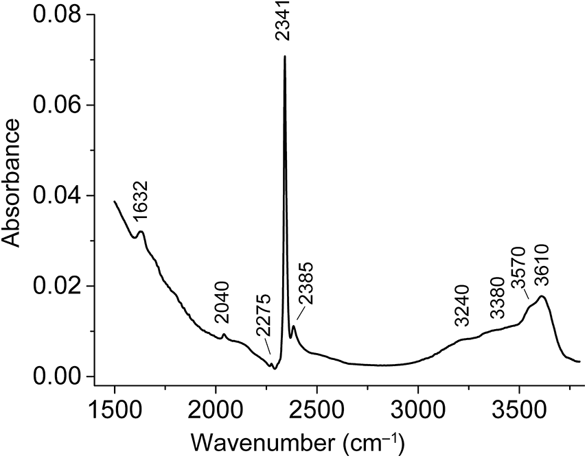
The COS (carbonyl sulfide) molecule has strong IR absorption bands because of its polar bonds, which have large dipole moments. As a result, IR spectroscopy can be used to detect even trace amounts of this species in minerals. The wavenumber of the strongest IR absorption band of COS (C=O stretching mode) is ∼2040 cm–1 (Tubergen et al., Reference Tubergen, Lavrich and McCargar2000). This band is often observed in IR spectra of sodalite-group minerals containing polysulfide groups and/or the HS– anion, including slyudyankaite (Fig. 9), lazurite with a high content of carbon dioxide (0.44 CO2 molecules per formula unit) from the Malo-Bystrinskoe deposit (Chukanov et al., Reference Chukanov, Vigasina, Zubkova, Pekov, Schäfer, Kasatkin, Yapaskurt and Pushcharovsky2020a), an intermediate member of the haüyne–lazurite solid-solution series with the empirical formula Na6.90K0.05Ca0.99(Al5.92Si6.08O24)(CO2)0.15(SO4)1.16S3•–0.22S2–0.12(COS)xCl0.21 (x ≪ 1) from the Badakhshan lapis lazuli province, Afghanistan (Chukanov et al., Reference Chukanov, Sapozhnikov, Shendrik, Vigasina and Steudel2020b), and sapozhnikovite from the Lovozero massif, Kola Peninsula (Chukanov et al., Reference Chukanov, Zubkova, Pekov, Shendrik, Varlamov, Vigasina, Belakovskiy, Britvin, Yapaskurt and Pushcharovsky2022b).
IR spectrum of slyudyankaite in the high-frequency region. The bands at 3240–3610, 2385, 2341, 2275 and 2040 cm–1 correspond to stretching vibrations of H2O, 12CO2 coordinated by H2O, 12CO2 coordinated by cations only, 13CO2 and COS, respectively. The band at 1632 cm–1 is related to bending vibrations of H2O molecules.

Figure 9 Long description
Absorbance A single line graph is shown. The x-axis is labeled Wavenumber (cm superscript minus 1), with tick labels at 1500, 2000, 2500, 3000 and 3500. The y-axis is labeled Absorbance, ranging from 0.00 to 0.08, with tick labels at 0.00, 0.02, 0.04, 0.06 and 0.08. A single curve starts near wavenumber 1500 at an absorbance a little above 0.03, then decreases toward about 0.01 by around wavenumber 2000.
The role of polysulfide groups in structure modulations and twinning of sodalite-group minerals
The tendency of extra-framework species to order in sodalite-type structures often leads to commensurate structure modulations, accompanied by symmetry lowering and n-fold multiplication of the period c* of the reciprocal basic cubic lattice characterized by the wave vector q = n –1c*, or incommensurate modulations with the wave vector q = p c* where k is an irrational modulation parameter (Bolotina et al., Reference Bolotina, Sapozhnikov, Chukanov and Vigasina2023). As a rule, the simultaneous presence of high amounts of sulfate anions and larger polysulfide groups (trisulfide radical anions, tetrasulfide molecules or cyclic hexasulfide molecules) in a sodalite-group mineral results in modulation of the crystal structure due to differences in the sizes of sodalite cages hosting these groups (Hassan and Buseck, Reference Hassan and Busek1989; Sapozhnikov, Reference Sapozhnikov1992; Bolotina, Reference Bolotina2006; Evsyunin et al., Reference Evsyunin, Sapozhnikov, Kashaev and Rastsvetaeva1997, Reference Evsyunin, Rastsvetaeva, Sapozhnikov and Kashaev1998; Kaneva et al., Reference Kaneva, Cherepanov, Suvorova, Sapozhnikov and Levitsky2011; Sapozhnikov et al., Reference Sapozhnikov, Kaneva, Cherepanov, Suvorova, Levitsky, Ivanova and Reznitsky2012; Kuribayashi et al., Reference Kuribayashi, Aoki and Nagase2018; Chukanov et al., Reference Chukanov, Sapozhnikov, Shendrik, Vigasina and Steudel2020b, Reference Chukanov, Shchipalkina, Shendrik, Vigasina, Tauson, Lipko, Varlamov, Shcherbakov, Sapozhnikov, Kasatkin, Zubkova and Pekov2022e; Sapozhnikov et al., Reference Sapozhnikov, Tauson, Lipko, Shendrik, Levitskii, Suvorova, Chukanov and Vigasina2021, Reference Sapozhnikov, Bolotina, Chukanov, Shendrik, Kaneva, Vigasina, Ivanova, Tauson and Lipko2023; Bolotina et al., Reference Bolotina, Sapozhnikov, Chukanov and Vigasina2023, Reference Blumentritt, Notari and Caplan2024). The modulation parameter of lazurite and related members of the lazurite–haüyne series varies from 0.169 to 0.218.
Commensurability or incommensurability of the superstructure is estimated by the value of the satellite displacement from the main reflection along the axis of the reciprocal lattice. If the displacement fits between the main reflections an integer number of times, the superstructure is commensurate; otherwise, it is incommensurate. Simple (hkl) symbols in the single-crystal and powder X-ray diffraction patterns correspond to the indices of basic reflections; symbols like (h±n –1 k l), (h±n –1 k±n –1 l) or (h±n –1 k±n –1 l±n –1) with integer n, refer to satellite reflections of commensurate superstructure whereas symbols like (h±p k l), (h±p k±p l) or (h±p k±p l±p) with irrational p correspond to satellite reflections of an incommensurate superstructure.
In some cases, both commensurate and incommensurate modulations take place. For example, the neotype specimen of lazurite with the empirical formula (Na6.97Ca0.88К0.10)(Si6.04Al5.96O24)(SO4)1.09(S3•–)0.55S2–0.05Cl0.04·0.72Н2О is characterized by commensurate and incommensurate modulations with n = 2 and p = 0.147 (Sapozhnikov et al., Reference Sapozhnikov, Tauson, Lipko, Shendrik, Levitskii, Suvorova, Chukanov and Vigasina2021).
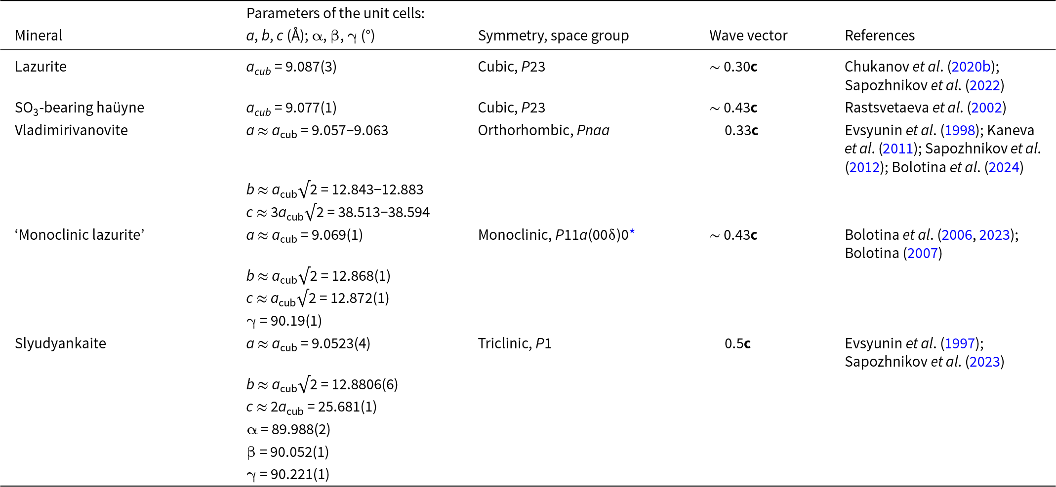
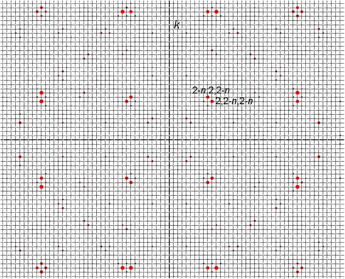
In a commensurately modulated pink cubic haüyne analogue with the modulation parameter of 0.2 and unit-cell parameter of 45.3629(3) Å and the simplified formula Na6Ca2–x(Si6Al6O24)(SO42–,HS–,S2•–,S4,S3•–,S52–)2–y from the Malo-Bystrinskoe lazurite deposit (‘haüyne-45Å’: Chukanov et al., Reference Chukanov, Zubkova, Shendrik, Sapozhnikov, Pekov, Vigasina, Chervonnaya, Varlamov, Bolotina, Ksenofontov and Pushcharovsky2025) modulations are caused by regular alternation of small sodalite cages with the volume of 380–390 Å3 and large sodalite cages with the volume of ∼ 417 Å3, presumably hosting large S4 molecules. It is important that the anomalous large cages in the structure of ‘haüyne-45Å’ do not host cations and anions. A similar situation takes place in the case of slyudyankaite, a triclinic sodalite-group mineral with the unit-cell parameters a ≈ a cub, b ≈ a cub√2, c ≈ 2a cub, and α, β and γ values close to 90° (Table 1). The analysis of the reciprocal space revealed the presence of additional weak satellite reflections which additionally triple the ∼45Å parameter of the cubic unit cell (Fig. 10). These reflections were not taken into account during the crystal structure refinement. A specific case is cubic lilac haüyne characterized by incommensurate modulations with p = 0.222 caused by alternating SO42– and S4 groups (Chukanov and Aksenov, Reference Chukanov and Aksenov2024).
Data on sodalite-group minerals with modulated structures
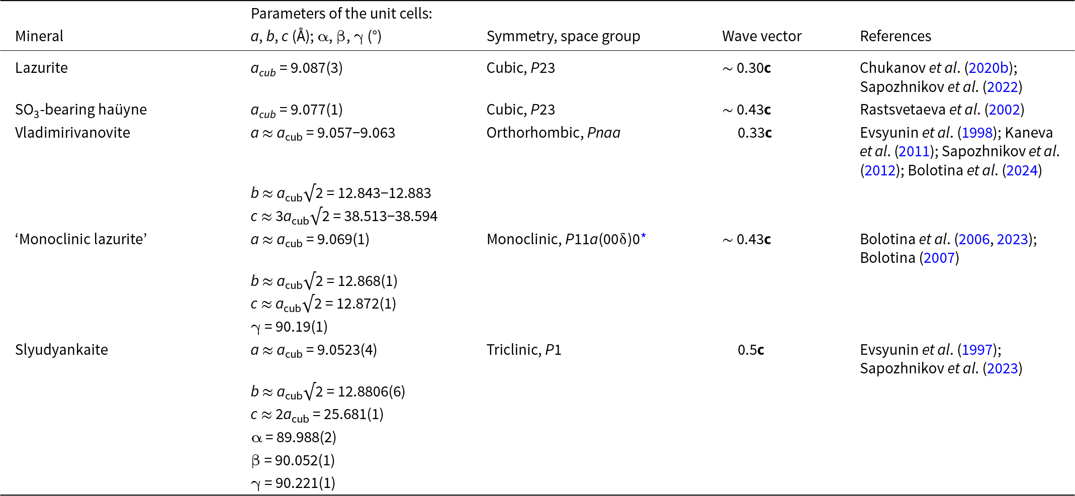
Table 1 Long description
The table lists unit-cell parameters, crystal symmetry and space group, modulation wave vector direction along the c axis, and literature sources for several sodalite-group minerals with modulated structures. Lazurite and sulfur trioxide bearing haüyne are cubic with cell edges about 9.09 and 9.08 angstroms, and wave vectors near 0.30 and about 0.43 along c. Vladimirivanovite is orthorhombic with a near 9.06 angstroms, b near 12.84 to 12.88 angstroms, and c near 38.51 to 38.59 angstroms, with a wave vector of 0.33 along c. Monoclinic lazurite has a near 9.07 angstroms, b and c near 12.87 angstroms, and a gamma angle slightly above 90 degrees, with a wave vector about 0.43 along c. Slyudyankaite is triclinic with a near 9.05 angstroms, b near 12.88 angstroms, c near 25.68 angstroms, angles close to 90 degrees, and the largest listed wave vector at 0.50 along c. Across minerals, the a dimension stays close to 9.05 to 9.09 angstroms while symmetry lowers from cubic to triclinic and the modulation wave vector varies within roughly 0.30 to 0.50 along c. Some values are approximate or given as ranges, so comparisons should be treated as indicative rather than exact.
Notes: * δ ≈ 0.43; symmetry operators: (1) x1, x2, x3, x4; (2) x1+½, x2, –x3, –x4.
Modelled fragment of the diffraction pattern of ‘haüyne-45Å’ in the plane h = 0 of the reciprocal lattice. The smallest dots correspond to additional weak satellites triple the ∼45 Å parameter of the cubic unit cell.

Figure 10 Long description
An abstract, diagram-like graphic made of a square grid of small cells. A vertical line and a horizontal line intersect at the center, forming a crosshair that divides the grid into four regions. Multiple small dot clusters are placed at various positions across the grid, including near the corners and along the outer areas. Near the upper-right area, a small group of dots has a short text label next to it; the label is too small to read accurately.
In the crystal structure of slyudyankaite (Sapozhnikov et al., Reference Sapozhnikov, Bolotina, Chukanov, Shendrik, Kaneva, Vigasina, Ivanova, Tauson and Lipko2023), commensurate modulations are related to alternation of small sodalite cages hosting SO42– anions and metal cations and larger sodalite cages which do not contain ions and are populated by neutral molecules (S6, S4, CO2 and H2O).
Data on some other sodalite-group minerals with modulated structures are given in Table 1. It was supposed that commensurate and incommensurate structure modulations of lazurite and related sodalite-group minerals arose during recrystallization of their early generations under mild conditions (Sapozhnikov et al., Reference Sapozhnikov, Ivanov, Levitsky and Piskunova1993, Reference Sapozhnikov, Tauson, Lipko, Shendrik, Levitskii, Suvorova, Chukanov and Vigasina2021, Reference Sapozhnikov, Bolotina, Chukanov, Shendrik, Kaneva, Vigasina, Ivanova, Tauson and Lipko2023; Chukanov et al., Reference Chukanov, Shendrik, Vigasina, Pekov, Sapozhnikov, Shcherbakov and Varlamov2022c). Relative contents of different S-bearing species in these minerals depend on the temperature of recrystallization and redox conditions as well as charge-balance requirement. After annealing at 600°C, modulation vectors of 0.42c – 0.44c shorten to 0.30c – 0.33c due to disordering of S-bearing extra-framework species. Further heating at higher temperatures results in the disappearance of modulations as a result of the complete disordering of S-bearing groups (Bolotina et al., Reference Bolotina, Sapozhnikov, Chukanov and Vigasina2023).
The tendency of sodalite-group minerals and synthetic materials with the sodalite-type structure to form growth and penetration twins has long been known.
Crystal twinning can be a consequence of a phase transition from a high-temperature cubic phase with symmetry lowering, as is the case with aluminate sodalites (Depmeier, Reference Depmeier1984). The twin components are connected by reflection in the (100) plane of a sodalite-type cubic lattice. Three phase transitions were observed for the sodalite-type compound Ca8[A112O24](CrO4)2 with decreasing temperature due to the ordering of the [Ca4CrO4]6+ clusters: from the cubic to cubic modulated phase at 610 K, into the tetragonal phase at 453 K and into the twinned orthorhombic phase at 432 K (Hassan, Reference Hassan1996). The twinning plane is (110) in the cubic setting. These kinds of twinning have not been observed in minerals belonging to the sodalite group.
Among sodalite-group minerals, nosean, sulfide-free haüyne, sodalite and bolotinaite are most disposed to form twins, whereas no simple twins are known for lazurite, sulfide-bearing haüyne, vladimirivanovite and slyudyankaite because modulated crystal structures of these minerals contain sodalite cages of different sizes. However, Bolotina et al. (Reference Bolotina, Rastsvetaeva, Sapozhnikov, Kashaev, Shönleber and Chapuis2003a) described the structure of lazurite as a polysynthetic twin composed of three orthorhombic components connected by the threefold axis in the (3+2)D space. A similar approach was applied to the description of twinning of haüyne from Sacrofano (Italy) in the orthorhombic Pba2 group (Bolotina et al., Reference Bolotina, Rastsvetaeva, Sapozhnikov and Kashaev2003b).
Simple twins of sodalite-group minerals are usually considered as twins on the [111] axis. However, application of two twinning operations [rotation by 180° around the [111] axis or reflection in any of the planes,  $(\overline 2 11)$, $(1\overline 2 1)$, or $(11\overline 2 )$] leads to almost the same result with identical single-crystal diffraction patterns. Moreover, both kinds of twins would be morphologically undistinguishable, with an angle of 109.5° between the (111) axes of the twin components. Analysis of the boundary between twin components has shown that twinning by a plane is accompanied by fewer distortions of the interatomic distances compared to twinning by the [111] axis and, consequently, is energetically more favourable (Bolotina et al., Reference Bolotina, Chukanov and Schäfer2022).
Tounkite, ideally (Na,K)30Ca18[Al36Si36O144](SO4)12Cl6·6H2O (Z = 1), is a multilayer cancrinite-group mineral whose aluminosilicate framework is formed by the САСАСВСВСАСВ stacking sequence and hosts cancrinite, Losod and liottite cages. It was shown (Chukanov et al., Reference Chukanov, Bolotina, Shendrik, Sapozhnikov, Zubkova, Pekov, Vigasina, Sandalov and Ksenofontov2024a) that tounkite samples from lazurite deposits of the Baikal Lake region with the simplified general formula (Na+3.89–5.18K+0.15–1.64Ca2+2.30–2.58(Al6Si6O24)(SO42–,S52–,S4)2–x(Cl–,HS–)1+y·nH2O (x, y, n < 1) are polysynthetic twins with twin components connected by a 180° rotation around the [001] axis. A large 10-layer cage (unknown in the structures of other cancrinite-group minerals) formed at the border between twin components, connected by a rotation of 180° around the [001] axis, hosts the large S52– anion.
In accordance with structural data, the IR spectrum of tounkite contains bands corresponding to liottite cages (at 543–546 cm–1) and columns of cancrinite cages (at 586–593, 663–665 and 680–683 cm–1) (Chukanov et al., Reference Chukanov, Aksenov and Pekov2023a) whereas bands corresponding to sodalite and giuseppettite cages are not observed. Additional bands at 500 and 650 cm–1, which are absent in IR spectra of other cancrinite-group minerals, may be related to the 10-layed cage at the border between twin components.
S-bearing extra-framework groups in feldspathoids as markers of their crystallization or transformation conditions
Numerous experiments have demonstrated complex mechanisms of thermal and radiation-induced transformations of S-bearing species occurring in minerals belonging to the cancrinite and sodalite groups under reducing and oxidizing conditions. These data can be used for the estimation of conditions under which these minerals crystallized or conditions of their post-crystallization transformations.
The concentration of sulfide sulfur in blue S3•–-bearing haüyne increased on heating under reducing conditions (in the presence of the Fe + FeS buffer) at 550°C and 800° from 1.34 wt.% to 2.10 and 5.44 wt.%, respectively (Sapozhnikov et al., Reference Sapozhnikov, Tauson and Matveeva2001). The parameter of incommensurate modulations changed on heating from 0.217 to 0.147, which corresponds to the transformation to lazurite, a sodalite-group mineral with species-defining S3•–.
The thermal transformation of SO42– to polysulfide radical anions in sodalite cages is possible even under oxidizing conditions. For example, heating sulfide-free haüyne at 750°C in air leads its crystals to acquire a blue colour and the appearance of Raman bands corresponding to S3•– and S2•– (Caggiani et al., Reference Caggiani, Mangone and Acquafredda2022). During gradual heating haüyne with the empirical formula Na6.39K0.06Ca1.57(Si6.08Al5.92O24)(SO4)1.78(S4)0.03(S2–)0.02Cl0.07(CO2)0.15·nH2O in air, an ESR signal of S3•– appears above 200°C and its intensity reaches maximal value at 700°C (Chukanov et al., Reference Chukanov, Shendrik, Vigasina, Pekov, Sapozhnikov, Shcherbakov and Varlamov2022c).
The product of transformations of a pink S4- and CO2-bearing haüyne sample at 700°C under reducing conditions (in the presence of an Fe–FeS buffer) has brown colour and contains HS– and S2– anions as well as subordinate amounts of S2•– and S4•– radical anions, identified on the basis of Raman and NIR-Vis-UV spectroscopy data (Chukanov et al., Reference Chukanov, Shendrik, Vigasina, Pekov, Sapozhnikov, Shcherbakov and Varlamov2022c, Reference Chukanov, Shchipalkina, Shendrik, Vigasina, Tauson, Lipko, Varlamov, Shcherbakov, Sapozhnikov, Kasatkin, Zubkova and Pekov2022e; Sapozhnikov et al., Reference Sapozhnikov, Tauson, Lipko, Danilov and Chukanov2025). Transformations of extra-framework components in this reaction can be described by the following scheme (e = electron):
S4 + e – → S4•– + e – → 2S2•–,
S4•– + S2•– → 2S3•–,
3S4 + 3SO42– → 5S3•– + 6O2 + 3e –,
SO42– → S2– + 2O2(gas),
CO2 + 2SO42– + H2O → 2HS– + CO32– + 4O2(gas),
3SO42– → S3•– +5e – + 6O2(gas),
2S3•– → S2•– + S4•–,
2CO2 + 2e – → C2O42–.
Subsequent annealing of the preheated sample in air at 800°C results in a change of the colour, first to green and then to blue because of the restoration of the S3•– radical anion (a blue chromophore) as a result of the following conversions:
S2•– + S2– + 2O2(gas) → SO42–,
S4•– + S2•– → 2S3•–,
S3•– + 5e– + 6O2(gas) → 3SO42–,
C2O42– → 2CO2(gas) + 2e –.
Annealing of S4-containing haüyne at 400°C decreases the relative intensities of Raman bands of both the S4 molecules and the SO42– anions. Furthermore, mass spectroscopic analysis reveals that intense oxygen evolution occurs at temperatures below 500°C (Sapozhnikov et al., Reference Sapozhnikov, Tauson, Lipko, Danilov and Chukanov2025). These two facts suggest the existence of a second pathway for the transformation of neutral tetrasulfide groups: 3S4 + 3SO42– → 5S3•– + 6O2 + 3e –. This reaction may serve as an alternative source of electrons required for the S4 transformation pathway without the participation of SO42–.
Heating of slyudyankaite at 700°C under reducing conditions (Chukanov et al., Reference Chukanov, Shendrik, Vigasina, Pekov, Sapozhnikov, Shcherbakov and Varlamov2022c) results in the transformation of S-containing groups, including a species-defining S6 molecule, into HS–, S2•–, and S4•– and, possibly, into the monosulfide anion S2–. Further annealing of preheated slyudyankaite at 800°C in air results in the disappearance of HS–, S2•– and S4•– groups and the formation of the anionic groups SO42– and S3•–. These data are consistent with both the experimental results obtained in investigations (Pokrovski and Dubrovinsky, Reference Pokrovski and Dubrovinsky2011; Pokrovski and Dubessy, Reference Pokrovski and Dubessy2015) and with theoretical quantum-chemical calculations of the mechanisms of thermal transformations of the S6 molecule (Fedyaeva et al., Reference Fedyaeva, Lepeshkin, Chukanov and Oganov2024), according to which the S3•– groups are the most thermally stable polysulfide radical anions. In particular, using density-functional simulations, it has been shown (Fedyaeva et al., Reference Fedyaeva, Lepeshkin, Chukanov and Oganov2024) that in the presence of an electron donor the main decay pathways of a cyclic S6 molecule with the chair-like conformation involve initial capture of one electron, which leads to the opening of the S6 cycle accompanied by the capture of a second electron and formation of pairs of radical anions, S3•– + S3•– or S2•– + S4•–. Both processes proceed with the same energy barrier of ∼0.4 eV, but the former channel, leading to the formation of two S3•– groups, is thermodynamically more favourable (the enthalpy difference between the S3•–+ S3•– and S2•– + S4•– pairs is ∼0.5 eV).
The product of annealing of pale green commensurately modulated cubic haüyne analogue with the general formula Na6Ca2–x(Si6Al6O24)(SO42–,H2S,HS–,S2•–,S4,S3•–,S52–)2–y, modulation parameter of 0.2 and unit-cell parameter of 45.3629(3) Å (‘haüyne-45Å’) in a vacuum at 400°C has a deep green colour. When compared to the initial sample, the product of annealing is characterized by weaker luminescence with a vibrational structure of S2•–, the appearance of an intense band at 284 cm–1, which can be attributed to the S4•– radical anion or S52– anion, a weaker band of HS–, and more intense bands of SO42– and S3•– (Chukanov et al., Reference Chukanov, Zubkova, Shendrik, Sapozhnikov, Pekov, Vigasina, Chervonnaya, Varlamov, Bolotina, Ksenofontov and Pushcharovsky2025). Annealing of this sample in a vacuum at 600°C and 800°C resulted in the colour change from green to blue, restoration of strong luminescence related to S2•– and further enhancement of intensities of Raman bands corresponding to S3•–, whereas the band at 284 cm–1 disappeared and the intensity of the SO42– band (at 990 cm–1) decreased. On heating of ‘haüyne-45Å’ in air at 400°C, the luminescence with the vibrational structure of S2•– disappears. Heating at higher temperatures resulted in a change in colour, first to light blue and then dark blue, an increase in the intensities of Raman bands related to S3•– and a gradual weakening of the bands of other polysulfide groups and SO42–.
Based on these observations, the following mechanisms of thermal transformations of S-bearing groups in ‘haüyne-45Å’ in the absence of oxygen have been proposed (Chukanov et al., Reference Chukanov, Zubkova, Shendrik, Sapozhnikov, Pekov, Vigasina, Chervonnaya, Varlamov, Bolotina, Ksenofontov and Pushcharovsky2025). The appearance of S4•– in a sample heated at 400°C may be a result of the reaction 2S52– + H2S → S5•– + S4•– + HS–. At 600°C, the S52– anion decomposes into a pair of thermally stable radical anions, S2•– + S3•–. At 800°C, partial reduction of sulfate sulfur occurs in accordance with the scheme SO42– → S2– + 2O2(gas). In the presence of air, S52–, S4 and, to a lesser extent, S2•–, are already unstable at 400°C. However, minor amounts of S2•– and SO42– occur, along with S2•–, in the product of the annealing of ‘haüyne-45Å’ in air 800°C. Transformations of S-bearing groups in ‘haüyne-45Å’ under oxidizing conditions at 800°C can be described by the following scheme:
S52– → S2•– + S3•–;
3S4 + 4e – → 4S3•–;
2S2•– + 2O2 + e – → SO42– + S3•–.
The preceding reaction 3H2O → 2H3O+ + 0.5O2 + 2e – could serve as a source of electrons.
Fragments of a rock containing a mineral of the sodalite–sapozhnikovite solid-solution series, heated in a fire at a tourist camp on the western shore of Lake Seidozero, Lovozero alkaline massif, were studied by Pekov et al. (Reference Pekov, Chukanov, Shcherbakov, Vigasina, Shendrik, Sandalov, Vyatkin and Turchkova2025). Grains located in the near-surface portions of the fired fragments are blue or bright green. Toward the interior of the fragments, the colouration of the sodalite group minerals gradually fades to pale green and then yellow, with the largest grains having colourless cores. The colourless and pale-coloured (yellowish, greenish) zones exhibit orange luminescence in the long-wavelength (λ = 330 nm) ultraviolet range, characteristic of sodalite and sapozhnikovite, as well as a broad absorption band at 375 nm and a broad ESR signal with g = 2.044, both related to S2•– radical anions. According to ESR and Raman spectroscopy data, when moving from colourless zones through yellow and green to blue, the content of the HS– anions gradually decreases and the content of S3•– increases, while the content of S2•– passes through a maximum. These observations confirm the earlier conclusion that the S3•– radical anion is the most stable polysulfide group (Pokrovski and Dubrovinsky, Reference Pokrovski and Dubrovinsky2011, Pokrovski and Dubessy, Reference Pokrovski and Dubessy2015; Jacquemet et al., Reference Jacquemet, Guillaume, Zwick and Pokrovski2014). Based on these data, the thermal transformations of extra-framework components in minerals of the sodalite–sapozhnikovite series under oxidizing conditions can be described by the reactions 2HS– → S22– + 2H+ + 2e– and 2HS– + ½O2 → S22– +H2O (Steudel, Reference Steudel and Steudel2003), completed by the elementary processes ½O2 + 2HS– → S2•– + OH− and 3S2•– → 2S3•– + e – (Pekov et al., Reference Pekov, Chukanov, Shcherbakov, Vigasina, Shendrik, Sandalov, Vyatkin and Turchkova2025). However, firing in the presence of charcoal could have taken place under reducing conditions. Transformations of HS– under reducing conditions can be described by the following scheme:
2HS– → S2•– + H2 + e–;
HS– → H+ + S2−;
2H+ + 2e – → H2; 3S2•– → 2S3•– + e – (Pekov et al., Reference Pekov, Chukanov, Shcherbakov, Vigasina, Shendrik, Sandalov, Vyatkin and Turchkova2025).
According to Shchipalkina et al. (Reference Shchipalkina, Vereshchagin, Chukanov, Gorelova, Pekov and Bocharov2023), a part of HS– anions in sapozhnikovite remain stable up to 800°C, unlike the synthetic analogue of sapozhnikovite whose annealing at 800°C under reducing and oxidizing conditions for 6 h results in the complete transformation of HS– anions to SO42– and S3•–, accompanied by drastic increase of the cubic unit cell a parameter above this temperature (from a = 8.91(1) Å for the initial sample at room temperature and a = 9.03(1) Å at 800°C to a ≈ 9.2 Å at 1000°). The proportion of the SO42– and S3•– groups in the products of annealing at 800°C depends on the redox conditions. The presence of residual HS– anions in sapozhnikovite annealed at 800°C can be explained by larger sizes of its particles compared to its synthetic analogue and, as a result, deceleration of diffusion of volatile species.
For the fire-roasted feldspathoids, an increase in the proportion of the blue colour component is accompanied by an increase in the cubic unit cell parameter from 8.910(7) Å for colourless zones to ∼8.95 and ∼9.07 Å for green and blue zones, respectively. The Raman spectrum of the blue roasting product is close to that of lazurite, but all bands related to S3•– are shifted to higher frequencies compared to similar bands of lazurite. The strongest shift of the band of symmetric stretching vibrations of S3•– (up to 557 cm−1) relative to the similar band of lazurite (at 546 cm−1) occurs for green zones, which is associated with the presence of S3•– groups in a small sodalite cage (Pekov et al., Reference Pekov, Chukanov, Shcherbakov, Vigasina, Shendrik, Sandalov, Vyatkin and Turchkova2025). The infrared spectra of the yellow and green zones do not contain sulfate bands, but the IR spectra of the blue zones exhibit a shoulder at 1135 cm−1, indicating partial oxidation of sulfide sulfur in the late stages of thermal transformations. Oxidation of sulfide sulfur may be a consequence of charge balance requirements and is thus not necessarily related to high oxygen activity.
The scheme of high-temperature transformations of the HS– anion in synthetic sapozhnikovite analogue under reducing conditions is 2HS– (solid) + 2.5O2 (gas) → SO42– (solid) + 0.25S4 (solid) + H2O (gas) whereas under oxidizing and moderately reducing conditions the S4 molecule is unstable and sulfide sulfur oxidizes in according with the following scheme:
2HS– (solid) + 3.5O2 (gas) → SO42– (solid) + SO2 (gas) + H2O (gas),
6HS– (solid) → S3•– (solid) + 3H2S (gas) (Chukanov et al. Reference Chukanov, Shchipalkina, Shendrik, Vigasina, Tauson, Lipko, Varlamov, Shcherbakov, Sapozhnikov, Kasatkin, Zubkova and Pekov2022e).
When the S3•–-, S4•–- and S4-bearing sodalite-group mineral vladimirivanovite is heated in air above 600°C, the intensity of the absorption band of S3•– (in the range of 480–850 nm) increases, and the step in the absorption spectrum, corresponding to the radical anion S4•– and its ESR signal with g = 2.021 disappear (Chukanov et al., Reference Chukanov, Shendrik, Vigasina, Pekov, Sapozhnikov, Shcherbakov and Varlamov2022c,e).
The oxidation of sulfide sulfur in S4- and CO2-bearing haüyne preheated over the Fe–FeS buffer was studied in air at 600 and 700°C by varying the heating time (Chukanov et al., Reference Chukanov, Shchipalkina, Shendrik, Vigasina, Tauson, Lipko, Varlamov, Shcherbakov, Sapozhnikov, Kasatkin, Zubkova and Pekov2022e). The composition of the formed phases was determined based on the value of the cubic unit cell parameter. The initial sulfide sample had a rather large a parameter of 8.944 Å due to the presence of S3•– and minor amounts of S4•–. Its transformation into a haüyne-type phase with a = 9.03–9.08 Å started after two days at 600°C and almost immediately at 700°C.
Thermal transformations of S-bearing extra-framework anions in sodalite- and cancrinite-type compounds synthesized under hydrothermal conditions at temperatures of 180°C to 230°C in the presence of Na2S·10H2O, Na2SO3·5H2O or Na2S2O3 were studied under different redox conditions by heating initial powders at 700–800°C for 6 hours (Chukanov et al., Reference Chukanov, Shchipalkina, Shendrik, Vigasina, Tauson, Lipko, Varlamov, Shcherbakov, Sapozhnikov, Kasatkin, Zubkova and Pekov2022e). Synthesis in the presence of Na2S2O3 resulted in the formation of the thiosulfate cancrinite analogue whereas S-bearing products of reactions with Na2S·10H2O and Na2SO3·5H2O belong to the sodalite type. In the reaction with Na2S·10H2O, sapozhnikovite, the HS– analogue of sodalite was formed. Heating of this sample under oxidizing conditions (in air) resulted in the disappearance of the Raman band of the HS– anion (at 2666 cm–1) and appearance of the Raman bands at 433, 616, 980 and 1130 cm–1, corresponding to sulfate group, as well as the band at 2159 cm–1 presumably corresponding to O–H stretching vibrations of HSO4–. Annealing of the synthetic sapozhnikovite analogue under reducing conditions resulted in the appearance of Raman bands of the SO42– and S3•– groups.
The sodalite-type phase synthesized in the presence of Na2SO3·5H2O (Chukanov et al., Reference Chukanov, Shchipalkina, Shendrik, Vigasina, Tauson, Lipko, Varlamov, Shcherbakov, Sapozhnikov, Kasatkin, Zubkova and Pekov2022e) has a very small unit-cell parameter of 8.8867 Å which indicates that a major part of the sulfur belongs to a small species (presumably, S2– because the characteristic Raman band of HS– was not detected). Heating of this sample in air resulted in the appearance of Raman bands of sulfate groups and enhancement of the unit-cell parameter to 9.0928 Å as a result of partial transformation of initial S-bearing species into S3•– and SO42–.
Radiation-induced transformations of a mineral belonging to the sodalite–sapozhnikovite series in a pegmatite hosted by naujaite at Karnasurt Mountain, Lovozero massif, Kola Peninsula are described by Pekov et al. (Reference Pekov, Chukanov, Shcherbakov, Vigasina, Shendrik, Sandalov, Vyatkin and Turchkova2025). In contact with grains of Th-bearing steenstrupine-(Ce) (with 10–15 wt.% of ThO2), this mineral acquires a bright blue colour due to a partial radiation-induced transformation of HS– to S3•–. This transformation is accompanied by an increase in the cubic unit cell parameter from 8.92 to 8.93 Å.
Irradiation of sulfide-bearing balliranoite with the general formula Na5.4K0.1Ca2.4(Si6Al6O24)Cl2[(CO3)0.7(SO4)0.18S*0.95Cl0.1(H2O)0.16] (where S* is total sulfide sulfur in the S2•–, cis- and trans-S4, S52–, minor S3•– and HS– groups) from the Tultuy deposit by X-rays results in the transformations of polysulfide groups other than S3•– into S3•– in accordance with the scheme:
S52− → S2•– + S3•–;
3S2•– → 2S3•– + e −;
S4 + S2•– + e − → 2S3•–;
S4 + S2•– + e − → 2S3•–;
S4 + S52– + e − → 3S3•– (Chukanov et al., Reference Chukanov, Sapozhnikov, Shendrik, Zubkova, Vigasina, Potekhina, Ksenofontov and Pekov2023b).
After irradiation of kyanoxalite with ultraviolet radiation with an energy of more than 40,000 cm–1, a shoulder near 15,000 cm–1 appears in the absorption spectrum, the ESR signal with g 1 = 2.002, g 2 = 2.050 and g 3 = 2.038, related to S3•– radical anions, disappears, and the ESR signal with g 1 = 2.009, g 2 = 2.004, and g 3 = 1.996 associated with of CO3•– (Angelov et al., Reference Angelov, Stoyanova, Dafinova and Kabasanov1986) increases (Chukanov et al., Reference Chukanov, Vigasina, Shendrik, Varlamov, Pekov and Zubkova2022d).
As noted above, the presence of the S52– anion was detected in six feldspathoids: bystrite, sulfhydrylbystrite, the K-analogue of bystrite, tounkite, sulfide-bearing balliranoite and a commensurately modulated haüyne analogue. In all these minerals, S52– is accompanied by the HS– anion which is considered as a marker of reducing crystallization conditions (Chukanov et al., Reference Chukanov, Zubkova, Pekov, Shendrik, Varlamov, Vigasina, Belakovskiy, Britvin, Yapaskurt and Pushcharovsky2022b; Pekov et al., Reference Pekov, Chukanov, Shcherbakov, Vigasina, Shendrik, Sandalov, Vyatkin and Turchkova2025). However, unlike HS– occurring in sapozhnikovite, which is stable at high temperatures (at least, up to 800°C: Shchipalkina et al., Reference Shchipalkina, Vereshchagin, Chukanov, Gorelova, Pekov and Bocharov2023), the S52– anion in commensurately modulated haüyne decomposes at temperatures between 400° and 600°C even in the absence of oxygen (Chukanov et al., Reference Chukanov, Zubkova, Shendrik, Sapozhnikov, Pekov, Vigasina, Chervonnaya, Varlamov, Bolotina, Ksenofontov and Pushcharovsky2025).
The geochemical role of sulfur-bearing species in feldspathoids cannot be considered separately from the role of carbon-bearing groups among which the neutral CO2 molecule is most common in sodalite-group minerals and multilayer members of the cancrinite croup (Bellatreccia et al., Reference Bellatreccia, Della Ventura, Piccinini, Cavallo and Brilli2009; Balassone et al., Reference Balassone, Bellatreccia, Mormone, Biagioni, Pasero, Petti, Mondillo and Fameli2012; Cámara et al., Reference Cámara, Bellatreccia, Della Ventura, Gunter, Sebastiani and Cavallo2012; Chukanov et al., Reference Chukanov, Vigasina, Zubkova, Pekov, Schäfer, Kasatkin, Yapaskurt and Pushcharovsky2020a, Reference Chukanov, Sapozhnikov, Shendrik, Vigasina and Steudel2020b, Reference Chukanov, Aksenov and Rastsvetaeva2021a, Reference Chukanov, Shendrik, Vigasina, Pekov, Sapozhnikov, Shcherbakov and Varlamov2022c, Reference Chukanov, Vigasina, Shendrik, Varlamov, Pekov and Zubkova2022d, Reference Chukanov, Zubkova, Schäfer, Pekov, Shendrik, Vigasina, Belakovskiy, Britvin, Yapaskurt and Pushcharovsky2022f).
Based on the above data, the extra-framework species CO2 and HS– (or H2S) in sodalite-group minerals can be considered as indicators of oxidizing and reducing conditions, respectively. The only known exception is slyudyankaite which contains CO2 and H2S molecules along with COS (Sapozhnikov et al., Reference Sapozhnikov, Bolotina, Chukanov, Shendrik, Kaneva, Vigasina, Ivanova, Tauson and Lipko2023). In most cases, the simultaneous presence of these species in minerals of the sodalite group was not observed. All feldspathoids containing the S52– anion simultaneously contain HS– or H2S groups, which are considered markers of reducing conditions of mineral formation (Chukanov et al., Reference Chukanov, Zubkova, Pekov, Shendrik, Varlamov, Vigasina, Belakovskiy, Britvin, Yapaskurt and Pushcharovsky2022b, Reference Chukanov, Sapozhnikov, Kaneva, Varlamov and Vigasina2023c, Reference Chukanov, Bolotina, Shendrik, Sapozhnikov, Zubkova, Pekov, Vigasina, Sandalov and Ksenofontov2024a, Reference Chukanov, Zubkova, Shendrik, Sapozhnikov, Pekov, Vigasina, Chervonnaya, Varlamov, Bolotina, Ksenofontov and Pushcharovsky2025). The presence of these groups is easily detected by Raman spectroscopy, based on the presence of a narrow band in the range of 2550–2580 cm–1. At the same time, the IR spectra of these minerals lack the narrow band of neutral CO2 molecules characteristic of cancrinite and sodalite-group minerals, the position of which depends on the host cage sodalite (2340–2343, 2338, 2351–2353 and 2351 cm–1 in the sodalite, Losod and loittite cages and a wide channel in the cancrinite-type framework, respectively: Chukanov et al., Reference Chukanov, Aksenov and Pekov2023a).
Regarding extra-framework CO2 molecules, it should be noted that, although the presence of this species indicates high carbon dioxide activity in the mineral-forming environment, carbonate anions are generally absent in sodalite-group minerals. Furthermore, reliable data on the possibility of direct hydrothermal synthesis of carbonate-containing sodalite-type compounds are lacking, due to steric constraints on the incorporation of the CO32– group into the sodalite cage.
Numerous experimental data show that HS–-bearing sodalite-group minerals crystallized under reducing conditions and CO32–-bearing sodalite-group minerals can be formed only as a result of thermal transformation of an initial CO2-bearing mineral (Chukanov and Aksenov, Reference Chukanov and Aksenov2024). In particular, CO32–-bearing haüyne occurring in a thermally metamorphosed rock (Ballirano and Maras, Reference Ballirano and Maras2005) could be formed as a result of high-temperature transformation of CO2 molecules in sodalite cages. The sodalite-type compound Na8(Al6Si6O24)(CO3) was synthesized in a two-step anion exchange reaction at 700–800°C in a CO2 atmosphere using basic nitrite sodalite as a starting material (Buhl, Reference Buhl1993). Carbonatization of nitrite sodalite can occur at high temperatures by the mechanism Na8(Al6Si6O24)(NO2)2 + CO2 → Na8(Al6Si6O24)(CO3) + NO + NO2 (Šehović et al., Reference Šehović, Robben and Gesing2015). The C2O42– and CO32– anions formed in sodalite cages as a result of transformations of CO2 molecules in the presence of S-bearing groups remain stable up to 700° and 800°C, respectively (Chukanov et al., Reference Chukanov, Sapozhnikov, Shendrik, Vigasina and Steudel2020b, Reference Chukanov, Shendrik, Vigasina, Pekov, Sapozhnikov, Shcherbakov and Varlamov2022c, Reference Chukanov, Shchipalkina, Shendrik, Vigasina, Tauson, Lipko, Varlamov, Shcherbakov, Sapozhnikov, Kasatkin, Zubkova and Pekov2022e) whereas, for example, pure sodium oxalate starts to decompose at 290°C (Yoshimori et al., Reference Yoshimori, Asano, Toriumi and Shiota1978).
Under moderately oxidizing conditions CO2 can coexist with COS molecules in the structures of minerals belonging to the haüyne–lazurite solid-solution series (Chukanov et al., Reference Chukanov, Sapozhnikov, Shendrik, Vigasina and Steudel2020b). A corresponding dynamic equilibrium can be described with the scheme 2CO2(solid) + S3•– (solid) + O2(gas) ↔ 2COS + SO42–.
Conclusions
Based on the above-described results of the study of natural tectosilicates and laboratory experiments, the following conclusions can be drawn.
(1) The extra-framework CO2 and HS– (or H2S) species in sodalite-group minerals are indicators of oxidizing and reducing conditions, respectively.
(2) The COS molecule may occur in equilibrium with CO2 under intermediate redox conditions.
(3) The trisulfide radical anion, S3•–, is the most stable polysulfide species when exposed to high temperature, radiation and oxygen. In the sodalite structure, S3•– remains stable at least up to 800°C.
(4) The radical anions S2•– and S4•– can be metastable up to 800°C in the absence of oxygen, but readily transform into S3•– and SO42– under oxidizing conditions. The S3•–: SO42– proportion in the transformation products is regulated by charge-balance requirements.
(5) The neutral S4 and S6 neutral molecules in feldspathoids are unstable at temperatures above 400°C.
(6) Under reducing conditions, the S52– anion decomposes at 600°C into a pair of thermally stable radical anions, S2•– + S3•–. In the presence of air, S52– is unstable already by 400°C.
(7) Direct incorporation of the CO32– group into the sodalite cage during crystallization of sodalite-group minerals is energetically unfavourable. Most probably, CO32– anions in these minerals formed as a result of thermal transformations of extra-framework CO2 molecules.
(8) Among feldspathoids, the sulfite group SO32–, partly substituting SO42–, is known only in some varieties of multilayer cancrinite-group minerals, which can be an indication of more reducing conditions of crystallization conditions of these varieties compared to their SO32–-free counterparts. In all cases, the sulfite group occurs in the liottite cage.
(9) Irradiation of feldspathoids containing sulfide sulfur by X-rays or gamma-radiation results in the transformations of polysulfide groups other than S3•– and the HS– anion into S3•–.
(10) Raman and NIR-Vis-UV absorption bands of relatively large species (CO32– and S3•–) formed as a result of transformation of smaller groups (CO2 and HS–, respectively) and occurring in small sodalite cages inherited from the initial phases are shifted towards a high-energy (i.e. high-frequency) region compared to these species, included in sodalite-group minerals during their crystallization and occurring in larger cages.
(11) As a rule, the simultaneous presence of sulfate anions and larger polysulfide groups in a sodalite-group mineral is a cause of commensurate and/or incommensurate modulations of its crystal structure. Crystallization of sodalite-group minerals containing sulfide sulfur under low-temperature conditions may result in the regular alternation of SO42– anions and polysulfide groups which results in the formation of commensurately modulated structures. The polysulfide groups causing such modulations are S4 + S3•– in vladimirivanovite (orthorhombic, with a ≈ 9.1, b ≈ 12.9 and c ≈ 38.6 Å), S6 in slyudyankaite (triclinic, with a ≈ 9.05, b ≈ 12.88, c ≈ 25.68 Å, α ≈ 89.99°, β ≈ 90.05° and γ ≈ 90.22°), S3•– in “monoclinic lazurite” (with a ≈ 9.07, b ≈ 12.87, c ≈ 12.87 Å and γ ≈ 90.2°), and S4 + S52– in haüyne-45Å (cubic, with a ≈ 45.36 Å).
(12) Simple and polysynthetic twins are typical for sulfide-free sodalite-group minerals crystallized under relatively low temperatures. Twins on any of the planes, $(\overline211)$,
$(1\overline 2 1)$, or
$(11\overline 2 )$, are energetically more favourable than twins on the [111] axis, which show identical single-crystal diffraction patterns.
$(\overline211)$,
$(1\overline 2 1)$, or
$(11\overline 2 )$, are energetically more favourable than twins on the [111] axis, which show identical single-crystal diffraction patterns.


Acknowledgements
This review was prepared in accordance with the state task, registration number 124013100858-3. The mineralogical studies were conducted under the state assignment of Lomonosov Moscow State University (state task No. 121061600049-4).
Competing interests
The authors declare none.