

Introduction
Schizophrenia is a severe psychiatric disorder with a typical onset in late adolescence or early adulthood, progressive deterioration, and a poor long-term outcome (Kahn et al., Reference Kahn, Sommer, Murray, Meyer-Lindenberg, Weinberger, Cannon and Insel2015; Marder, Ropper, & Cannon, Reference Marder, Ropper and Cannon2019). Schizophrenia could be staged from a high-risk phase including individuals with an at-risk mental state (clinical risk) and unaffected relatives of schizophrenia patients (genetic risk), to full-blown psychosis and eventually a chronic condition (Davis, Moylan, Harvey, Maes, & Berk, Reference Davis, Moylan, Harvey, Maes and Berk2014; Mcgorry, Hickie, Yung, Pantelis, & Jackson, Reference Mcgorry, Hickie, Yung, Pantelis and Jackson2016; Ortiz et al., Reference Ortiz, Eden, De Souza, Teciano, De Lima, Noto and Gadelha2017; Wood, Yung, Mcgorry, & Pantelis, Reference Wood, Yung, Mcgorry and Pantelis2011). The clinical staging model is paralleled by prominent pathophysiological paradigms that have conceptualized schizophrenia as a neurodevelopmental disease with progressive brain abnormalities (Andreasen, Reference Andreasen2010; Buoli, Serati, Caldiroli, Cremaschi, & Altamura, Reference Buoli, Serati, Caldiroli, Cremaschi and Altamura2017; Velakoulis, Wood, Mcgorry, & Pantelis, Reference Velakoulis, Wood, Mcgorry and Pantelis2000). From a clinical perspective, identification and characterization of the neurobiological markers across different stages of schizophrenia may allow for more targeted and effective intervention strategies.
As an emerging discipline at the intersection of psychiatry and radiology, psychoradiology applies neuroimaging technologies to psychiatry, which not only yields insight into brain abnormalities in patients with psychiatric disorders but also has potential clinical utility (Luo, You, Delbello, Gong, & Li, Reference Luo, You, Delbello, Gong and Li2022). A number of neuroimaging studies have documented brain structural changes in schizophrenia at different stages of the illness, including clinical high-risk (cHR) (Collins et al., Reference Collins, Ji, Chung, Lympus, Afriyie-Agyemang, Addington and Cannon2022; Jalbrzikowski et al., Reference Jalbrzikowski, Hayes, Wood, Nordholm, Zhou, Fusar-Poli and Hernaus2021; Luna et al., Reference Luna, Radua, Fortea, Sugranyes, Fortea, Fusar-Poli and Carvalho2022; Niznikiewicz, Reference Niznikiewicz2019), genetic high-risk (gHR) (Cattarinussi, Kubera, Hirjak, Wolf, & Sambataro, Reference Cattarinussi, Kubera, Hirjak, Wolf and Sambataro2022; De Zwarte et al., Reference De Zwarte, Brouwer, Agartz, Alda, Aleman, Alpert and Van Haren2019, Reference De Zwarte, Brouwer, Agartz, Alda, Alonso-Lana, Bearden and Van Haren2020; Luna et al., Reference Luna, Radua, Fortea, Sugranyes, Fortea, Fusar-Poli and Carvalho2022; Zhang et al., Reference Zhang, Sweeney, Yao, Li, Zeng, Xu and Nery2020), first-episode schizophrenia (FES) (Brugger & Howes, Reference Brugger and Howes2017; Hoang et al., Reference Hoang, Xu, Lutz, Bannai, Zeng, Bishop and Lizano2022; Moon et al., Reference Moon, Park, Lee, Lee, Lho, Kim and Kwon2023; Remiszewski et al., Reference Remiszewski, Bryant, Rutherford, Marquand, Nelson, Askar and Kraguljac2022; Shah et al., Reference Shah, Zhang, Xiao, Yao, Zhao, Gao and Lui2016), and chronic schizophrenia (ChS) (Brandl et al., Reference Brandl, Avram, Weise, Shang, Simões, Bertram and Sorg2019; Cui et al., Reference Cui, Li, Liu, Sui, Song, Chen and Jiang2022; Kuo & Pogue-Geile, Reference Kuo and Pogue-Geile2019; Picó-Pérez et al., Reference Picó-Pérez, Vieira, Fernández-Rodríguez, De Barros, Radua and Morgado2022; Van Erp et al., Reference Van Erp, Walton, Hibar, Schmaal, Jiang, Glahn and Orhan2018). Moreover, a selective review of longitudinal magnetic resonance imaging (MRI) studies suggests that schizophrenia is associated with progressive gray matter abnormalities from cHR to FES to ChS (Dietsche, Kircher, & Falkenberg, Reference Dietsche, Kircher and Falkenberg2017). A recent neuroimaging meta-analysis of cortical thickness changes at different schizophrenia stages reveals no significant differences between cHR and FES as well as greater cortical thinning in ChS compared to FES, highlighting progressive brain structural alterations following illness onset (Zhao et al., Reference Zhao, Zhang, Shah, Li, Sweeney, Li and Gong2022). A comprehensive meta-analysis of voxel-based morphometry (VBM) studies demonstrates a cortical–subcortical, left-to-right homotopic progression of gray matter loss from cHR to FES to ChS, but no significant gray matter variations in gHR relative to healthy controls (HC) (Liloia et al., Reference Liloia, Brasso, Cauda, Mancuso, Nani, Manuello and Rocca2021). These prior findings jointly work to support the neuroprogressive models of schizophrenia from the viewpoint of regional morphology.
A coordinate-based meta-analysis can summarize the extensive body of neuroimaging literature in schizophrenia and identify the convergent neuroanatomical alterations linked to this illness across studies (Brugger & Howes, Reference Brugger and Howes2017; Eickhoff et al., Reference Eickhoff, Laird, Grefkes, Wang, Zilles and Fox2009). Nonetheless, there is growing awareness that neuropathological processes associated with a disease fail to map to a specific brain region but do map to a specific brain network (Fornito, Zalesky, & Breakspear, Reference Fornito, Zalesky and Breakspear2015; Fox, Reference Fox2018; Taylor, Siddiqi, & Fox, Reference Taylor, Siddiqi and Fox2021). Accordingly, localization of a disease has recently shifted from a conventional region-based method to an updated network-based approach. Functional connectivity network mapping (FCNM) is a novel and well-validated technique that can map a disease, symptom, or psychological process to a common brain network, which is achieved through a combined analysis of brain locations of interest (e.g. lesion, structural damage, functional abnormality, and neural activation) and large-scale functional connectome data (Darby, Joutsa, & Fox, Reference Darby, Joutsa and Fox2019; Joutsa et al., Reference Joutsa, Moussawi, Siddiqi, Abdolahi, Drew, Cohen and Fox2022; Peng, Xu, Jiang, & Gong, Reference Peng, Xu, Jiang and Gong2022; Taylor et al., Reference Taylor, Lin, Talmasov, Ferguson, Schaper, Jiang and Fox2023). This connectome localization approach has been widely applied to neuropsychiatric disorders and has enjoyed considerable success in enhancing our understanding of disease neuropathology from a network perspective (Cheng et al., Reference Cheng, Cai, Liu, Yang, Pan, Zhang and Zhu2024; Cotovio et al., Reference Cotovio, Talmasov, Barahona-Corrêa, Hsu, Senova, Ribeiro and Fox2020; Cotovio, Faro Viana, Fox, & Oliveira-Maia, Reference Cotovio, Faro Viana, Fox and Oliveira-Maia2022; Darby et al., Reference Darby, Joutsa and Fox2019; Jones et al., Reference Jones, Zhukovsky, Hawco, Ortiz, Cipriani, Voineskos and Husain2023; Mo et al., Reference Mo, Zhao, Li, Cai, Song, Wang and Zhu2024; Padmanabhan et al., Reference Padmanabhan, Cooke, Joutsa, Siddiqi, Ferguson, Darby and Fox2019; Taylor et al., Reference Taylor, Siddiqi and Fox2021, Reference Taylor, Lin, Talmasov, Ferguson, Schaper, Jiang and Fox2023; Tetreault et al., Reference Tetreault, Phan, Orlando, Lyu, Kang, Landman and Darby2020; Trapp et al., Reference Trapp, Bruss, Manzel, Grafman, Tranel and Boes2023; Zhang, Xu, Ma, Qian, & Zhu, Reference Zhang, Xu, Ma, Qian and Zhu2024; Zhukovsky et al., Reference Zhukovsky, Anderson, Coughlan, Mulsant, Cipriani and Voineskos2021). Despite these promising findings, investigations of brain structural damage networks of different stages of schizophrenia are still lacking, leaving a research gap in this field.
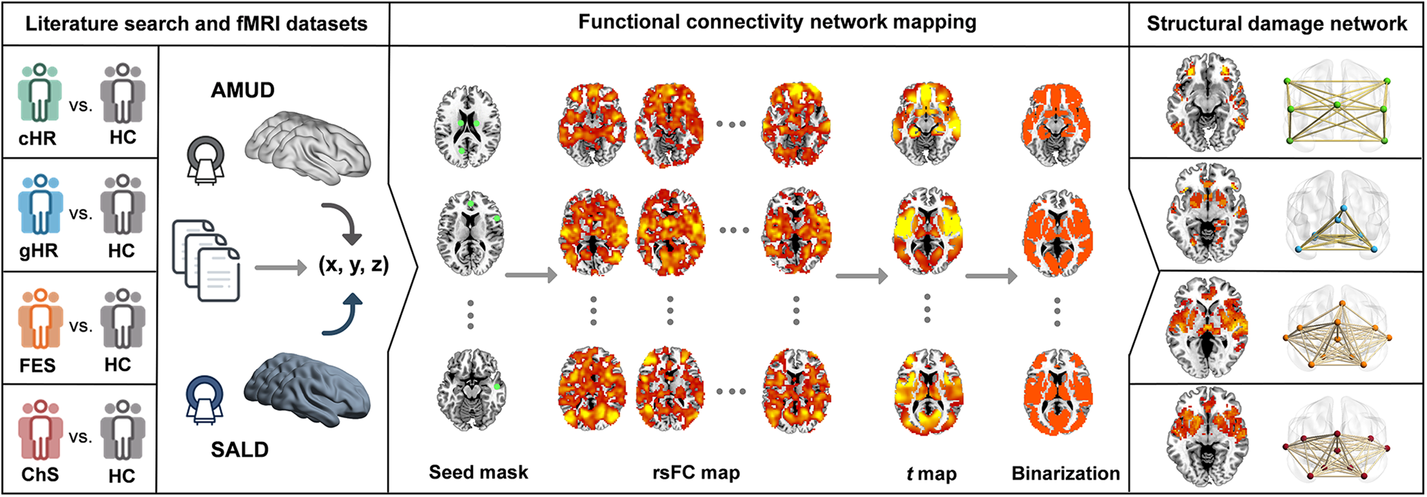
The objective of this exploratory study was to examine brain structural damage networks at different stages of schizophrenia. To achieve this goal, we initially synthesized published literature to identify gray matter alterations in cHR, gHR, FES, and ChS individuals relative to HC. By combining these affected brain locations with large-scale discovery and validation resting-state functional MRI (fMRI) datasets, we then used the FCNM approach to construct four brain structural damage networks corresponding to different stages of schizophrenia. Schematic representation of the study design and analytical procedure is shown in Fig. 1. Additionally, we assessed the spatial similarity of network patterns between different schizophrenia stages to investigate differences and commonalities in neuropathology across disease progression.
Study design and analytical procedure. We initially synthesized the published literature to identify gray matter alterations in cHR, gHR, FES, and ChS individuals relative to HC. By combining these affected brain locations with large-scale discovery (AMUD) and validation (SALD) resting-state fMRI datasets, we then used the FCNM approach to construct four brain structural damage networks corresponding to different stages of schizophrenia in the following way. Specifically, spheres centered at each coordinate of a contrast were first created and merged together to generate a contrast-specific combined seed mask. Second, based on the resting-state fMRI data, we computed a contrast seed-to-whole brain rsFC map for each subject. Third, the subject-level rsFC maps were entered into a voxel-wise one-sample t test to identify brain regions functionally connected to each contrast seed. Fourth, the resulting group-level t maps were thresholded and binarized at p < 0.05 corrected for multiple testing using a voxel-level FDR method. Finally, the binarized maps were overlaid to produce four network probability maps, which were thresholded at 60% to yield brain structural damage networks of cHR, gHR, FES, and ChS respectively. AMUD, Anhui Medical University Dataset; cHR, clinical high-risk; ChS, chronic schizophrenia; FDR: false-discovery rate; FES, first-episode schizophrenia; fMRI, functional magnetic resonance imaging; gHR, genetic high-risk; HC, health controls; rsFC, resting-state functional connectivity; SALD, Southwest University Adult Lifespan Dataset.
Materials and methods
Study search and selection
We conducted a systematic literature search on the PubMed, Brainmap, and Web of Science databases to identify studies examining gray matter alterations in cHR, gHR, FES, or ChS individuals relative to HC, published before March 1, 2023. We used the following terms to search in title/abstracts: (‘voxel-based morphometry’ OR ‘VBM’ OR ‘voxel-wise’) AND (‘schizophrenia’ OR ‘chronic schizophrenia’ OR ‘SZ’ OR ‘first episode schizophrenia’ OR ‘first episode psychosis’ OR ‘high risk schizophrenia’ OR ‘siblings schizophrenia’ OR ‘first degree relatives’ OR ‘genetic risk schizophrenia’ OR ‘at risk of mental state’ OR ‘ARMS’ OR ‘ultra-high risk’). We also screened the reference lists of the identified studies, review articles, and meta-analyses to search for additional qualified studies. Note that cHR individuals met the criteria of either the Personal Assessment and Crisis Evaluation (Yung et al., Reference Yung, Phillips, Mcgorry, Mcfarlane, Francey, Harrigan and Jackson1998), the Comprehensive Assessment of At-Risk Mental States (Yung et al., Reference Yung, Yuen, Mcgorry, Phillips, Kelly, Dell'olio and Buckby2005), or the Structured Interview for Prodromal Symptoms (Miller et al., Reference Miller, Mcglashan, Rosen, Cadenhead, Cannon, Ventura and Woods2003). The cHR factors mainly include early clinical symptoms (e.g. cognitive deficits) and detrimental environmental factors (e.g. adverse life experiences) (Catalan et al., Reference Catalan, Salazar De Pablo, Aymerich, Damiani, Sordi, Radua and Fusar-Poli2021; Jauhar, Johnstone, & Mckenna, Reference Jauhar, Johnstone and Mckenna2022; Radua et al., Reference Radua, Ramella-Cravaro, Ioannidis, Reichenberg, Phiphopthatsanee, Amir and Fusar-Poli2018). The gHR factors primarily include monozygotic twins, siblings, and first-/second-degree relatives of schizophrenia patients. FES referred to an illness duration of less than 2 years, and ChS referred to an illness duration of more than 2 years.
Since there are some cases where a single study contains multiple neuroimaging contrasts (i.e. cHR, gHR, FES, or ChS individuals v. HC), we focused our analysis on contrasts rather than studies. Inclusion criteria for neuroimaging contrasts were: (a) to be included in a research article published in a peer-review journal; (b) to use a specified whole-brain VBM analysis; (c) to report gray matter alterations in cHR, gHR, FES, or ChS individuals by means of a between-group comparison with HC; (d) to report results in the form of stereotactic space (i.e. x, y, z coordinates in Talairach or Montreal Neurological Institute [MNI] space); (e) experimental groups without other medical comorbidities; and (f) diagnosis of schizophrenia based on fulfilling International Classification of Diseases or Diagnostic and Statistical Manual of Mental Disorders criteria. Exclusion criteria were: (a) experimental groups with a sample size <10 subjects; (b) use of region of interest analysis or small volume correction; and (c) mixed experimental sample (e.g. FES and ChS). Study search and selection were independently performed by two investigators. A flow diagram of the study selection process is shown in Fig. S1 in the online Supplementary materials. This protocol was registered on PROSPERO (https://www.crd.york.ac.uk/PROSPERO/, registration number: CRD42023481658). Coordinates of peak voxels of significant clusters reported in each contrast were extracted, with coordinates in Talairach space converted to MNI space. It is notable that subsequent analysis was based on the peak coordinates extracted from the published studies rather than MRI images from our patient and control groups.
Discovery and validation datasets
Our study used Anhui Medical University Dataset (AMUD) as a discovery dataset and Southwest University Adult Lifespan Dataset (SALD) (Wei et al., Reference Wei, Zhuang, Ai, Chen, Yang, Liu and Qiu2018) as a cross-scanner validation dataset. AMUD included 656 healthy adults of Chinese Han and right handedness (396 female, mean 26.57 ± 8.57 years), who were enrolled from local universities and communities through poster advertisements. Participants with neuropsychiatric or severe somatic disorders, a history of head injury with consciousness loss, MRI contraindications, or a family history of psychiatric diseases among first-degree relatives were excluded. SALD included 329 healthy adults (207 female, mean 37.81 ± 13.79 years) and full details regarding the sample have been described in the data descriptor publication (Wei et al., Reference Wei, Zhuang, Ai, Chen, Yang, Liu and Qiu2018). For the SALD, the exclusion criteria included MRI contraindications, current psychiatric or neurological disorders, use of psychiatric drugs within 3 months, pregnancy, or a history of head trauma. It is noteworthy that all included participants were restricted to an age range of 18–60 years to exclude the potential influences of neurodevelopment and neurodegeneration. Demographic information of the discovery and validation datasets is provided in Table S1 in the online Supplementary materials.
The authors assert that all procedures contributing to this work comply with the ethical standards of the relevant national and institutional committees on human experimentation and with the Helsinki Declaration of 1975, as revised in 2008. All procedures involving human subjects in the AMUD were conducted following approval by the ethics committee of The First Affiliated Hospital of Anhui Medical University (approval number 20200094), and all participants provided written informed consent after being given a complete description of the study. As for the SALD, it is a publicly available resource and detailed ethical information can be found at http://fcon_1000.projects.nitrc.org/indi/retro/sald.html.
fMRI data acquisition and preprocessing
Resting-state fMRI data of AMUD were collected on a 3.0-Tesla General Electric Discovery MR750w scanner, and those of SALD on a 3.0-Tesla Siemens Trio scanner. The fMRI parameters of the two datasets are provided in Table S2 in the online Supplementary materials. Participants with poor image quality (e.g. visible artifacts) and incomplete brain coverage were excluded.
Resting-state fMRI data were preprocessed using Statistical Parametric Mapping software (SPM12, http://www.fil.ion.ucl.ac.uk/spm) and Data Processing & Analysis for Brain Imaging (DPABI, http://rfmri.org/dpabi) (Yan, Wang, Zuo, & Zang, Reference Yan, Wang, Zuo and Zang2016). The first 10 volumes for each participant were discarded to allow the signal to reach equilibrium and the participants to adapt to the scanning noise. The remaining volumes were corrected for the acquisition time delay between slices. Then, realignment was performed to correct the motion between time points. Head motion parameters were computed by estimating the translation in each direction and the angular rotation on each axis for each volume. All participants' data were within the defined motion thresholds (i.e. maximal translational or rotational motion parameters less than 2 mm or 2°). We also calculated frame-wise displacement (FD), which indexes the volume-to-volume changes in head position. Several nuisance covariates (the linear drift, the estimated motion parameters based on the Friston-24 model, the spike volumes with FD > 0.5 mm, the global signal, the white matter signal, and the cerebrospinal fluid signal) were regressed out from the data. Since global signal regression can enhance the detection of system-specific correlations and improve the correspondence to anatomical connectivity, we included this step in the preprocessing of resting-state fMRI data (Murphy & Fox, Reference Murphy and Fox2017). Then, the datasets were band-pass filtered using a frequency range of 0.01–0.1 Hz. In the normalization step, individual structural images were first co-registered with the mean functional images; the transformed structural images were then segmented and normalized to MNI space using a high-level nonlinear warping algorithm, i.e. the diffeomorphic anatomical registration through exponentiated Lie algebra technique (Ashburner, Reference Ashburner2007). Next, each filtered functional volume was spatially normalized to MNI space using the deformation parameters estimated during the above step and resampled into 3 mm isotropic voxel. Finally, all data were spatially smoothed with a Gaussian kernel of 6 × 6 × 6 mm3 full-width at half maximum.
Functional connectivity network mapping
We adopted the FCNM approach to construct brain structural damage networks of different schizophrenia stages based on the extracted coordinates of gray matter alterations in cHR, gHR, FES, and ChS individuals relative to HC (Fig. 1). First, 4 mm radius spheres centered at each coordinate of a contrast were created and merged together to generate a contrast-specific combined seed mask (henceforth referred to as the contrast seed). Second, based on the preprocessed resting-state fMRI data of AMUD, we computed a contrast seed-to-whole brain functional connectivity (FC) map for each subject, by calculating Pearson's correlation coefficients between time courses of the contrast seed and each voxel within the whole brain, followed by Fisher's Z transformation to improve normality. Third, the 656 subject-level FC maps were entered into a voxel-wise one-sample t test to identify brain regions functionally connected to each contrast seed. It is noteworthy that we only considered positive FC as the biological meaning of negative FC is still a matter of debate (Murphy & Fox, Reference Murphy and Fox2017; Murphy, Birn, Handwerker, Jones, & Bandettini, Reference Murphy, Birn, Handwerker, Jones and Bandettini2009). Fourth, the resulting group-level t maps were thresholded and binarized at p < 0.05 corrected for multiple testing using a voxel-level false-discovery rate method. Finally, the binarized maps were overlaid to produce four network probability maps, which were thresholded at 60% to yield brain structural damage networks of cHR, gHR, FES, and ChS, respectively.
Association with canonical brain networks
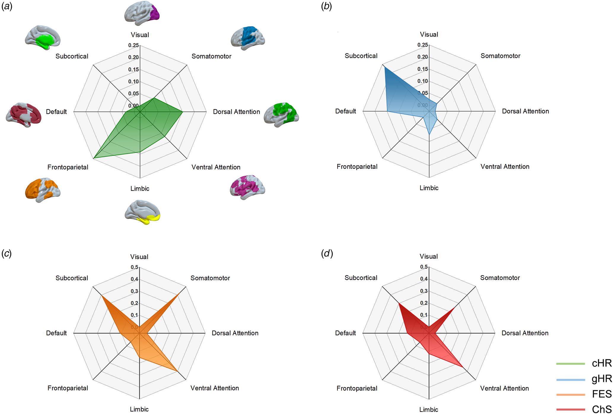
To ease interpretability, we tested the spatial associations between four schizophrenia brain structural damage networks and eight well-established canonical brain networks. The seven cortical networks were the visual, somatomotor, dorsal attention, ventral attention, limbic, frontoparietal, and default networks from the study by Yeo et al. (Reference Yeo, Krienen, Sepulcre, Sabuncu, Lashkari, Hollinshead and Buckner2011). The subcortical network was composed of the amygdala, hippocampus, basal ganglia, and thalamus from the Human Brainnetome Atlas (Fan et al., Reference Fan, Li, Zhuo, Zhang, Wang, Chen and Jiang2016). The proportion of overlapping voxels between each brain structural damage network and a canonical network to all voxels within the corresponding canonical network was calculated to estimate their spatial association.
Validation analyses
We carried out several validation analyses to examine the robustness of our results. First, we repeated our analyses by use of an independent validation SALD to assess the influence of dataset selection. Second, we repeated the FCNM procedure using 1 and 7 mm radius spheres to test the effect of seed size. Finally, we adopted 60% to threshold the network probability maps in our main analysis. To examine the impact of threshold selection, we repeated the FCNM procedure using 55% and 65% thresholds.
Results
Included studies
A total of 136 studies with 156 neuroimaging contrasts were included in our analyses. Specifically, 14 studies contained 14 contrasts from 523 cHR and 524 HC, 16 studies contained 17 contrasts from 855 gHR and 888 HC, 55 studies contained 58 contrasts from 2162 FES and 2488 HC, and 61 studies contained 67 contrasts from 2640 ChS and 3063 HC. Table S3 in the online Supplementary materials summarizes sample characteristics of the included studies.
Brain structural damage network of cHR
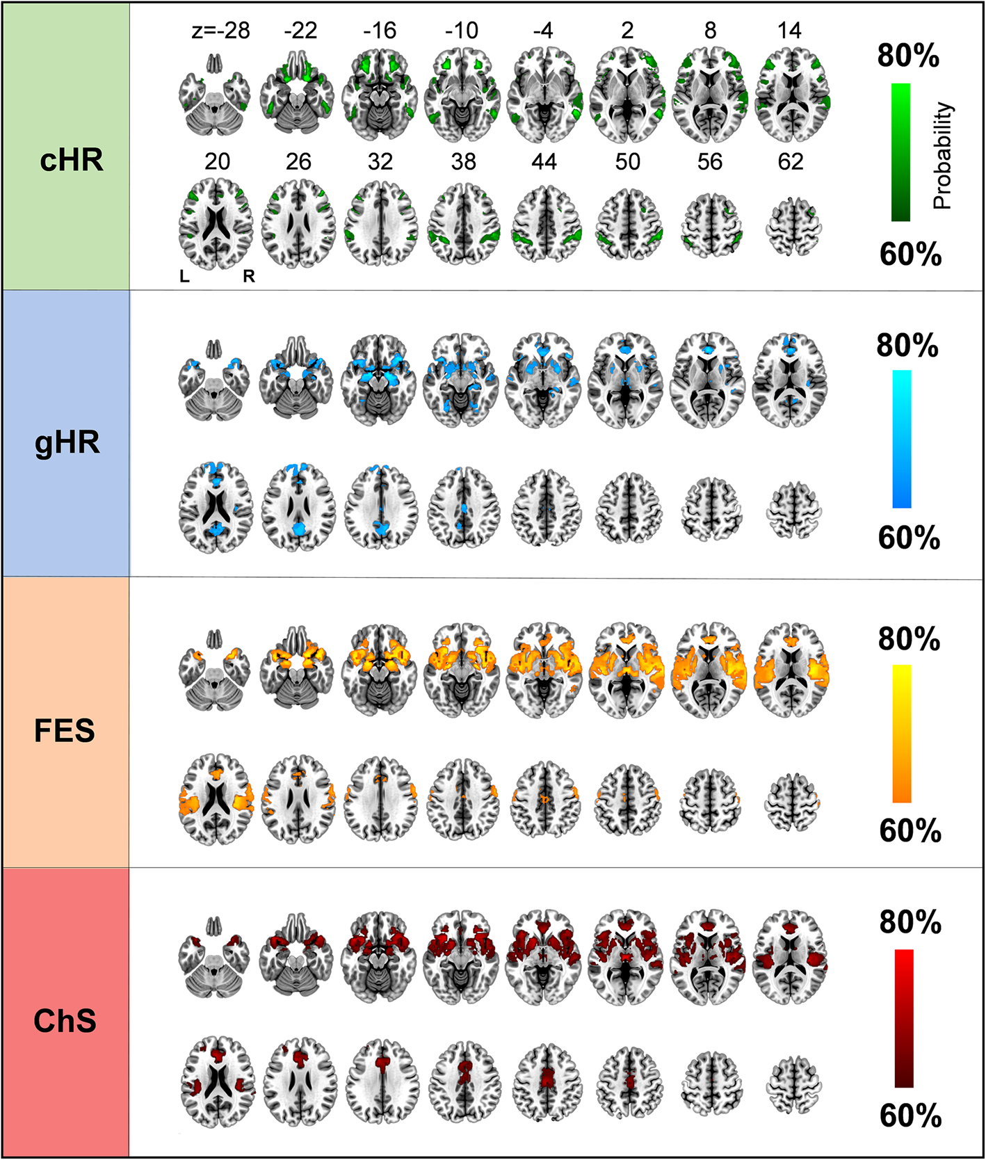
Brain structural damage network of cHR was primarily composed of the bilateral orbitofrontal cortex, lateral prefrontal cortex, posterior parietal cortex, and lateral temporal cortex (Fig. 2). With regard to canonical brain networks, brain structural damage network of cHR mainly involved the frontoparietal network (overlapping proportion: 24.5%) (Fig. 3a).
Brain structural damage networks of different schizophrenia stages. Brain structural damage networks of cHR, gHR, FES, and ChS are shown as network probability maps thresholded at 60%, showing brain regions functionally connected to more than 60% of the contrast seeds. cHR, clinical high-risk; ChS, chronic schizophrenia; FES, first-episode schizophrenia; gHR, genetic high-risk; L, left; R, right.
Associations of brain structural damage networks of cHR (a), gHR (b), FES (c), and ChS (d) with canonical brain networks. Polar plots display the proportion of overlapping voxels between each brain structural damage network and a canonical network to all voxels within the corresponding canonical network. cHR, clinical high-risk; ChS, chronic schizophrenia; FES, first-episode schizophrenia; gHR, genetic high-risk.
Brain structural damage network of gHR
Brain structural damage network of gHR chiefly consisted of the bilateral striatum, hippocampus, amygdala, thalamus, medial prefrontal cortex, posterior cingulate cortex/precuneus, and temporal pole (Fig. 2). As to canonical networks, brain structural damage network of gHR principally implicated the subcortical network (23.6%) (Fig. 3b).
Brain structural damage network of FES
Brain structural damage network of FES comprised of widespread brain areas mainly including the bilateral auditory cortex, middle cingulate cortex, ventrolateral prefrontal cortex, insula and operculum, temporal pole, medial prefrontal cortex, striatum, hippocampus, amygdala, and thalamus (Fig. 2). With respect to canonical networks, brain structural damage network of FES predominantly involved the somatomotor (42.1%), ventral attention (40.0%), and subcortical (39.5%) networks (Fig. 3c).
Brain structural damage network of ChS
Brain structural damage network of ChS was similar to but less extensive than structural damage network of FES, mainly comprising the bilateral auditory cortex, middle cingulate cortex, ventrolateral prefrontal cortex, insula and operculum, temporal pole, medial prefrontal cortex, striatum, amygdala, and thalamus (Fig. 2). Regarding canonical networks, brain structural damage network of ChS also primarily implicated the ventral attention (35.0%), subcortical (30.4%), and somatomotor (25.1%) networks (Fig. 3d).
Network similarity between schizophrenia stages
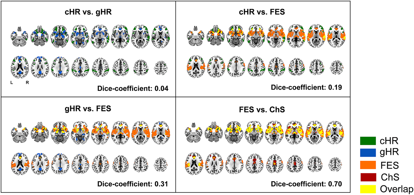
To quantify the similarity of network patterns between different schizophrenia stages, we estimated the spatial overlap between the networks of two schizophrenia stages by calculating a Dice coefficient, defined as 2 × (overlapping voxels)/(network #1 voxels) + (network #2 voxels). A higher Dice coefficient indicates more similar networks. As shown in Fig. 4, we observed a quite low spatial overlap between brain structural damage networks of cHR and gHR (Dice coefficient = 0.04), partial overlaps between cHR and FES (Dice coefficient = 0.19) and between gHR and FES (Dice coefficient = 0.31), and a high overlap between FES and ChS (Dice coefficient = 0.70).
Network similarity between schizophrenia stages. To quantify the similarity of network patterns between different schizophrenia stages, we estimated the spatial overlap between the networks of two schizophrenia stages by calculating a Dice coefficient, defined as 2 × (overlapping voxels)/(network #1 voxels) + (network #2 voxels). A higher Dice coefficient indicates more similar networks. cHR, clinical high-risk; ChS, chronic schizophrenia; FES, first-episode schizophrenia; gHR, genetic high-risk; L, left; R, right.
Validation analyses
First, brain structural damage networks of different schizophrenia stages derived from the validation SALD were similar to those from the discovery AMUD, with subtle differences largely attributed to variation in sample sizes (656 v. 329) (Fig. S2 in the online Supplementary materials). Second, when repeating the FCNM procedure using 1 and 7 mm radius spheres, we observed that the resulting brain structural damage networks of cHR, gHR, FES, and ChS were nearly identical to those using the 4 mm radius sphere (Figs S3 and S4 in the online Supplementary materials). Finally, using the 55% and 65% thresholds yielded brain structural damage networks that had larger and smaller spatial extents than those based on the 60% threshold (Figs S5 and S6 in the online Supplementary materials). However, the core areas of brain structural damage networks were still present across these thresholds. These findings verified the robustness of our results to distinct datasets and methodological differences.
Discussion
By a combination of the novel FCNM approach and large-scale brain functional connectome data, the present study interrogated brain structural damage networks of different schizophrenia stages based on gray matter alterations in cHR, gHR, FES, and ChS reported in previous literature. Results showed that brain structural damage networks of cHR and gHR had limited and non-overlapping spatial distributions, with the former mainly involving the frontoparietal network and the latter principally implicating the subcortical network, indicative of distinct neuropathological mechanisms underlying cHR and gHR. By contrast, brain structural damage networks of FES and ChS manifested as similar patterns of widespread brain areas predominantly involving the somatomotor, ventral attention, and subcortical networks, suggesting an emergence of more prominent brain structural abnormalities with illness onset that have trait-like stability over time. Our findings may not only provide a refined picture of schizophrenia neuropathology from a network perspective, but also potentially contribute to more targeted and effective intervention strategies for individuals at different schizophrenia stages.
Although neuroimaging has strengthened its position as the most widely used tool in the study of brain abnormalities in psychiatric illnesses, results are often irreproducible in this area (Elliott, Knodt, & Hariri, Reference Elliott, Knodt and Hariri2021; Etkin, Reference Etkin2019; Westlin et al., Reference Westlin, Theriault, Katsumi, Nieto-Castanon, Kucyi, Ruf and Barrett2023) such that neuroimaging findings are considered of limited clinical and translational value (Poldrack et al., Reference Poldrack, Baker, Durnez, Gorgolewski, Matthews, Munafò and Yarkoni2017). This lack of reproducibility may be due to a series of factors, such as limited sample sizes, clinical heterogeneity of patient populations, varying experimental designs, and different analytic techniques (Etkin, Reference Etkin2019; Poldrack et al., Reference Poldrack, Baker, Durnez, Gorgolewski, Matthews, Munafò and Yarkoni2017). One potent approach to mitigating these concerns is to perform a coordinate-based meta-analysis, which is able to synthesize the multitude of results from different neuroimaging studies and identify more consistently affected anatomical regions in psychiatric disorders (Eickhoff et al., Reference Eickhoff, Laird, Grefkes, Wang, Zilles and Fox2009; Wager, Lindquist, & Kaplan, Reference Wager, Lindquist and Kaplan2007). Nonetheless, researchers recently caution against mapping diseases to single brain regions and instead emphasize network localization, as it is largely accepted that neuropathological processes by no means act in isolation, but rather are interconnected via distributed brain networks (Fornito et al., Reference Fornito, Zalesky and Breakspear2015; Van Den Heuvel & Sporns, Reference Van Den Heuvel and Sporns2019). In this context, localization of a disease has increasingly shifted from a traditional region-based method to an updated network-based approach. The conceptual and methodological advances have been facilitated by the development of the FCNM approach that can map a disease, symptom, or psychological process to a common network through a combined analysis of brain locations of interest (e.g. lesion, structural damage, functional abnormality, and neural activation) and large-scale brain functional connectome data (Darby et al., Reference Darby, Joutsa and Fox2019; Joutsa et al., Reference Joutsa, Moussawi, Siddiqi, Abdolahi, Drew, Cohen and Fox2022; Peng et al., Reference Peng, Xu, Jiang and Gong2022; Taylor et al., Reference Taylor, Lin, Talmasov, Ferguson, Schaper, Jiang and Fox2023). By leveraging FCNM, investigators have mapped many neuropsychiatric diseases, some of which have eluded traditional region-based localization, to specific brain networks (Cheng et al., Reference Cheng, Cai, Liu, Yang, Pan, Zhang and Zhu2024; Cotovio et al., Reference Cotovio, Talmasov, Barahona-Corrêa, Hsu, Senova, Ribeiro and Fox2020, Reference Cotovio, Faro Viana, Fox and Oliveira-Maia2022; Darby et al., Reference Darby, Joutsa and Fox2019; Jones et al., Reference Jones, Zhukovsky, Hawco, Ortiz, Cipriani, Voineskos and Husain2023; Mo et al., Reference Mo, Zhao, Li, Cai, Song, Wang and Zhu2024; Padmanabhan et al., Reference Padmanabhan, Cooke, Joutsa, Siddiqi, Ferguson, Darby and Fox2019; Taylor et al., Reference Taylor, Siddiqi and Fox2021, Reference Taylor, Lin, Talmasov, Ferguson, Schaper, Jiang and Fox2023; Tetreault et al., Reference Tetreault, Phan, Orlando, Lyu, Kang, Landman and Darby2020; Trapp et al., Reference Trapp, Bruss, Manzel, Grafman, Tranel and Boes2023; Zhang et al., Reference Zhang, Xu, Ma, Qian and Zhu2024; Zhukovsky et al., Reference Zhukovsky, Anderson, Coughlan, Mulsant, Cipriani and Voineskos2021). With respect to brain structural alterations across different stages of schizophrenia, previous coordinate-based neuroimaging meta-analyses and reviews have demonstrated progressive brain abnormalities in schizophrenia from the viewpoint of regional morphology (Dietsche et al., Reference Dietsche, Kircher and Falkenberg2017; Liloia et al., Reference Liloia, Brasso, Cauda, Mancuso, Nani, Manuello and Rocca2021; Zhao et al., Reference Zhao, Zhang, Shah, Li, Sweeney, Li and Gong2022). Complementing and expanding these pilot efforts, we used the FCNM approach to characterize the neuropathological progression in schizophrenia from a network perspective. The present work, in concert with other FCNM investigations, suggests that the network-based framework holds substantial potential for refining our understanding of disease neuropathology.
We found that brain structural damage networks of cHR and gHR had distinct spatial patterns, possibly reflective of underlying neuropathological differences. The current observation of neuropathological differences between cHR and gHR is consistent with previous findings (Luna et al., Reference Luna, Radua, Fortea, Sugranyes, Fortea, Fusar-Poli and Carvalho2022; Smieskova et al., Reference Smieskova, Marmy, Schmidt, Bendfeldt, Riecher-Rӧssler, Walter and Borgwardt2013). A plausible explanation lies in the fact that the former may be a consequence of complex interactions between genetic architecture and environmental factors, whereas the latter may reflect a genetic effect. Brain structural damage network of cHR mainly involved the frontoparietal network, which is engaged in high-order cognitive processes (Lorenz et al., Reference Lorenz, Violante, Monti, Montana, Hampshire and Leech2018; Uddin, Reference Uddin2021; Zuo, Yang, Liu, Li, & Jiang, Reference Zuo, Yang, Liu, Li and Jiang2018). Behavioral evidence has shown that cHR state for psychosis is associated with significant and widespread impairments in neurocognitive functioning and social cognition (Carrión et al., Reference Carrión, Goldberg, Mclaughlin, Auther, Correll and Cornblatt2011; Catalan et al., Reference Catalan, Salazar De Pablo, Aymerich, Damiani, Sordi, Radua and Fusar-Poli2021; Pedruzo et al., Reference Pedruzo, Aymerich, Pacho, Herrero, Laborda, Bordenave and Catalan2023), highlighting the importance of frontoparietal network dysfunction in the pathophysiology of prodromal psychosis. Brain structural damage network of gHR principally implicated the subcortical network, which was also found to be strongly affected in FES and ChS, suggesting that subcortical network abnormalities may be at the root of pathogenesis of schizophrenia. Indeed, gray matter alterations in subcortical structures including the striatum, hippocampus, amygdala, and thalamus have been frequently reported in gHR, FES, and ChS individuals (Brosch et al., Reference Brosch, Stein, Schmitt, Pfarr, Ringwald, Thomas-Odenthal and Kircher2022; Cattarinussi et al., Reference Cattarinussi, Kubera, Hirjak, Wolf and Sambataro2022; Cooper, Barker, Radua, Fusar-Poli, & Lawrie, Reference Cooper, Barker, Radua, Fusar-Poli and Lawrie2014; Cui et al., Reference Cui, Li, Liu, Sui, Song, Chen and Jiang2022; Ganzola, Maziade, & Duchesne, Reference Ganzola, Maziade and Duchesne2014; Nenadic et al., Reference Nenadic, Dietzek, Schönfeld, Lorenz, Gussew, Reichenbach and Smesny2015; Van Erp et al., Reference Van Erp, Hibar, Rasmussen, Glahn, Pearlson, Andreassen and Turner2015). The dopamine hypothesis is the longest standing pathoetiologic theory of schizophrenia (Howes et al., Reference Howes, Kambeitz, Kim, Stahl, Slifstein, Abi-Dargham and Kapur2012; Mccutcheon, Abi-Dargham, & Howes, Reference Mccutcheon, Abi-Dargham and Howes2019; Mccutcheon, Krystal, & Howes, Reference Mccutcheon, Krystal and Howes2020; Purves-Tyson et al., Reference Purves-Tyson, Owens, Rothmond, Halliday, Double, Stevens and Shannon Weickert2017). Altered striatal dopamine synthesis and release are present in schizophrenia, with the changes to a lesser degree in individuals at risk of schizophrenia and more prominent in patients with established schizophrenia (Howes, Mccutcheon, Owen, & Murray, Reference Howes, Mccutcheon, Owen and Murray2017; Kesby, Eyles, Mcgrath, & Scott, Reference Kesby, Eyles, Mcgrath and Scott2018). Earlier work indicates that hippocampal pathology in schizophrenia may be due to genetic factors, aberrant neurodevelopment, and/or abnormal neural plasticity; hippocampal pathology is likely to be associated with the neuropsychological impairments of schizophrenia rather than with its psychotic symptoms (Harrison, Reference Harrison2004; Szeszko et al., Reference Szeszko, Strous, Goldman, Ashtari, Knuth, Lieberman and Bilder2002). Evidence from functional neuroimaging suggests that abnormal amygdala activation in response to emotional stimuli might contribute to emotional dysfunction, a critical clinical feature of schizophrenia (Anticevic et al., Reference Anticevic, Van Snellenberg, Cohen, Repovs, Dowd and Barch2012; Guimond, Mothi, Makowski, Chakravarty, & Keshavan, Reference Guimond, Mothi, Makowski, Chakravarty and Keshavan2022; Mukherjee et al., Reference Mukherjee, Sabharwal, Kotov, Szekely, Parsey, Barch and Mohanty2016). The thalamus is a key hub of cortical–subcortical circuitry and plays a vital role in the coordination of information within the brain; given that thalamic abnormalities have been observed early in the progression of schizophrenia, one may postulate that a disruption of the thalamocortical network might give rise to some cardinal symptoms of schizophrenia (Benoit, Canetta, & Kellendonk, Reference Benoit, Canetta and Kellendonk2022; Cronenwett & Csernansky, Reference Cronenwett and Csernansky2010; Giraldo-Chica & Woodward, Reference Giraldo-Chica and Woodward2017).
Brain structural damage networks of FES and ChS manifested as similar patterns of widespread brain areas, suggesting an emergence of more prominent brain structural abnormalities with illness onset that have trait-like stability over time. Strikingly, structural damage network of FES was slightly more extensive than that of ChS, which might seem counter-intuitive at first sight. Treatment-induced or experience-dependent neuroplasticity during the chronic condition may provide a potentially mechanistic account for this finding. An alternative methodological explanation is that variation in the number of the included studies on FES and ChS (55 v. 61) may lead to the subtle difference. Aside from the subcortical network, brain structural damage networks of FES and ChS prominently involved the somatomotor and ventral attention networks. The somatomotor network is implicated in auditory, visual, tactile, and direct pain perception (Keysers, Kaas, & Gazzola, Reference Keysers, Kaas and Gazzola2010; Lee et al., Reference Lee, Kim, Čeko, Park, Lee, Park and Woo2021). Empirical findings have shown a strong link between robotically controlled alterations in somatomotor processing and auditory misattribution in psychosis, providing evidence for the role of somatomotor processes in altered self-monitoring in psychosis (Salomon et al., Reference Salomon, Progin, Griffa, Rognini, Do, Conus and Blanke2020). In addition, recent finding of abnormal cortical hierarchy organization in schizophrenia suggests that cascading impairments from the disruption of the somatosensory-motor system and inefficient integration of bottom-up sensory information with attentional demands and executive control processes partially account for high-level cognitive deficit characteristics of schizophrenia (Dong et al., Reference Dong, Yao, Wang, Hong, Genon, Xin and Luo2021; Kaufmann et al., Reference Kaufmann, Skåtun, Alnæs, Doan, Duff, Tønnesen and Westlye2015). With respect to the ventral attention network, prior data have demonstrated its prominent engagement in reorienting attention to salient stimuli (Corbetta, Patel, & Shulman, Reference Corbetta, Patel and Shulman2008; Decety & Lamm, Reference Decety and Lamm2007). Intense research has reported structural, functional, and connectivity changes in the ventral attention network in schizophrenia (Dong, Wang, Chang, Luo, & Yao, Reference Dong, Wang, Chang, Luo and Yao2018; Jimenez et al., Reference Jimenez, Lee, Wynn, Cohen, Engel, Glahn and Green2016; Shafiei et al., Reference Shafiei, Markello, Makowski, Talpalaru, Kirschner, Devenyi and Mišić2020). This invites the speculation that faulty assignment of salience to internally generated mental events forms the basis of positive symptoms, such as delusions and hallucinations, in patients suffering from schizophrenia. Echoing our findings, there is also evidence from functional neuroimaging that patients with schizophrenia exhibit functional abnormalities in the frontoparietal, somatomotor, ventral attention, and subcortical networks (Li et al., Reference Li, Yao, You, Liu, Deng, Li and Gong2023; Luo et al., Reference Luo, Li, Wang, He, Wang, You and Li2023; You et al., Reference You, Luo, Yao, Zhao, Li, Wang and Li2022).
Several caveats should be considered when interpreting the current findings. First, it is important to point out that our analyses cannot determine causality of illness progression due to the lack of longitudinal design. However, robust cross-sectional analyses are also essential to clarify this issue and build a more integrative view. Second, we cannot rule out the influence of antipsychotic medications on our results, particularly those of ChS individuals who were treated using different types of antipsychotics. Third, we utilized resting-state fMRI data from healthy subjects to construct brain structural damage networks of schizophrenia across different stages of the illness. It appears more reasonable to utilize fMRI data from individuals at different stages of schizophrenia. However, there is evidence showing that sample selection has a negligible effect on network localization results (Boes et al., Reference Boes, Prasad, Liu, Liu, Pascual-Leone, Caviness and Fox2015; Fox et al., Reference Fox, Buckner, Liu, Chakravarty, Lozano and Pascual-Leone2014; Horn et al., Reference Horn, Reich, Vorwerk, Li, Wenzel, Fang and Fox2017). Fourth, the present study did not perform any correlation analysis due to the study design. Exploring the associations between brain MRI characteristics and clinical profiles has been a focus of psychiatric research (Li et al., Reference Li, Zhao, Hu, Liu, Wang, Zhang and Gong2024; Vieira et al., Reference Vieira, Bolton, Schöttner, Baecker, Marquand, Mechelli and Hagmann2024). Investigation into the relationship between the identified brain structural damage networks and clinical characteristics in schizophrenia will be part of our future work. Likewise, subsequent research is needed to determine whether the brain structural damage networks found in clinical and gHR individuals could constitute effective biomarkers to predict the onset or the treatment outcome and prognosis in the long-term course of schizophrenia (Jauhar et al., Reference Jauhar, Johnstone and Mckenna2022; Long et al., Reference Long, Chen, Zhang, Li, Wang, Wang and Li2024). Fifth, we did not consider each study's sample size and effect size in our FCNM analysis, since there has been no consensus yet on how to account for these factors. Further investigation of their influences, in concert with analytical advances in the future, will help address this issue. Finally, we adopted an overlapping threshold of 60% to identify brain regions functionally connected to 60% of the contrast seeds. This less stringent threshold was selected due to the fact that many sources of variance may impede the identification of a common brain network at a more rigorous threshold.
In summary, this study combined the novel FCNM approach and large-scale brain functional connectome data to localize gray matter alterations in cHR, gHR, FES, and ChS to four brain structural damage networks corresponding to different stages of schizophrenia. Our findings may provide a refined picture of schizophrenia neuropathology from a network perspective. More broadly, the current identification of these brain structural damage networks may expose more effective prevention and treatment targets for individuals at different schizophrenia stages, laying the foundation for precision medicine in psychiatry.
Supplementary material
The supplementary material for this article can be found at https://doi.org/10.1017/S0033291724003088
Data availability statement
The data and analysis codes used in the preparation of this article are publicly available in the study's Open Science Framework repository (https://osf.io/pm3x5/).
Acknowledgments
The study was supported by the National Natural Science Foundation of China (grant number: 82471952), the Anhui Provincial Natural Science Foundation (grant numbers: 2308085MH277 and 2208085MH257), the Scientific Research Key Project of Anhui Province Universities (grant number: 2022AH051135), and the Scientific Research Foundation of Anhui Medical University (grant number: 2022xkj143).
Competing interests
None.


 Open access