


Introduction
The over-use of antibiotics leads to bacterial resistance, which complicates the treatment of infectious diseases and makes them difficult to control. Therefore, studies on new antibacterial materials are an active area of research. When developing new antibacterial materials, they should not only have improved antibacterial efficacy but also be less irritating and less toxic. To overcome these problems, many researchers have focused on the immobilization of antibacterial agents on mineral surfaces. Immobilization on suitable substrates reduces their toxicity to the host and facilitates their eventual elimination (Saito et al., Reference Saito, Takatsuka, Kato, Ishihara and Okuda1997).
The use of montmorillonite-type (Mnt) clay minerals in the synthesis of antibacterial materials has attracted great interest due to their outstanding structural properties such as large surface area, expansion capacity, cation and anion exchange capacity, as well as ease of preparation, non-toxicity, and chemical inertness (Saha et al., Reference Saha, Butola and Joshi2014; Malek and Ramli, Reference Malek and Ramli2015; De Oliveira et al., Reference De Oliveira, Trigueiro, Souza, de Carvalho, Osajima, da Silva-Filho and Fonseca2022; Şahiner et al., Reference Şahiner, Özdemir, Bulut and Yapar2022). Quaternary ammonium-type surfactants (QACs) are used as disinfectants in many areas such as food, cosmetics and personal care products, and medicine. They also represent one of the most effective classes of disinfectants in dentistry, dental materials, and resin composites (Makvandi et al., Reference Makvandi, Jamaledin, Jabbari, Nikfarjam and Borzacchiello2018). The incorporation of an immobilized antibacterial agent in dental formulations as an excipient can promote the oral elimination of a variety of pathogens. QACs and their mixtures with other types of surfactants are immobilized on Mnt and O-Mnt surfaces/interlayers by a variety of interactions such as electrostatic, hydrogen bonding, van der Waals, π/π, OH/π, and NH/π, and form stable O-Mnt structures. Many different surface configurations can arise, depending on the chemical structure and amount of QAC and other surfactants used in the modification. In addition to single, bilayer, and/or multi-layer formation, QACs can also be distributed between the external surface and the interlayer of Mnt or O-Mnt platelets. Many researchers have also revealed that a large part of the antibacterial activity of cationic monomers in non-agent-releasing type antibacterial materials is influenced by their surface-active properties and hence by the length of the hydrophobic chains (Marcotte et al., Reference Marcotte, Barbeau and Lafleur2005; Caillier et al., Reference Caillier, de Givenchy, Levy, Vandenberghe, Géribaldi and Guittard2009; Imazato et al., Reference Imazato, Chen, Ma, Izutani and Li2012), their configuration on the material, and their chemical structure, respectively. Despite the fact that the antibacterial activity of O-Mnt is widely attributed to the process of contact killing (Makvandi et al., Reference Makvandi, Jamaledin, Jabbari, Nikfarjam and Borzacchiello2018), there is insufficient research connecting the configuration of the surfactants on the external surface to the killing mechanism. In the present study, the surfactants used as immobilized agents were a benzethonium chloride (BZC) and cetylpyridinium (CP)–N-lauroylsarcosinate (SR) pair. The presence of a linear alkyl chain, an anionic amino acid group, and double aromatic rings originating from these surfactants are the structural factors that confer greater antibacterial activity on the O-Mnt samples studied (Şahiner et al., Reference Şahiner, Özdemir, Bulut and Yapar2022). Subsequently, X-ray photoelectron spectroscopy (XPS) analyses were performed to elucidate the configuration of the surfactants on the external surface of O-Mnt and thus obtain information on the interactions between the surfactant molecules and the cell wall of the bacteria.
The proposed mechanism for QACs with immobilized long lipophilic alkyl chains is that the lipophilic chains penetrate the cell membrane and bind to bacterial cell wall components, leading to cytoplasmic leakage, autolysis, and bacterial cell death (Kawabata and Nishiguchi, Reference Kawabata and Nishiguchi1988; Ahlström et al., Reference Ahlström, Thompson and Edebo1999; Kenawy et al., Reference Kenawy, Abdel-Hay, El-Shanshoury and El-Newehy2002). However, this mechanism does not take into account the contribution of groups other than long hydrophobic chains, such as double benzene rings, anionic amino acid groups, etc., which could lead to other interactions and thus to a different mechanism of action. For example, a recent study reported that inhibition of SARS-CoV-2 viruses is the result of the interaction of spike proteins with the amino acid group of SR molecules (Tateyama-Makino et al., Reference Tateyama-Makino, Abe-Yutori, Iwamoto, Tsutsumi, Tsuji, Morishita, Kurita, Yamamoto, Nishinaga and Tsukinoki2021).
Exclusion of pathogenic bacteria is beneficial in the control of infections such as periodontal disease, endocarditis, and lung and intestinal infections. The gram-positive bacterium investigated, Actinomyces viscosus (A. viscosus), causes periodontal inflammations, endocarditis, and lung infections. Actinomyces species are inhabitants of mucosal surfaces. However, they can cause opportunistic infections because of tissue disruption on these surfaces for any reason. This strain is difficult to culture because it grows slowly and requires specific conditions, and its microscopic detection is challenging due to its thin, filamentous shape. This often prevents infections caused by it from being diagnosed (Eng et al., Reference Eng, Corrado, Cleri, Cherubin and Goldstein1981; Mardis and Many, Reference Mardis and Many2001; Shimada et al., Reference Shimada, Kataoka, Miyazawa, Yamamoto and Igarashi2012). The gram-negative bacterium Bacteroides fragilis (B. fragilis) is part of the normal microbiota of the human colon, but is often associated with intra-abdominal infections, intracavitary abscesses, and complicated skin/soft tissue infections. It is also the most common anaerobe in bacteremia. Species of the B. fragilis group account for about a quarter of all anaerobes isolated from clinical specimens and are associated with the maximum virulence among all pathogenic anaerobes and the capacity to develop resistance mechanisms (Gupta et al., Reference Gupta, Shenoy, Kumar and Chawla2021). As representatives of both a gram-positive and a gram-negative strain, these opportunistic species were selected as control bacteria.
Various types of O-Mnt were prepared and tested against gram-positive and -negative bacteria and the efficiency of O-Mnt samples is reported to depend on the amount of antibacterial material as well as the cell wall structure of the bacteria (Yapar et al., Reference Yapar, Ateş and Özdemir2017). The O-Mnt samples prepared by immobilizing benzethonium chloride (Mnt-BZT) and cetyl pyridinium chloride-sodium lauroyl sarcosinate pair (Mnt-CP-SR) were used in previous studies to test their antibacterial properties against Streptococcus mutans (Şahiner et al., Reference Şahiner, Özdemir, Bulut and Yapar2022). Many of the characterization methods of O-Mnt such as Fourier transform infrared spectroscopy (FT-IR), X-ray diffraction (XRD), thermogravimetric analysis (TGA), and zeta potential were tested in previous studies (Özdemir et al., Reference Özdemir, Hoşgör-Limoncu and Yapar2010; Özdemir et al., Reference Özdemir, Yapar and Hoşgör-Limoncu2013; Türker et al., Reference Türker, Yarza, Sánchez and Yapar2017; Özdemir and Yapar, Reference Özdemir and Yapar2020; Şahiner et al., Reference Şahiner, Özdemir, Bulut and Yapar2022; Yapar et al., Reference Yapar, Ateş and Özdemir2017). The aim of the present study was to investigate Mnt-BZT and Mnt-CP-SR as antibacterial materials against A. viscosus and B. fragilis and to support and evaluate the proposed killing mechanism by elucidating the external surface structure.
The goals of the present study were original in several ways. There are only a few examples in the literature of the use of the BZC and CPC-SR pair for the production of O-Mnt with antibacterial activity. BZC is used as a topical anti-infective agent in medicaments, deodorants, mouthwashes, etc., and is used in the disinfection of apparatus in the food processing and pharmaceutical industries and in surgery. BZC exhibits a broad spectrum of microbiocidal activity against bacteria, fungi, mold, and viruses (Basaran, Reference Basaran2011; Czerwinski et al., Reference Czerwinski, Cozean and Cozean2014). It is highly effective against pathogens such as methicillin-resistant Staphlococcus aureus (Bearden et al., Reference Bearden, Allen and Christensen2008) and human immunodeficiency virus (HIV) (Sattar and Springthorpe (Reference Sattar and Springthorpe1991). Lee et al. (Reference Lee, Park, Song, Kim, Han, Park, Jo, Hwang, Sim, Kang and Tark2023) studied the use of BZC as a single compound or as a mixture in disinfectant during the coronavirus disease (COVID-19) pandemic in 2019. They reported that the disinfectants containing 0.05–0.4% BZC exhibited 90–100% virucidal efficacy against SARS-CoV-2 after 5–10 min of exposure.
Cetylpyridinium chloride (CPC) is found to be safe for human use over a wide concentration range in solution. It is commonly found at concentrations ranging from 0.05 to 0.1% in products such as mouthwashes, toothpastes, aphthous treatment products, lozenges, throat sprays, breath sprays, nasal sprays, and deodorants, and is effective against bacteria, fungi, and enveloped viruses. Some of these products also mediate antiseptic activity and offer a protective effect against dental plaque and reduce gingivitis (Riveira-Muñoz et al., Reference Riveira-Muñoz, Garcia-Vidal, Bañó-Polo, León, Blanc, Clotet and Ballana2023). Takeda et al. (Reference Takeda, Sawa, Sasaki, Orba, Maishi, Tsumita, Ushijima, Hida, Sano, Kitagawa and Hida2022) further concluded that CPC showed anti-SARS-CoV-2 effects without disrupting the virus envelope.
N-Lauroyl sarcosinate sodium salt (SR) on the other hand, is an anionic green surfactant. Fosdick et al. (Reference Fosdick, Calandra, Blackwell and Burrill1953) reported that SR inhibited enzymatic glycolysis in dental plaque. SR is currently being utilized in some dentifrices in an effort to prevent tooth decay (Englander et al., Reference Englander, Hoerman and Shklair1957). In a recent study, Tateyama-Makino et al. (Reference Tateyama-Makino, Abe-Yutori, Iwamoto, Tsutsumi, Tsuji, Morishita, Kurita, Yamamoto, Nishinaga and Tsukinoki2021) examined the inhibitory effects of common ingredients of toothpastes and mouthwashes on spike protein–ACE2 interaction and TMPRSS2 protease activity using an in vitro assay. They showed that toothpaste and mouthwash ingredients, including SR, may help prevent SARS-CoV-2 infection.
Another unique feature of this study is that, to our knowledge, there are no antibacterial studies with O-Mnt for A. viscosus and B. fragilis in the literature. Moreover, XPS analyses were performed to detail comprehensively the interaction between the external surface of the Mnt and the surfactants and thus to support and evaluate the proposed killing mechanism. At this point, it should be emphasized that there are few XPS studies on O-Mnt in the literature, which were prepared using mostly hexadecyltrimethylammonium bromide (HDTMA) or its homologs to characterize their external surfaces. However, the present study characterizes the external surface of two different modified montmorillonites prepared with a single surfactant (BZC) and with two surfactants (CP and SR), thus having a more complex structure. In the case of Mnt-CP-SR, the interaction characteristics were evaluated using Mnt-CP to highlight the effect of SR addition.
The antibacterial tests were performed to indicate that Mnt-BZT and Mnt-CP-SR are effective antibacterial agents. Finally, the results of in vitro cytotoxicity tests and in vivo animal tests corroborated the suggestion that the O-Mnts synthesized have the potential for both topical and oral applications at safe levels.
Materials and methods
Reagents and materials
Na-Mnt samples were obtained from various mines in Middle Anatolia and labeled as Mnt-1 and Mnt-2. Coarse impurities such as iron oxide and silica were separated by repeated sedimentation. The cation exchange capacities (CEC) were determined as 87 mmol 100 g–1 of clay (Mnt-1) and 68 mmol 100 g–1 of clay (Mnt-2) by the [Cu(trien)]2+ method. The quaternary ammonium surfactants and anionic surfactant were of analytical grade and were used without further purification. The surfactants were cetylpyridinium chloride monohydrate (C21H38ClN·H2O, MW = 358.01 g mol–1) (Merck, Darmstadt, Germany), benzethonium chloride (C27H42ClNO2, MW = 448.08 g mol–1) (Sigma Aldrich, St Louis, MO, USA), and N-lauroylsarcosine sodium salt (C15H28NNaO3, MW = 293.38 g/mol) (Sigma Aldrich, USA). The cultures, A. viscosus ATCC 15987 and B. fragilis ATCC 23745, were purchased from Kwik-Stik (Microbiologics Inc., St Cloud, MN, USA).
Preparation of the organo-montmorillonites
The benzethonium montmorillonite (Mnt-BZT) was prepared by a microwave irradition method (Türker et al., Reference Türker, Yarza, Sánchez and Yapar2017) using Mnt-1. First, aqueous dispersions containing 10 g of Mnt-1 and BZT equivalent to 100% CEC were prepared and then stirred at 700 rpm for 5 min. The final dispersion was subjected to 360 W microwave irradiation for 5 min. The cetylpyridinium and N-lauroylsarcosinate immobilized montmorillonite (Mnt-CP-SR) was prepared by the wet method in two steps. In the first step, cetylpyridinium montmorillonite (Mnt-CP) was synthesized by adding 10 g of Mnt-2 to a solution containing CP ions equivalent to 70% CEC of Mnt-2 and the dispersions were shaken at 25°C for 4 h. N-Lauroyl sarcosinate (SR) was then added to the Mnt-CP dispersions at 100% CEC of Mnt-2 and shaken for a further 24 h at 25°C (Özdemir et al., Reference Özdemir, Yapar and Hoşgör-Limoncu2013; Şahiner et al., Reference Şahiner, Özdemir, Bulut and Yapar2022).
Characterization
Scanning electron microscopy (SEM) and XPS analyses were used to characterize the samples. The SEM analyses were carried out to study the morphology of pristine and O-Mnt samples using a Thermo Scientific Apreo S scanning electron microscope (Waltham, MA, USA) equipped with BSE (back-scattered electron), EBIC (electron beam-induced current), STEM (scanning transmission electron microscopy), (low-voltage) (LVD), and energy-dispersive spectroscopy (EDS) detectors at 15 keV. The analyses were conducted using gold-plated samples.
The XPS spectra of Mnt-Na and O-Mnt were recorded on a Thermo Scientific SSX 100/206 spectrophotometer (XPS, Thermo Fisher Scientific, Loughborough, UK) with a monochromated microfocused AlKα X-ray source (1486.68 eV and 300 μm spot size), equipped with a multi-channel detector. The pressure in the analysis chamber was ~10−8 mbar. The binding energy scale of the spectrometer was calibrated with respect to the Au4f 7/2 peak at 83.94 eV (Seah, Reference Seah1989). Survey and high-resolution spectra of the samples were obtained with pass energies of 50 eV and 200 eV, respectively. The data were acquired and processed using the Avantage XPS software (5.9915) package.
Antibacterial susceptibility tests
The bacteria were cultured in accordance with their growth conditions: A. viscosus was cultured using Brain Heart Infusion (BHI) broth/agar (Merck Millipore, Darmstadt, Germany) and incubated microaerophilically (95% air, 5% CO2) at 37°C for 48–72 h; B. fragilis was cultured under anaerobic conditions using modified chopped meat medium/Brucella agar supplemented with 1 mg mL–1 menadione, 5 mg mL–1 hemin, and 0.01 mg mL–1 Vit K1 for 24–48 h at 37°C. The turbidity of the microbial suspension was adjusted to 0.5 McFarland units using a densitometer (Grant Instruments, Cambridge, UK) and the suspension was diluted to the desired concentration according to the procedure of each test method. The pristine Mnt was used as a negative control, because it exhibited no inhibition activity and this was also corroborated by other authors (He et al., Reference He, Yang, Yuan, Shen and Frost2006; Yang et al., Reference Yang, Yuan, Zhu and He2007; Meng et al., Reference Meng, Zhou, Zhang and Shen2009a; Meng et al., Reference Meng, Zhou, Zhang and Shen2009b; Özdemir et al., Reference Özdemir, Hoşgör-Limoncu and Yapar2010). On the other hand, 0.2% solution of chlorhexidine (CHX) digluconate was used as a positive control.
Agar well diffusion method
The agar well diffusion method was used to evaluate the antibacterial efficacy of the samples against the bacteria tested. In each run of the tests, the initial concentration of the microorganism was adjusted to 108 CFU mL–1. The BHI agar was inoculated by spreading 100 μL of bacterial suspension over the entire agar surface and allowed to dry; 10 mg of sample was placed into 8 mm-diameter wells punched on the agar using a sterile pipette tip (Güven et al., Reference Güven, Ustun, Tuna and Aktoren2019). The sample amount was determined according to the size of the wells and ~10 mg of each sample was used to fill the wells. Before the incubation period, the plates were kept at room temperature for 2 h. After 48 h of incubation at 37°C, the inhibition zones formed were observed and the zone diameters were recorded in millimeters. The tests were performed in triplicate.
Determination of MIC and MBC
The MIC and MBC values of the samples were determined by modifying the Clinical and Laboratory Standards Institute (CLSI) 2015 guidelines. Aqueous suspensions of the samples were prepared at a concentration of 100 mg mL–1 and 80 μL of the suspension was poured into the first well of 96-well plates, each containing 80 μL of BHI broth; 80 μL of the well-mixed sample in the first well was transferred to the second well. A twofold dilution was made from the second well to the eighth well. Wells 9 and 10 were used as negative and positive controls and were kept untreated. A 0.2% CHX digluconate solution and distilled water were used as positive and negative controls, respectively. Bacterial suspension (20 μL) was added to the wells to obtain a final bacterial inocula of 106 CFU mL–1. Experiments were carried out in duplicate for each sample and controls. After incubation (24 h and 48 h, 37°C), microbial growth was determined visually and the lowest concentration without turbidity (visible growth) was taken as the MIC value. The MBC was determined using the plate-dripping method. Samples (10 μL) from the wells were inoculated onto plates containing BHI agar and the minimum concentration at which no bacterial growth was observed was determined as the MBC value.
Time-kill assay
To determine the time-dependent antibacterial activity of the samples, a time-kill assay was performed using ASTM 2149 (standard test method for determining the antimicrobial activity of immobilized antimicrobial agents under dynamic contact conditions; ASTM International, 2013). To determine the effect of amount, O-Mnt suspensions were prepared at concentrations corresponding to three and 10 times the MIC values. They were then transferred to 10 mL buffer solutions (PBS pH = 7.4) and inoculated with bacterial suspensions (0.1 mL), to a final concentration of ~105 CFU mL–1. The samples were then incubated at 37°C and 100 rpm on an orbital shaker.
After 0, 2, 3, 5, and 15 min, and 1, 3, 6, 12, and 24 h of incubation, tenfold serial dilutions were made and 0.1 mL aliquots were inoculated onto the BHI agar plates by the spread plate count technique. After 48 h of incubation at 37±1°C, the numbers of colonies on agar plates were counted and log10 reductions were calculated. The tests were accomplished in triplicate. Minimum contact time was determined by taking the time as the basis when log10 reached the value <1.
In vitro cytotoxicity tests
Cytotoxicity tests were performed at Ege University Central Research Test and Analysis Laboratory Application and Research Center (EGEMATAL) according to the standard procedure (Maines, Reference Maines2002). The cytotoxicity test of Mnt, Mnt-CP-SR, and Mnt-BZT was conducted on HaCaT cell line by tetrazolium dye (3-(4,5-dimethylthiazol-2-yl)-2,5-diphenyltetrazolium bromide) (MTT) based assay. Because the samples were in powder form, they were first diluted with distilled water in ratios of 1:1, 1:2, 1:4, and 1:8 and extracted for 24 h. Samples extracted from O-Mnt with water were then taken as supernatants. The supernatant and MTT were added to a HaCaT cell seeded in 96-well culture followed by an additional 4 h incubation at 37°C under a CO2 atmosphere. In the case of a cytotoxic effect, the yellow MTT was expected to be converted to purple formazan because living cells metabolize MTT to produce this purple color. There is a direct correlation between the absorption value and cell viability. The resulting intracellular formazan crystals were solubilized in acidic isopropanol and the absorbance of the solution was measured at 570 nm using a UV-vis spectrophotometer. In the present study, media containing only HaCaT was used as a control. Data were obtained in three independent biological replicates performed on separate passages of cells and on separate days with a total number of six replicates.
In vivo acute toxicity tests
The study was approved by the Ege University Animal Experiments Local Ethics Committee (29.06.2022, 2022-049). Ethical guidelines for investigations of experimental pain in conscious animals were considered for all in vivo experiments (Zimmermann, Reference Zimmermann1983). The antibacterial clay to be used in the the study does not possess any toxic properties. Therefore, no adverse effects related to the application were anticipated. Mice were purchased from the Ege University Center for Research on Laboratory Animals (Izmir, Turkey). The study was conducted in accordance with the 3Rs principle. The number of animals was kept to a minimum, and no procedures causing pain or distress were performed. The animals were housed individually in separate cages.
The up-and-down-procedure (UDP) was conducted on 15 animals in accordance with OECD Guideline No: 425 (OECD, 2022). Mnt-CP-SR and Mnt-BZT were administered orally at doses of 175, 500, and 2000 mg kg–1 per day. Over the course of a 14-day exposure period, body weight information and signs of toxicity were recorded.
Results and Discussion
Characterization of the organo-montmorillonite samples
The key structural properties and thermal behavior of Mnt, Mnt-CP-SR, and Mnt-BZT were reported previously (Özdemir et al., Reference Özdemir, Hoşgör-Limoncu and Yapar2010; Özdemir et al., Reference Özdemir, Yapar and Hoşgör-Limoncu2013; Türker et al., Reference Türker, Yarza, Sánchez and Yapar2017; Yapar et al., Reference Yapar, Ateş and Özdemir2017; Özdemir and Yapar, Reference Özdemir and Yapar2020; Şahiner et al., Reference Şahiner, Özdemir, Bulut and Yapar2022). In the present study, SEM and XPS analyses were performed to obtain further information about and new insights into the structures of the O-Mnt samples.
The XRD analyses performed in previous studies showed that the samples Mnt-1 (with d 001 = 1.26 nm and d 060 = 0.15 nm) and Mnt-2 (with d 001 = 1.29 nm; d 002 = 0.62 nm; d 003 = 0.42 nm) consist mostly of smectite (Yapar, Reference Yapar2009; Özdemir et al., Reference Özdemir, Yapar and Hoşgör-Limoncu2013). Additionally, a calcite peak with d = 0.30 nm for Mnt-1 and quartz peaks with d = 0.506 nm and d = 0.33 nm, kaolinite peak with d = 0.71 nm, and cristobalite peaks with d = 0.41 nm and d = 0.31 nm for Mnt-2 were observed.
The interlayer spacings of the O-Mnt synthesized were calculated by substracting a d 001 value of dehydrated Mnt (0.97 nm) from those of O-Mnt samples as 0.83 nm and 1.22 nm for Mnt-CP-SR and Mnt-BZT, respectively (Şahiner et al., Reference Şahiner, Özdemir, Bulut and Yapar2022). These values corresponded to a bilayer and paraffin-like monomolecular arrangement at the interface (Şahiner et al., Reference Şahiner, Özdemir, Bulut and Yapar2022).
The onset and offset decomposition temperature ranges of Mnt-CP-SR and Mnt-BZT were determined by thermal analysis to be 200–556°C and 173–492°C, respectively (Şahiner et al., Reference Şahiner, Özdemir, Bulut and Yapar2022). The amount of cationic surfactants remaining on montmorillonite were calculated to be 93% for Mnt-BZT and 69% for Mnt-CP-SR (Türker et al., Reference Türker, Yarza, Sánchez and Yapar2017; Yapar et al., Reference Yapar, Özdemir, Solarte and Sanchez2015).
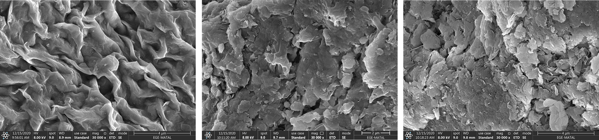
A set of SEM images (Fig. 1) revealed that the Mnt-Na has typical smectite morphology with curly margins forming a rather compact, net-like structure. However, both Mnt-CP-SR and Mnt-BZT had a rather distinct surface morphology attributed to the quaternary ammonium cations used in the modification (Vaia et al., Reference Vaia, Teukolsky and Giannelis1994; Hackett et al., Reference Hackett, Manias and Giannelis1998; Paul et al., Reference Paul, Zeng, Yu and Lu2005; Lagaly et al., Reference Lagaly, Ogawa and Dékány2013; Wójcik-Bania and Matusik, Reference Wójcik-Bania and Matusik2021).
SEM images of Mnt-Na, Mnt-CP-SR, and Mnt-BZT.

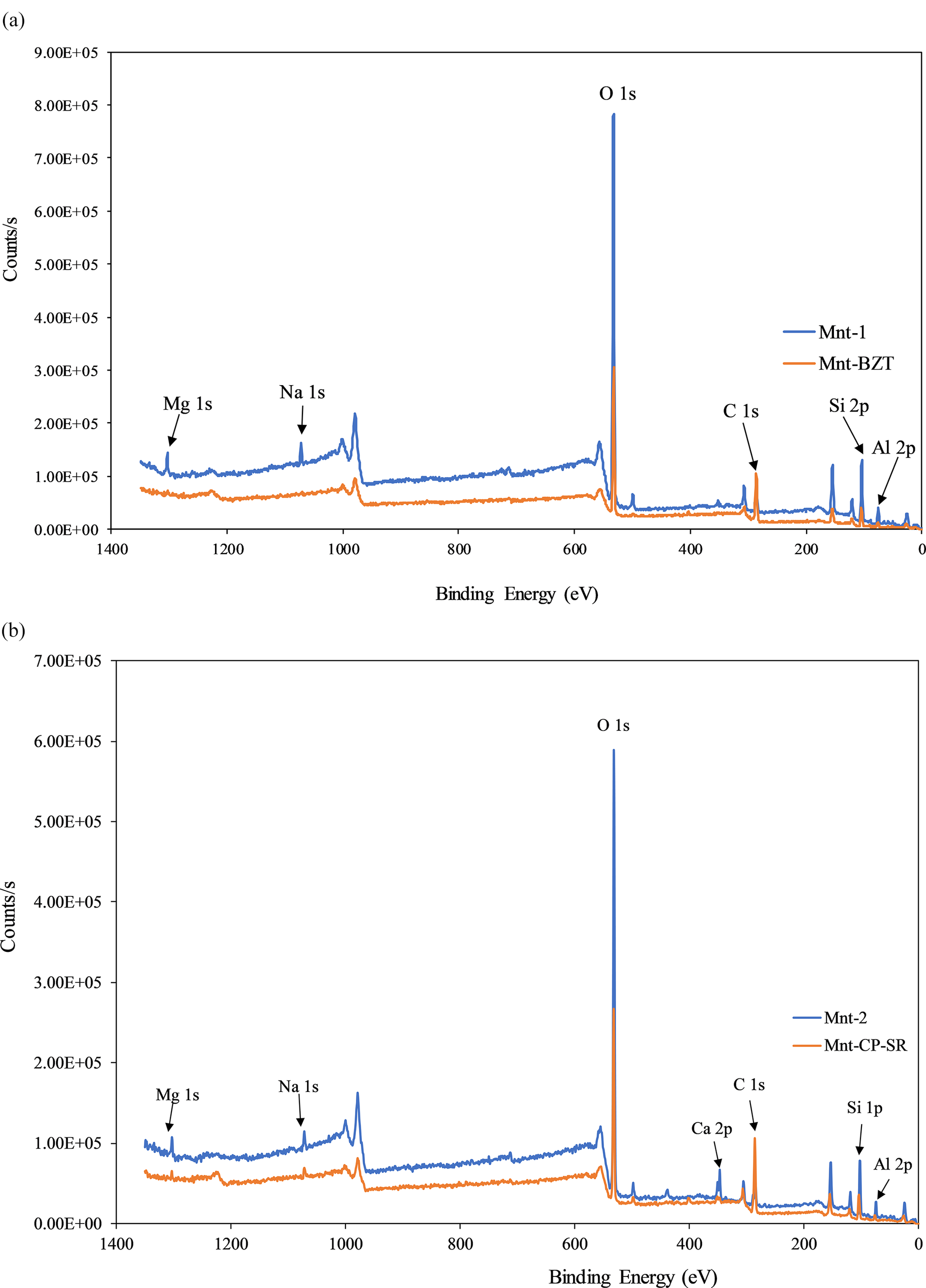
Analysis by XPS was used to obtain information about the uppermost 10 nm of the Mnt and O-Mnt surface by measuring the kinetic energy and quantity of electrons that escape from the top 0–10 nm of the samples. The XPS spectra of Mnt-1 and Mnt-2 (Fig. 2a,b) indicated the presence of O, Si, Al, Mg, Na, Ca, and C. The observation of a C 1s signal with greater intensity in Mnt-CP-SR and Mnt-BZT was assigned to the adsorption and intercalation of surfactants. The Al 2p peak was visible at ~75 eV, while Na 1s vanished after modification with BZT. However, a lower-intensity Na 1s peak appeared in the XPS spectrum of the Mnt-CP-SR, indicating the successful adsorption/intercalation of N-lauroyl sarcosinate Na salt into the Mnt-CP structure.
XPS spectra of: (a) Mnt-1 and Mnt-BZT; (b) Mnt-2 and Mnt-CP-SR.
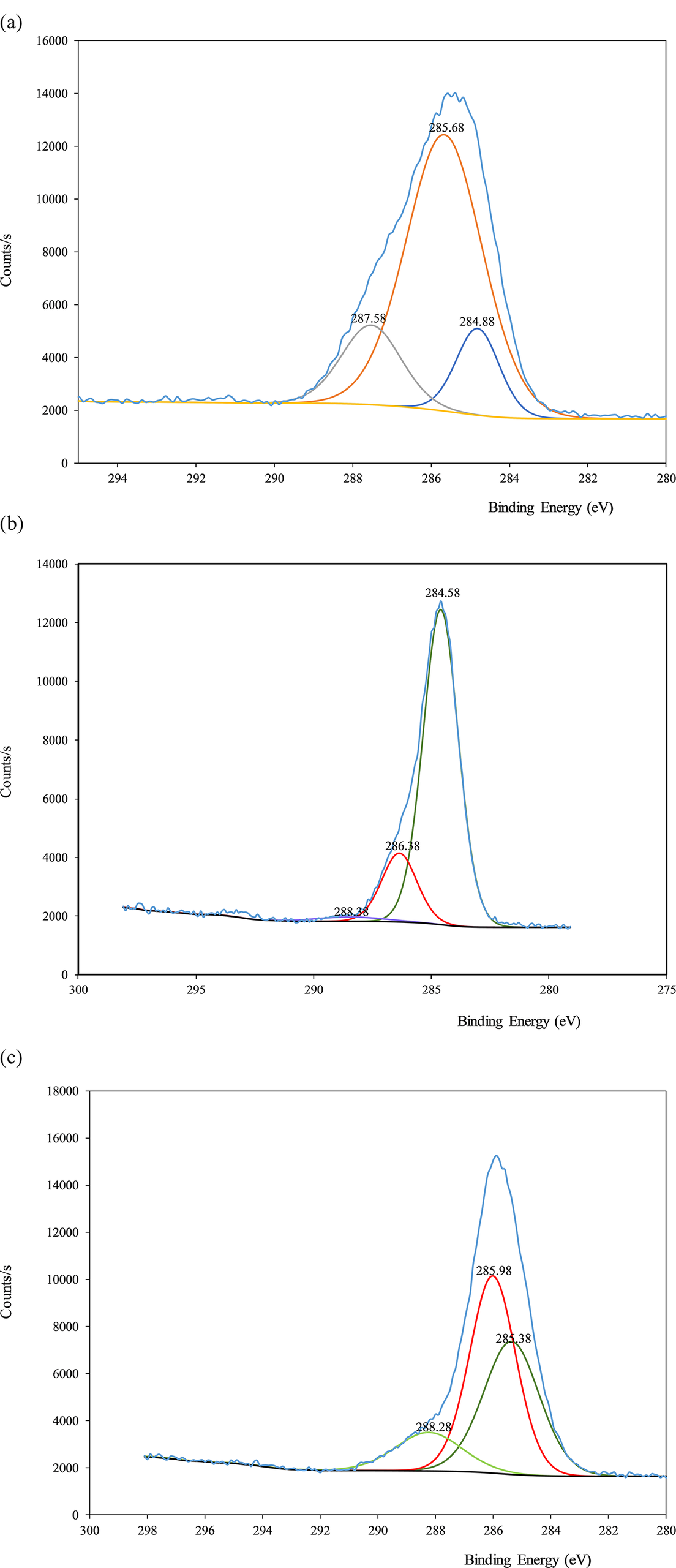
The high-resolution C 1s spectrum of Mnt-BZT (Fig. 3a) is characterized by three transitions centered at 287.58 eV, 285.68 eV, and 284.88 eV, corresponding to the C–N+ bond, C–N/C–H bonds, and C–C bond in the long chain and/or benzene ring, respectively (He et al., Reference He, Zhou, Frost, Wood, Duong and Kloprogge2007; Liu et al., Reference Liu, Gu, Yalin, Asif, Xiong, Zhang and Liu2017).
High-resolution XPS spectra of: (a) C 1s for Mnt-BZT; (b) C 1s region for Mnt-CP; (c) C 1s region for Mnt-CP-SR.

The XPS characterization of the Mnt-CP-SR was compared with that of the Mnt-CP samples to clarify the structures. The C 1s region of Mnt-CP showed three peaks at 288.38 eV, 286.38 eV, and 284.58 eV, assigned to the C–N bond, C–C bond in the long chain, and C–C/C–N bonds, respectively (Fig. 3b) (He et al., Reference He, Zhou, Frost, Wood, Duong and Kloprogge2007; Liu et al., Reference Liu, Gu, Yalin, Asif, Xiong, Zhang and Liu2017). On the other hand, the C 1s region of Mnt-CP-SR showed three transitions at 288.28 eV (C–N bond), 285.98 eV (C=O bond centered), and 285.38 eV (C–C bond) (Fig. 3c).
The change in binding energies and their corresponding structures in the XPS spectra of the C 1s region of Mnt-CP and Mnt-CP-SR samples indicated that SR molecules were adsorbed successfully into the Mnt-CP structure (Table 1). The variation of the binding energy assigned to C–C bonds in the range 284.58–285.38 eV was attributed to the interaction between the alkyl chains of the surfactants. As shown by the molecular modeling studies, the head groups (nitrogen) of alkyl chains are believed to be close to the Mnt surface due to the strong electrostatic interaction between the negatively charged Mnt surface and the positive (quaternary ammonium) head groups (Čapková et al., Reference Čapková, Burda, Weiss and Schenk1999; Zeng et al., Reference Zeng, Yu, Lu and Standish2004; He et al., Reference He, Galy and Gerard2005; He et al., Reference He, Zhou, Frost, Wood, Duong and Kloprogge2007). Owing to the charge localization on the pyridinium ring, the head groups of the CP+ interact electrostatically with the negative charge delocalized on the Mnt surface (Schampera et al., Reference Schampera, Solc, Woche, Mikutta, Dultz, Guggenberger and Tunega2015).
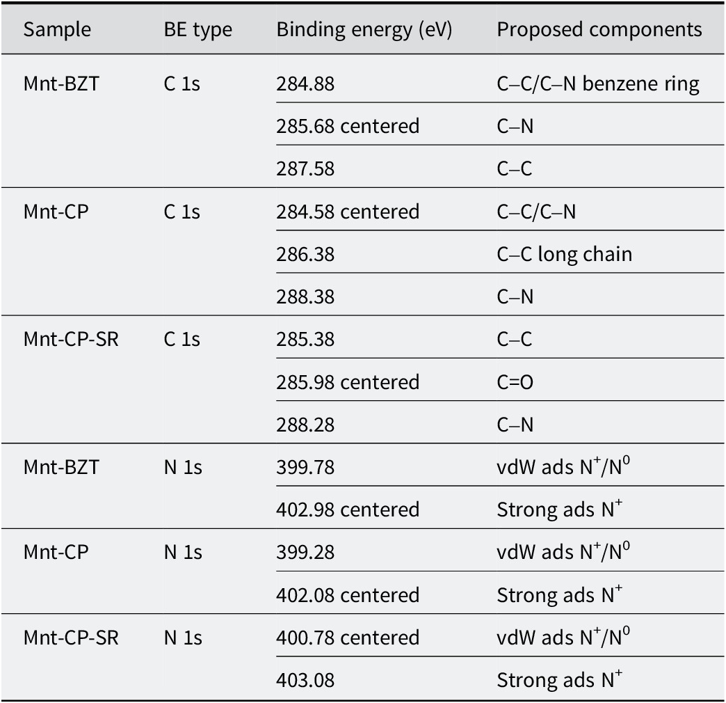
The binding energies and proposed components in XPS spectra of the C 1s and N 1s regions for the samples

vdW ads = van der Waals adsorption.
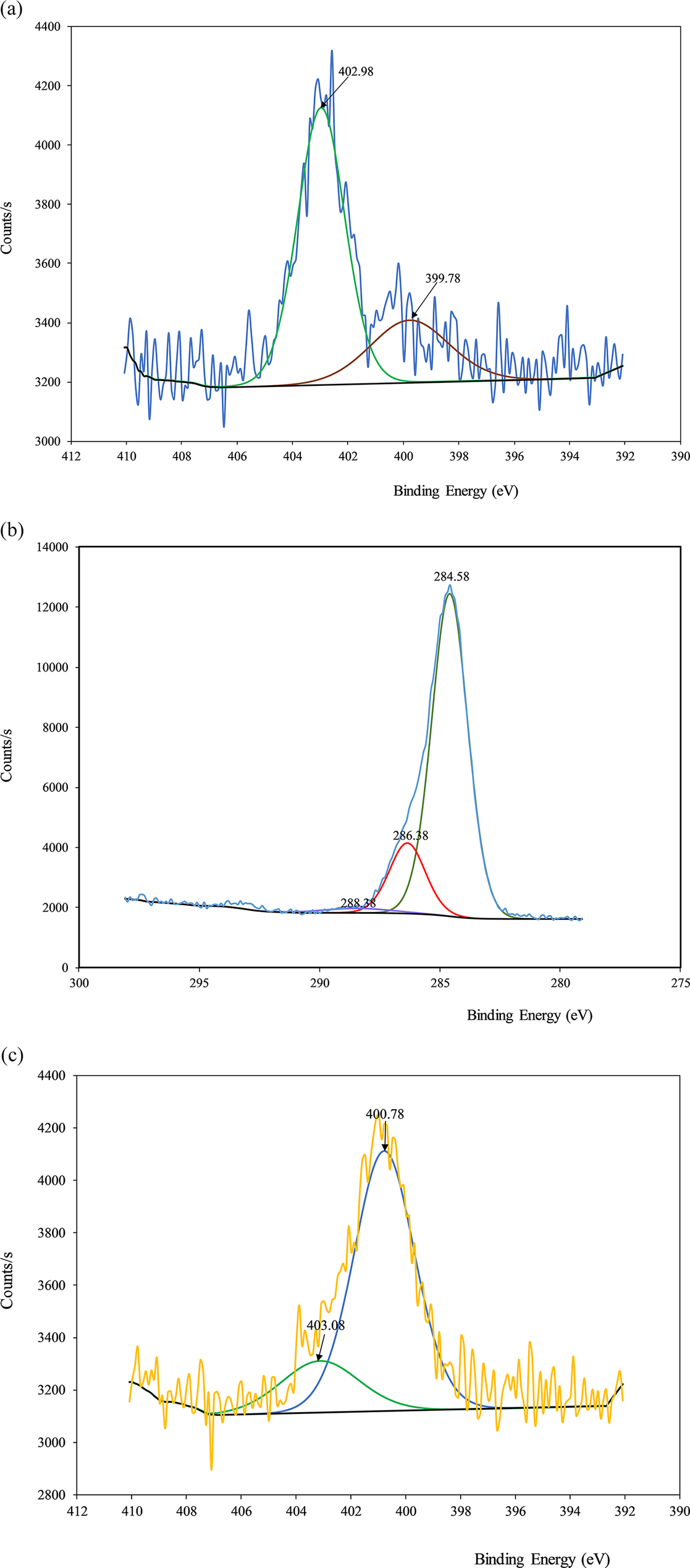
Most nitrogen atoms in molecules bound to carbon atoms, including nitrogen in amines, amides, nitriles, ureas, and aromatic rings, exhibit N 1s binding energies in the range 399–400 eV. However, consistent with the localized positive charge, quaternary nitrogen shows larger binding energies in the 401.5–402.5 eV region (Table 1) (Briggs, Reference Briggs1998; Liu et al., Reference Liu, Wu, van de Ven, Molenaar and Besamusca2010; Naranjo et al., Reference Naranjo, Sham, Rodriguez-Castellon, Torres-Sanchez and Farfan-Torres2013).
From the high-resolution XPS spectra of N 1s for Mnt-BZT and Mnt-CP (Fig. 4a,b), two peaks were obtained with binding energies of 399.78 eV (N+/N0) and 402.1 eV (Mnt-BZT) and similarly 399.28 eV and 402.08 eV (Mnt-CP), respectively. The peak centered at 400 eV was attributed to N+ in the quaternized nitrogen atom in BZT and CP (Melinte et al., Reference Melinte, Buruianã and Buruianã2015). The existence of the peaks showed that two different types of adsorption of surfactant molecules to the surface occurred; one with low binding energy with van der Waals (vdW) interactions and the main part with greater binding energy and electrostatic interactions. Considering Mnt-CP and Mnt-CP-SR (Fig. 4b,c), the binding energy of nitrogen changed from 399.28 eV to 400.78 eV (centered peak) with SR adsorption on Mnt-CP. On the other hand, in the second peak, the strong binding energy changed from 402.08 eV (centered peak) to 403.08 eV with SR adsorption. The change in the centered peaks with SR adsorption on Mnt-CP indicated that part of the N 1s behavior of nitrogen in Mnt-CP-SR is vdW in nature. In samples with greater cation/anion loadings, peaks with lower binding energies suggest that some positively/negatively charged heads do not interact directly with the clay surface, but are embedded between flexible aliphatic chains, emphasizing a more complex surface arrangement (Schampera et al., Reference Schampera, Solc, Woche, Mikutta, Dultz, Guggenberger and Tunega2015). This result showed that the alkyl chains of CP and SR were directed outward and proved the contact killing mechanism based on the interaction between alkyl chains and bacterial cell walls.
High-resolution XPS spectra of the N 1s region of: (a) Mnt-BZT; (b) Mnt-CP; (c) Mnt-CP-SR.

The XPS spectrum of the Al 2p region of Mnt-BZT showed a slight shift to a greater binding energy value and a decrease in the intensity of the signal compared with that of Mnt-1 (Table 2). This observation could be explained by the electrostatic interaction between the organic cations and the external Mnt surface. The decrease in intensity is attributed to the external surface being covered by benzene rings and tetrahedrally structured tetramethylbutyl moiety. The findings indicated that interactions between the benzene rings and cell walls were possible in addition to interactions with the tetramethylbutyl moiety covering the external surface.
The peaks and corresponding binding energies (BE) at the Al 2p and O 1s regions of Mnt-1, Mnt-BZT, Mnt-2, Mnt-CP, and Mnt-CP-SR spectra
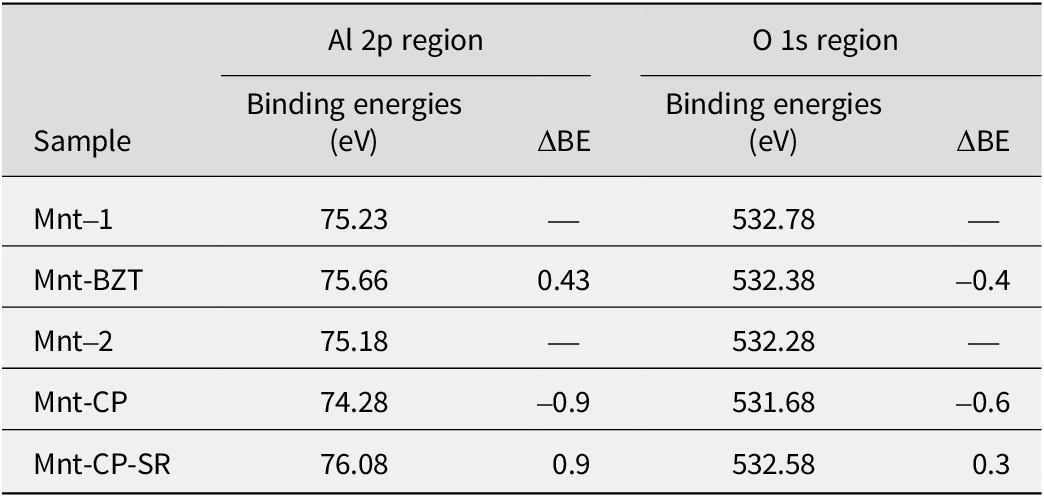
On the other hand, when the XPS spectrum of Mnt-CP in the Al 2p and O 1s regions was compared with that of Mnt-2, a decrease in binding energies in both Al 2p and O 1s spectral regions was observed (Table 2). In the comparison of the Mnt-CP-SR spectrum with that of Mnt-2, a larger shift toward a higher-energy value for Al 2p (from 75.15 to 76.08) and a decrease in electron density were observed. This suggested that the quaternary ammonium N+ of the pyridinium head group binds more strongly to the Mnt surface and the C-N group of SR has a weak effect on the electrostatic interaction between the pyridinium ring and negatively charged surface.
Because oxygen is the element that is most exposed on the Mnt surface, the interaction between Mnt and surfactants primarily affects the binding energy of O. The binding energy of O 1s in Mnt-1 and Mnt-2 was ~532 eV, representing oxygen in Si-O(H) and Al(Mg, Fe)-O(H) groups (Zhuang et al., Reference Zhuang, Zhang, Peng, Gao, Pereira and Jaber2019). The lower binding energies of Mnt-BZT and Mnt-CP indicate the existence of a greater electron density around the O atoms, while the greater binding energy level in the O 1s region of Mnt-CP-SR shows the decline of electron density around the O atoms.
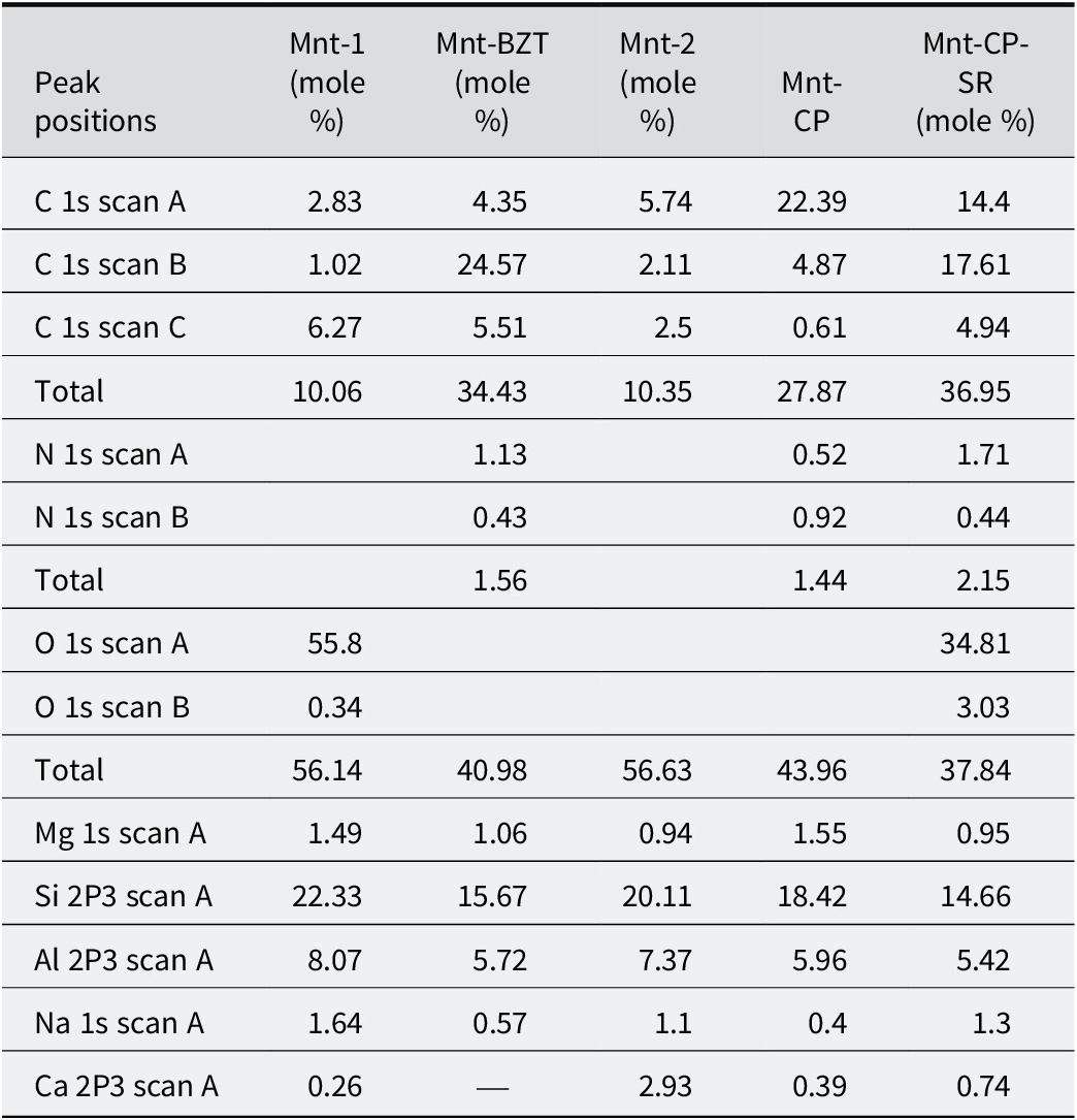
Comparing the surface XPS values of Mnt-1 and Mnt-BZT, an increase in carbon mole fractions (mole %) was observed along with a decrease in Na+ and Ca2+ cations in addition to the existence of nitrogen (Table 3). This observation confirmed the exchange of quaternary ammonium cations, which was also corroborated by other authors (Türker et al., Reference Türker, Yarza, Sánchez and Yapar2017).
Surface XPS values of Mnt-1, Mnt-2, Mnt-BZT, Mnt-CP, and Mnt-CP-SR (mole fractions in %)

The presence of C in Mnt-1 and Mnt-2 was attributed to calcite (Zhuang et al., Reference Zhuang, Zhang, Peng, Gao, Pereira and Jaber2019) and trace amounts of contamination from the plastic vessels used in storage. The increase in mole fractions of carbon and the presence of 2.15 mol% nitrogen in Mnt-CP-SR (Table 3) further confirmed that the modification was successful. Moreover, the variation in the mole fractions of Na+ and Ca2+ was ascribed to the exchange of CP+ and SR– on the external surface. This observation is in agreement with the previous work reporting the distribution of CP+ and SR– between external and interlayer surfaces (Yapar et al., Reference Yapar, Özdemir, Solarte and Sanchez2015). In addition, the same Si/Al ratios (~2.7) for both Mnt and O-Mnt samples indicated that ion exchange did not cause a change in the structure of aluminosilicate nanolayers.
Antibacterial susceptibility tests
To the authors’ knowledge, the opportunistic gram-positive A. viscosus and gram-negative B. fragilis bacteria were subjected, here, for the first time, to an agar well diffusion test, MIC and MBC determinations, and time-kill assay with two different O-Mnts.
Agar well diffusion test results showed that Mnt-CP-SR samples exhibited zone diameters of 26 mm (A. viscosus) and 16.7 mm (B. fragilis), while Mnt-BZT samples exhibited zone diameters of 20 mm (A. viscosus) and 15.2 mm (B. fragilis). CHX used as a positive control had clear zone diameters of 25.5 mm for both bacteria, whereas Mnt-Na used as a negative control showed no zone diameter.
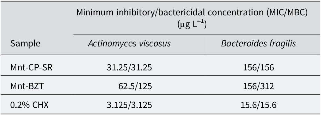
For A. viscosus, a gram-positive and aerobic bacterium, smaller MIC and MBC values were obtained for Mnt-CP-SR compared with Mnt-BZT (Table 4). This result was explained by the different external surface structures of O-Mnts. The XPS results revealed that the external surface of Mnt-BZT was covered with tetrahedral tetramethylbutyl moieties and benzene rings, while the surface of Mnt-CP-SR had double surfactant layers extending into solution.
MIC and MBC values

The larger MIC and MBC values determined for both Mnt-CP-SR and Mnt-BZT for B. fragilis indicated that the samples were less effective against gram-negative anaerobic bacteria. Although the MIC values were the same for both samples, the MBC value of Mnt-BZT was twice that of Mnt-CP-SR, indicating that the gram-negative bacterium cell-wall structures may have played a role. The differences in MIC and MBC values observed for A. viscosus and B. fragilis suggest that the structural characteristics of the bacterial cell walls influence the interaction with O-Mnt surfaces, where the outer membrane of the gram-negative B. fragilis probably limited the penetration or binding of Mnt-BZT and Mnt-CP-SR, whereas the gram-positive A. viscosus exhibited greater susceptibility due to its relatively more permeable peptidoglycan layer.
Comparison of the effects of the amount of O-Mnt used on the minimum contact time for log10 reduction (Table 5) revealed the minimum contact times required for bacterial counts to fall below the detection limit (<1 log10 CFU) for two different concentrations of O-Mnt (3× and 10× MIC). At 3× MIC, Mnt-CP-SR showed significantly shorter contact times than Mnt-BZT for both A. viscosus (6 h vs 12 h) and B. fragilis (3 h vs 12 h). When the concentration was increased to 10× MIC, the contact time decreased dramatically for both formulations. The time required by Mnt-CP-SR dropped to 5 min for A. viscosus and 15 min for B. fragilis. Similarly, Mnt-BZT showed shorter contact times, but remained less effective than Mnt-CP-SR. These results indicate that Mnt-CP-SR is more efficient, and that increasing the concentration improves antibacterial activity. The lower antibacterial activity of Mnt-BZT may be attributed to the presence of double aromatic rings, which could hinder its interaction with bacterial cell membranes.
Minimum contact time for (log10*) reduction
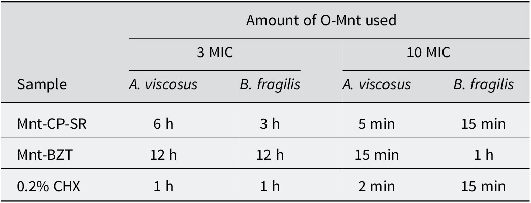
* Minimum contact time was defined as the shortest exposure time at which the bacterial count dropped below the detection limit (<1 log10 CFU).
In summary, comparison of the minimum contact time required for log10 reduction of gram-positive bacteria with that of gram-negative bacteria confirmed the conclusion that both O-Mnt samples were less effective against gram-negative anaerobic bacteria.
In vitro cytotoxicity tests
The supernatant obtained by leaching of O-Mnt with water was inoculated into the cell cultures and in vitro cytotoxicity tests were performed. Previous studies with Mnt-BZT and Mnt-CP-SR showed that Mnt-BZT and Mnt-CP-SR samples have a non-leaching character when washed with water (Yapar et al., Reference Yapar, Özdemir, Solarte and Sanchez2015; Özdemir and Yapar, Reference Özdemir and Yapar2020). However, supernatants from 1:1, 1:2, and 1:4 O-Mnt-water dilutions contained O-Mnt, which adsorbed cationic MTT and inhibited the formation of red formazan dye. This effect led to very low absorbance values in UV measurements, causing false positive results and making the samples appear cytotoxic. Some researchers also reported that this problem, observed in methods using colorimetric dyes to assess the cytotoxicity of nanomaterials, may result in false positive or negative toxic effects due to possible interactions between the material and colorimetric dyes (Herzog et al., Reference Herzog, Casey and Lyng2007; Chung et al., Reference Chung, Kim, Baek, Yu and Choi2011). As a result, O-Mnt samples diluted 1:8 were considered to ensure the correct results of the experiments and the samples were characterized to be non-cytotoxic. Baek et al. (Reference Baek, Lee and Choi2012) also reported that no significant toxicity was found in mice receiving up to 1000 mg kg–1 Mnt, but long-term exposure to large amounts of Mnt may lead to some cytotoxic effects.
In vivo animal toxicity tests
Considering that possible interactions of O-Mnt samples with MTT may lead to false positive/negative results, in vivo toxicity tests were also performed. In animal tests, the method allows the evaluation of acute tests for compounds with a lethal dose. According to the oral acute toxicity test results, no mortality was detected. In light of the OECD (2022) guidelines, the LD50 was found to be >2000 mg kg–1 for both O-Mnt samples, as more than three animals survived for each group. Furthermore, there were no abnormal clinical signs and no changes in body weight gain during the observation period. These findings are in agreement with the results of Baek et al. (Reference Baek, Lee and Choi2012) and healthy animals provide evidence that the antibacterial agents Mnt-CP-SR and Mnt-BZT are as safe as pristine Mnt when administered orally.
Conclusions
The XPS results revealed double surfactant layers extending into solution at the external surface of Mnt-CP-SR and the existence of tetrahedral tetramethylbutyl moieties and benzene rings covering that of the Mnt-BZT. They confirmed the contact killing mechanism by demonstrating the presence of outwardly oriented surfactant moieties on the external surface. The comprehensive examination of XPS analyses highlighted the potential of the study to contribute to the acquisition of new knowledge about different O-Mnt species. This research will pave the way for future studies involving XPS analyses of more complex O-Mnt samples. Antibacterial susceptibility tests showed that the antibacterial properties of both O-Mnts were superior for A. viscosus and slightly less effective for B. fragilis. Mnt-CP-SR was observed to be marginally more effective than Mnt-BZT on B. fragilis. In vitro cytotoxicity tests and in vivo animal toxicity studies showed that these substances were non-toxic. It was concluded that both O-Mnt samples have the potential to be used in topical and oral antibacterial treatments for short- or long-term use as promising antibacterial agents, depending on the field of application.
Author contributions
Conducting antibacterial susceptibility tests: A.Ş.; Conducting in vitro cytotoxicity tests and in vivo animal toxicity tests: C.K.K.; Preparation of O-Mnt and writing: G.Ö., S.Y.
Financial support
The authors gratefully acknowledge the support of Scientific Research Projects of Ege University (BAP) through the project number FGA-2019-20715.
Competing interests
The authors declare no competing interests.
Data availability statement
Data will be provided upon request.


 Open access