





Introduction
The ductal origin of the pulmonary artery is an exceedingly rare malformation, estimated to occur in approximately 1 in 200,000 cases. Reference Khoshhal, Al-Mutairi, Morsy, Kreary, Alnajjar and Abo-Haded1 While more commonly observed in association with complex congenital heart diseases, Reference Trivedi, Karamlou, Yoo, Williams, Freedom and McCrindle2 such as tetralogy of Fallot, pulmonary atresia with ventricular septal defect, and heterotaxia syndrome, it can also manifest as an isolated anomaly.
A hallmark of this anomaly is the contralateral location of the ectopic pulmonary artery relative to the aortic arch. Typically asymptomatic, this condition may frequently lead to the misdiagnosis of congenital “absence” of the pulmonary artery. Furthermore, bilateral patent ducti can be mistaken for aortopulmonary collaterals if a comprehensive full-colour and spectral Doppler assessment is not conducted. Reference Rozema, Ashwath and Snyder3
Closure of the ductus arteriosus results in a gradual decline in blood flow to the dependent pulmonary artery, with perfusion of the affected lung becoming reliant on either the bronchial or anomalous systemic collateral arteries, often leading to multisystem morbidity, predisposing patients to recurrent pneumonias, haemoptysis, and the eventual development of pulmonary hypertension.
Early intervention is imperative to support the growth of the disconnected pulmonary artery, but the frequent diminutive size of this vessel and its inadequate blood flow often make it unsuitable for immediate surgical correction. Reference Butera, Giusti and Pomè4
We present a unique case of a newborn with DiGeorge syndrome, no associated intracardiac defects, a right aortic arch with an aberrant left subclavian artery, and a discontinuous left pulmonary artery (LPA). The case highlights the diagnostic evolution from a prenatal suspicion of aortic origin to the postnatal confirmation of a carotid origin via a ductus arteriosus and discusses the subsequent hybrid management.
Case presentation
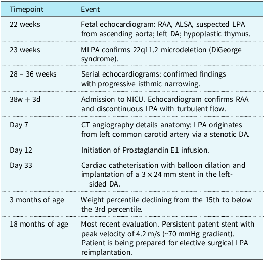
A male infant was born at 38 weeks and 3 days of gestation to a 40-year-old multiparous woman. The mother’s history included being a carrier of Duchenne muscular dystrophy and a prior pregnancy termination following a prenatal diagnosis of trisomy 21. Fetal echocardiography at 22 weeks had identified a right aortic arch with an aberrant left subclavian artery, left ductus arteriosus and raised the suspicion of a left pulmonary artery arising from the ascending aorta (Figure 1). Genetic testing via Multiplex Ligation-dependent Probe Amplification confirmed a 22q11.2 microdeletion, diagnosing DiGeorge syndrome. Serial fetal echocardiograms also demonstrated progressive isthmus narrowing, with a minimum Z-score −2.44 at 36 weeks of gestation. The timeline of diagnostic and therapeutic events is presented in Table 1.
Fetal echocardiography. ( a ) Four-chamber view demonstrating atrioventricular concordance and balanced ventricular dimensions; ( b ) Three-vessel and trachea (3VT) view showing a right-sided aortic arch and right-sided ductus arteriosus, both located to the right of the trachea; ( c ) and ( d ) Sequential images from the same sweep: the four-chamber view ( c ) confirming levocardia and a 3VT view ( d ) demonstrating a right-sided “V-sign” formed by the aortic arch and ductus arteriosus (DA); ( e ) Colour Doppler imaging in a modified view, showing a vessel arising from the aorta and directed towards the left lung, consistent with the left pulmonary artery (LPA); ( f ) Colour Doppler imaging demonstrating the right pulmonary artery (RPA) arising from the main pulmonary artery (MPA). The DA, which connects the MPA to the descending aorta, is also visible.

The patient was born weighing 2970g, did not require resuscitation, and was admitted to the Neonatal Intensive Care Unit for monitoring. Physical examination was unremarkable: the infant was haemodynamically stable, with normal peripheral oxygen saturation, arterial pressure, and serum calcium levels. No dysmorphic features or heart murmurs were noted.
Initial postnatal echocardiography confirmed a right aortic arch, with the left pulmonary artery appearing discontinuous from the main pulmonary artery and seemingly arising from the aortic arch. On subsequent echocardiographic evaluations, progressive turbulence developed across the left pulmonary artery, with continuous-wave Doppler demonstrating a peak velocity of 3.9 m/s (estimated peak gradient of 60 mmHg), with reduced left pulmonary venous return.
Definitive anatomical detail was provided by thoracic computed tomography (CT) angiography on day 7 (Figure 2). It confirmed a right aortic arch with mild isthmus narrowing and an aberrant left subclavian artery. Additionally, an anomalous vessel emerged from the left common carotid artery, proximal to its bifurcation, coursing leftward and posteriorly towards the left pulmonary hilum, where it continued as the left pulmonary artery. A stenotic portion measuring 2.8 mm (Z-score: −2.33) with an extent of 2 mm was present at its origin (Figure 2( a ) and ( b )).
Postnatal CT angiography - ( a ) Volume-rendered reconstruction showing the right aortic arch. An anomalous vessel arises from the left common carotid artery (LCCA); ( b ) Detailed view highlighting the stenotic ductal origin (circle) of the left pulmonary artery (LPA). LSA = left subclavian artery; RSA = right subclavian artery; RCCA, = right common carotid artery.

Figure 2 Long description
Panel A: A volume-rendered reconstruction showing the right aortic arch. An anomalous vessel arises from the left common carotid artery. The diagram includes labels for the left common carotid artery, right common carotid artery, left subclavian artery, and right subclavian artery. Panel B: A detailed view highlighting the stenotic ductal origin of the left pulmonary artery, marked by a red circle. The left pulmonary artery is also labeled.
Given the new onset of left pulmonary artery stenosis, in contrast to the laminar flow observed on both prenatal and immediate postnatal imaging, a ductal origin of the left pulmonary artery from a left-sided ductus arteriosus was presumed. Prostaglandin E1 infusion was initiated to maintain ductal patency, resulting in a reduction of the peak gradient to approximately 40–50 mmHg and improved left venous return. Given the duct-dependent, diminutive, and stenotic nature of the vessel, immediate surgical reimplantation was deemed.
On day 33, percutaneous cardiac catheterisation was performed. The left-sided ductus arteriosus was dilated, and a 3 × 24 mm Ultimaster Nagomi™ stent was successfully implanted (Figure 3).
Cardiac catheterisation and ductal stenting - ( a ) Anteroposterior angiogram demonstrating anomalous origin of the left pulmonary artery (LPA) from the left common carotid artery via a stenotic ductal connection; ( b ) Balloon angioplasty across the stenotic left ductus arteriosus (DA); ( c ) Deployment of a Ultimaster Nagomi™ stent; ( d ) Final angiography showing improved opacification of the left pulmonary artery and enhanced perfusion of the left lung following stent implantation.

The patient was discharged on day 37 on aspirin and oral diuretics. Follow-up demonstrated progressive changes over time. While the stent remained patent, serial echocardiograms showed a progressive increase in the peak velocity through the stent, from 3.2 m/s (peak gradient ∼40 mmHg) at one month to 4.2 m/s (peak gradient ∼70 mmHg) at the most recent evaluation at 18 months of age. Additionally, from 3 months of age, the patient demonstrated a progressive decline in weight, from the 15th to below the 3rd percentile. This decline was associated with an exacerbation of symptoms, manifesting as increased sweating and fatigue, following the mother’s independent decision to discontinue diuretics. These symptoms improved once the patient resumed the medication. Although other clinical signs of decompensated heart failure were absent, these findings, together with the echocardiographic progression, raised significant concern. He is currently undergoing active medical management and optimisation in preparation for elective surgical left pulmonary artery reimplantation, which has not yet taken place due to maternal refusal.
Discussion
The heterogeneous anomalies observed in the aortic arch can be explained by Edwards’ double aortic arch model, Reference Edwards5 where aberrant persistence or regression of different segments can result in a spectrum of different malformations. The presented anomaly arises from abnormal involution of the left fourth arch situated between the left subclavian and left common carotid artery. This leads to a right aortic arch and aberrant left subclavian artery, alongside regression of the proximal sixth branchial arch and persistence of its distal portion. Consequently, an ectopic pulmonary artery and a ductus, typically located on the right side, manifest without the formation of a vascular ring (Figure 4).
In patients with discontinuity of the pulmonary arteries and structurally normal hearts, the most prevalent configuration consists of a left aortic arch with a right-sided ductus arteriosus and an ectopic distal ductal origin of the right pulmonary artery. Reference Freedom, Moes and Pelech6 Conversely, alternative configurations such as a right aortic arch with a left-sided ductus arteriosus (or ductal remnant) and a distal ductal origin of the left pulmonary artery, with normal regression of the right-sided ductus, are more commonly encountered in patients with congenital heart disease. Reference Priya, Thomas, Nagpal, Sharma and Steigner7
Microdeletions in the locus 22q11.24 are frequently associated with various arch anomalies, Reference Hanneman, Newman and Chan8 suggesting a genetic predisposition as part of the CATCH 22 syndrome complex. Reference Prifti, Bonacchi and Murzi9 )
While a prior report described a similar ectopic pulmonary artery originating from the internal carotid artery in a patient with complex heart disease, Reference Thankavel, Martho and Zeltser10 our case is distinct. As highlighted in an article commentary by Bamforth et al.(11), an origin from the internal carotid implies a more superior, vertical path via the embryological “carotid duct,” a transient embryological structure that connects the third and fourth arches. In contrast, our case demonstrates an origin from the left common carotid artery with a more horizontal course, suggesting persistence of a connection between the distal sixth arch and the aortic sac or proximal fourth arch derivative.
This distinction underscores the diagnostic challenge and the imperative for precise imaging. A “disconnected” pulmonary artery must not be mislabelled as “absent.” Comprehensive echocardiography with spectral Doppler and cross-sectional imaging with CT or MRI is essential to trace the ectopic vessel to its systemic origin, which is most commonly a ductus arteriosus.
Management is dictated by anatomy. Early intervention is crucial to allow the normal development of the pulmonary vascular bed and mitigate the potential for late complications. However, as evidenced in the present case, the diminutive calibre of the ectopic vessel and its inadequate blood flow often preclude immediate surgical intervention. In such cases, palliative ductal stenting has emerged as a viable bridging strategy to maintain perfusion and allow for vessel growth, creating more favourable conditions for later surgery. Reference Khoshhal, Al-Mutairi, Morsy, Kreary, Alnajjar and Abo-Haded1, Reference Rozema, Ashwath and Snyder3 This hybrid approach was successfully applied in our case.
In a study by Marcos et al. in 2010, Reference Crespo Marcos, Adrián Gutiérrez, Alvarez Martín and Zunzunegui Martínez12 a case of ectopic right pulmonary artery originating from the first supraaortic trunk was documented. Similar to our case, treatment with PGE1 was initiated; however, the patient ultimately underwent reimplantation of the right pulmonary artery without prior interventions, resulting in post-operative stenosis requiring multiple percutaneous interventions.
To our knowledge, this represents the first reported case of a patient with DiGeorge syndrome exhibiting a structurally normal heart with a right aortic arch and an aberrant left subclavian artery concomitant with nonconfluent pulmonary arteries, originating from the common carotid artery.
Conclusion
Discontinuous pulmonary arteries represent a heterogeneous spectrum of embryologically complex malformations. This case of a carotid-origin left pulmonary artery underscores the essential role of a high clinical index of suspicion coupled with systematic multimodal imaging to differentiate a ductal-dependent vessel from a truly absent artery. It demonstrates that percutaneous ductal stenting provides effective temporary palliation, while also emphasising that vigilant serial monitoring for rising gradients or clinical compromise is critical to determine the optimal timing for definitive surgical repair. A tailored, multidisciplinary approach is fundamental for achieving optimal outcomes.
Schematic representation based on Edwards’ double aortic arch model. The diagram illustrates a right aortic arch with an aberrant left subclavian artery (LSA) arising from the descending aorta. Selective regression of the left fourth aortic arch between the left common carotid artery (LCCA) and LSA results in the right-sided aortic arch configuration. Regression of the proximal left sixth aortic arch leads to discontinuity of the left pulmonary artery (LPA) from the main pulmonary artery (MPA), while persistence of the distal left sixth aortic arch forms a left-sided ductus arteriosus (DA) connecting the LCCA to the LPA, rendering the LPA ductal-dependent. Arteries emerging from the aortic arch, in order: LCCA, LSA = right subclavian artery (RSA) = right common carotid artery (RCCA). Illustration created by Gonçalo Borges.

Figure 4 Long description
A schematic representation of the Edwards double aortic arch model. The diagram illustrates a right aortic arch with an aberrant left subclavian artery (LSA) arising from the descending aorta. The arteries emerging from the aortic arch, in order, are the left common carotid artery (LCCA), the left subclavian artery (LSA), the right subclavian artery (RSA), and the right common carotid artery (RCCA). Selective regression of the left fourth aortic arch between the LCCA and LSA results in the right-sided aortic arch configuration. Regression of the proximal left sixth aortic arch leads to discontinuity of the left pulmonary artery (LPA) from the main pulmonary artery (MPA), while persistence of the distal left sixth aortic arch forms a left-sided ductus arteriosus (DA) connecting the LCCA to the LPA, rendering the LPA ductal-dependent.
Timeline of diagnostic and therapeutic events

Learning points
-
• DiGeorge syndrome can be associated with extremely rare and complex aorticopulmonary connections. A detailed prenatal and postnatal arch evaluation is mandatory.
-
• A “disconnected” or “absent” pulmonary artery should prompt a search for an ectopic systemic origin, often via a ductus arteriosus. Comprehensive imaging is essential to define the anatomy and avoid misdiagnosis.
-
• In ductal-dependent discontinuous pulmonary arteries with a stenotic, diminutive vessel, percutaneous ductal stenting is an effective palliative strategy to maintain flow, promote growth, and serve as a bridge to definitive surgery.
Financial support
This research received no specific grant from any funding agency, commercial or not-for-profit sectors.
Competing interests
None declared.
Ethical standards
The authors confirm that written informed consent for publication of this case report and accompanying images was obtained from the patient’s parents in accordance with COPE guidelines.



 Open access

