

1. Introduction
In Germany alone, each year approximately 270,000 people suffer from a stroke. As a consequence, about 25% of the survivors develop long-term neurological damage, which subsequently leads to severe handicaps in daily life (Robert-Koch-Institut, 2015). A common handicap is the unilateral sensorimotor arm paresis - a disorder of the motor and sensory functions of the arm and hand limited to one side of the body (Reference NellesNelles, 2004) that causes lack of sensation and muscle control. Beyond the substantial impact on daily life, stroke also represents one of the costliest diseases for healthcare systems (Reference Höer, Schiffhorst and BerkemeierHöer et al., 2023). Therefore, early and effective treatment is crucial to improving patient outcomes and reducing long-term economic burdens.
Therapeutic approaches for recovering arm and hand functionality typically target the human ability to (re-)learn skills through practice and repetition (Reference Taub, Uswatte and ElbertTaub et al., 2002). An elementary prerequisite for success in this regard is an intact feedback loop, which enables the patient to assess the effect of their efforts and to adapt their strategy of problem solving accordingly. Even though some degree of voluntary activity of the muscles remains, patients with arm paresis often lack the ability to perform visible movements. As a result, they cannot assess the effectiveness of their efforts, and the feedback loop is impaired. An established approach to solve this problem is called biofeedback therapy. Here, biomedical sensors are used to measure imperceptible processes in the user's body and provide them with visual or auditory feedback, thus closing the feedback loop again (Reference Haus, Held, Kowalski, Krombholz, Nowak, Schneider, Strauß and WiedemannHaus et al., 2016). For the therapy of sensorimotor dysfunctions, surface electromyography (sEMG) based biofeedback is particularly suitable. Here, muscle activity in the targeted body region is recorded non-invasively with electrodes applied to the surface of the skin (Reference Giggins, Persson and CaulfieldGiggins et al., 2013). Even though the use of sEMG biofeedback therapy for the training of volitional motor activity is an established approach in the clinical treatment of stroke induced paresis, its applicability for arm paresis is currently limited to the training of rudimentary hand movements (see. e.g. the Brucker-biofeedback-method (Brucker, 2006)). As everyday activities, like grasping or manipulating small objects, involve the fine motor control of individual fingers though, a technical solution that enables the training of fine motor control on the level of individual fingers would be useful.
One of the main limitations of current sEMG biofeedback systems is that they rely on rather large electrodes, that are manually positioned based on palpation. As the anatomy of the forearm, with its many small, layered muscles, is rather complex (Reference KapandjiKapandji, 2016), and there is significant crosstalk (Reference Mogk and KeirMogk & Keir, 2003), these systems measure the combined activity of multiple muscles at once. A possible approach is therefore to develop a system with smaller, more sensitive electrodes and place them in areas, where they only capture the activity of individual muscles. This is no trivial task though, as it is not clear, if these areas exist and where they are, as standard literature for recommended placement of sEMG electrodes does not contain this information (compare (Reference Barbero, Merletti and RainoldiBarbero et al., 2012; Criswell & Cram, Reference Criswell and Cram2011; Reference Hermens, Freriks, Merletti, Stegeman, Blok, Rau, Klug, Hägg and BlokH. Hermens et al., 1999)) and differences in optimal placement due to interindividual variations of the anatomy are to be expected. While there has been research on the distribution of forearm sEMG amplitude during isolated finger movements, this research is incomprehensive with regard to the range of investigated finger movements and does not provide solutions on how to handle crosstalk during combined movements (Reference Beek, Stegeman, Noort, Veeger and MaasBeek et al., 2018; Gallina & Botter, Reference Gallina and Botter2013; Reference Hu, Suresh, Xue and RymerHu et al., 2015).
Our goal is therefore to develop a system that can differentiate individual extrinsic finger muscle activity based on carefully positioned, small electrodes. To account for requirements regarding technical complexity and cost, our goal is to use the minimal amount of electrodes necessary. In order to avoid the need for electrode redundancy to compensate for interpersonal variations in forearm anatomy, the system relies on a patient-specific forearm sleeve with individualized electrode positions. This paper documents our current development progress. We show that areas with high selectivity for most of the extrinsic finger muscles in the forearm exist and that their locations vary between individuals. We also present our concept for identifying these positions, testing their suitability and using this knowledge to derive an individualized forearm sleeve with a simplified electrode configuration. The development of this process is based on an extensive analysis of measurement data acquired in a small study involving seven subjects. Section 2 details our experimental procedure and describes, how we used custom hard- and software to gather the necessary data. Section 3 describes our measurement results and how we use them to derive suitable electrode positions for a simplified system. Section 4 discusses our current progress towards our overall objective and concludes the necessary steps regarding evaluation and further research.
2. Experimental approach
To generate the necessary data for our design process, we conducted an experimental study. In this study, seven healthy subjects performed isolated and combined isometric contractions of finger muscles with varying force, while the sEMG signals on the forearm were recorded.
2.1. Data acquisition with a custom-built experimental setup
For the study, we developed custom experimental hard- and software. On the hardware side, we built a universal forearm sleeve with 96 integrated electrodes to capture sEMG data across a wide portion of the forearm. As we wanted to have a reference for the degree of muscle activation, we developed a hand force meter and used it to monitor hand and finger forces. To automate the experimental process as good as possible, we developed custom measurement software. The following section describes the setup in detail.
2.1.1. Measuring hand and finger forces
The developed hand-force-meter uses load cells to measure hand- and finger forces. Its surface is formed in a way, that the subject's hand can comfortably be placed on top, with the fingertips resting on individual pressure plates, where they are fixated with Velcro. The pressure plates are connected to the internal load cells, that are arranged in a way that the device can measure the forces resulting from flexion and extension of each individual finger, abduction and adduction of the thumb, as well as flexion, extension, radial- and ulnar abduction of the wrist. Arm rests at the wrist and elbow location support the forearm in its position.
2.1.2. Measuring surface electromyographic signals
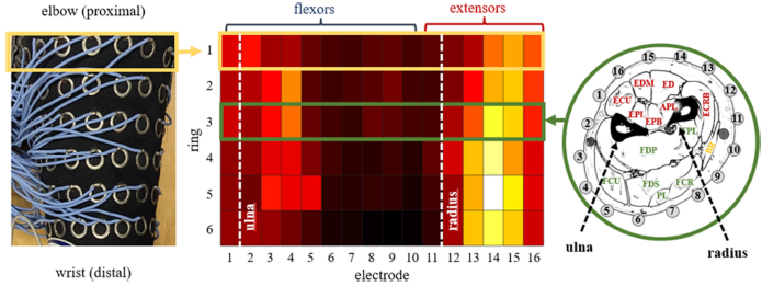
The 96 electrodes of the forearm sleeve are arranged in 6 rings, each containing 16 evenly spaced electrodes (Figure 1). The distance between each ring is 20 mm, resulting in a matrix of about 100 mm length. The sleeve is positioned in a way that electrode 1 of each ring is centred on a straight line between the subject's processus styloideus ulnae and the epicondylus lateralis humeri. Electrode 1 of ring 6 sits at 23% of the distance between those two reference points. This way, the electrode matrix covers between 35% and 44% of the subject's forearm, depending on its length, where most of the muscle bellies of extrinsic forearm muscles are located (Reference Hirt, Seyhan, Wagner and ZumhaschHirt et al., 2015). With our setup, we differ from other publications examining the distribution of sEMG amplitude during finger movements (Reference Beek, Stegeman, Noort, Veeger and MaasBeek et al., 2018; Gallina & Botter, Reference Gallina and Botter2013; Reference Hu, Suresh, Xue and RymerHu et al., 2015), where sEMG signals are recorded with rectangular electrode matrices that are positioned on the ventral or dorsal side of the proximal forearm. Based on anatomical considerations and the results of the studies, we feel that these setups omit potentially relevant data from the areas at the sides and the distal parts of the forearm, so we chose a continuous matrix and positioned it further towards the distal side of the forearm. Two Tower-of-Measurement (ToM) A/D-converters and six ToEM16G bio-amplifiers by German company DeMeTec were used to measure sEMG signals at the 96 electrode sites with a sample rate of 1024 Hz.
Forearm in measurement position with electrode sleeve applied and hand resting on the hand-force-meter. Sectional views from the literature (Reference Möller and EmilMöller & Emil, 2008) give an impression of the position of the electrodes in regard to the forearm muscles at different distances from the epicondylus lateralis humeri

2.2. Experimental protocol
Five male and two female subjects between the age of 30 and 40 (34,57 ± 3,55) performed isolated as well as combined isometric contractions of extrinsic finger muscles. BMI ranged from 22.1 to 28.1 (77,50 ± 10,09) kg/m². Forearm length varied between 224 and 280 (249,29 ± 20,44) mm. Participants had no known neuromuscular disorders or disabilities of the upper limb.
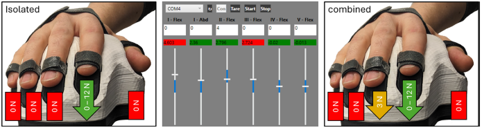
At the beginning of each session, the subject's forearm was cleaned. While the subject assumed measurement posture, the arm's reference points were identified and the necessary marks for positioning the sleeve were applied with a pen. Afterwards, the electrode sleeve was applied, and the subject's task was explained to them. During the experiment, subjects were to adopt and hold different states of hand- and finger-forces. The current state of their applied forces for wrist and fingers were measured with the hand force meter and shown to them in contrast to the target forces (Figure 2, center). sEMG data was measured as long as applied forces stayed within set boundaries. If the subject was able to hold the target state for 3 seconds, the measurement was deemed acceptable, automatically saved and the next target state was presented.
Depiction of subjects' tasks regarding isolated and combined application of finger force

In the first part of the session, subjects were instructed to only apply unidirectional force to a single finger, while applying as little force as possible with the other fingers and wrist (Figure 2, left). Starting at 0 N, target forces for the active finger were incrementally increased until either a predetermined threshold was reached, or subjects were unable to hold the state for 3 seconds even after a couple of tries. In this case, the session was advanced to the next finger. Near the end of the session, when subjects were more comfortable with the setup, task complexity increased, as now two non-zero forces had to be applied simultaneously (Figure 2, right). At the end of the experiment, the electrode sleeve was removed, and actual positioning of the rings was verified by inspecting electrode indentation marks. If there were inaccuracies during application of the sleeve and electrode 1 of a ring was not centred on the line, the distance between electrode and line was measured and put on record as a means for data corrections.
2.3. Data preparation
Each individual sEMG measurement file of 3 seconds was labelled (subject, target state) and the data filtered (bandpass between 10 and 500 Hz, 2 Hz wide notch at 50 Hz). For the 1024 samples between the 1 s and 2 s mark, root-mean-square (RMS) was calculated as a measure of sEMG activity. For the identification and evaluation of activity hotspots, the data was transferred to a heatmap plot and arranged according to the electrode's positions in the matrix, with the colour of each rectangle representing the RMS at one electrode (Figure 3). If necessary for the aspect investigated, normalization across the matrix was performed by subtracting the lowest RMS value of the 96 electrodes and then scaling RMS on all electrodes from 0 to 100 percent. For comparison of the hotspot position between subjects, electrodes with the highest amplitude in the relevant area of the matrix were identified. The position of the electrodes in relation to the reference line and the determined length of the forearm were then used to normalize the position of the peak in relation to the different arm geometries (Figure 4, bottom). For evaluation of the relationship between RMS and applied force, the course of the amplitude at one or more individual electrodes was plotted as line plots.
Relationship between the layout of the calculated sEMG heatmaps and the positions of the sleeve's electrodes derived from (Reference Möller and EmilMöller & Emil, 2008)

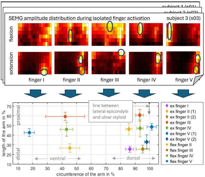
Overview of the peak positions of the identified activity hotspots for each finger and each subject(bottom), extracted from the evaluation of the sEMG amplitude distribution during isolated finger activity (top)

3. Results
The following sections present the results of our study on the identification of characteristic areas of high sEMG amplitude (hotspots) for isolated and combined finger movements. The findings serve as the basis for our concept of the process to derive an individual electrode sleeve that enables the precise recording of the activity of individual extrinsic finger muscles.
3.1. Evaluation of the measurement results
3.1.1. Localization and identification of amplitude hotspots from isolated finger flexion and extension
All finger movements led to an increase in sEMG amplitude across the electrode matrix. As expected, this increase was not limited to the area above agonist muscle bellies but, due to co-contractions of the antagonist muscles, generally extended across both sides of the forearm (Reference Charissou, Amarantini, Baurès, Berton and VigourouxCharissou et al., 2017). From the analysis of the sEMG amplitude distribution, we were able to identify distinct areas of high sEMG activity in the regions of agonist muscles for all isolated isometric contractions (Figure 4, top) except for flexion of finger I.
Compared to the literature (compare (Reference Beek, Stegeman, Noort, Veeger and MaasBeek et al., 2018; Gallina & Botter, Reference Gallina and Botter2013; Reference Hu, Suresh, Xue and RymerHu et al., 2015)), a higher number of distinct hotspots could be identified. Two additional hotspots were identified during individual extension of fingers II and V and can presumably be attributed to the fact, that those two fingers each possess two muscles responsible for extension. We were also able to reliably identify a distinct hotspot for finger I extension. All three newfound hotspots appear near the distal end of the matrix, suggesting that we were able to identify them due to extending our measurement area in this direction compared to previous studies. A comparison of peak positions of the identified activity hotspots for each movement between subjects shows the formation of movement specific clusters (Figure 4, bottom), suggesting that hotspots for the same movement generally form in the same areas of the forearm, even though standard deviation varies between 5 and 20 %. An exception is the hotspot for finger III flexion, where the position varies a lot more in radial direction.
3.1.2. Identification of suitable electrode positions for the differentiation of muscle activity during combined finger movement
For isolated as well as combined finger contraction, sEMG-amplitude across the electrode matrix increased with the amount of force applied. During isolated activation, the relationship between the applied finger force and sEMG amplitude in the finger-specific activity hotspots seems to be approximately proportional. As a tendency, sEMG amplitude per force decreases with distance from the hotspot's peak position and is higher on the extensor side. This is to be expected, as the finger extensors are located closer to the skin compared to the finger flexors.
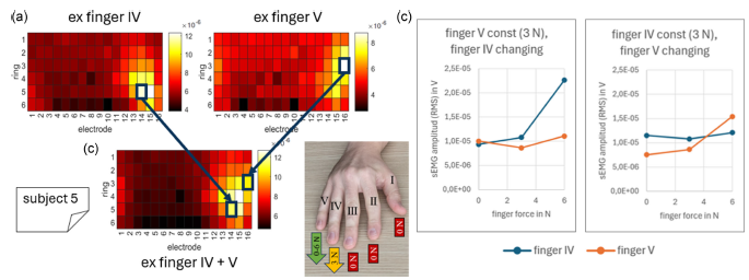
During combined activation of adjacent fingers, an increase of amplitude in the hotspots of both fingers can be observed. This can result in hotspots with high overlap fusing together. Comparing sEMG amplitude per applied force in edge areas of the fused hotspot still shows selectivity towards one single muscle. In Figure 5, this is demonstrated for the example of combined extension of fingers IV and V in the data of subject 5. Figure 5a shows heatmaps that result from individual extension of fingers IV and V, with two distinct hotspots forming in different parts of the map. As these hotspots overlap, they fuse together during combined movements, with the peak amplitude shifting towards the centre of the newly formed hotspot, which can be seen in Figure 5b. Due to our knowledge of the amplitude distribution during individual finger extension, we are able to identify electrodes in the non-overlapping area with potentially high selectivity towards either finger IV or finger V. Figure 5c shows that in these positions, the sEMG signal amplitude for the finger applying constant force stays nearly constant, while for the finger that increases the force, the amplitude at the corresponding electrode also increases.
Identification of suitable electrode positions for the differentiation of individual muscle activity during

3.2. Derivation of a subject individual electrode sleeve with a reduced number of electrodes
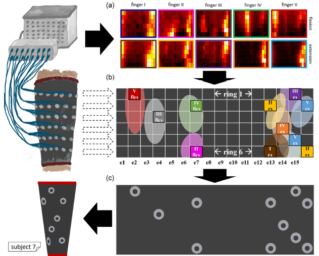
The results from section 3.1 show that a differentiation of individual finger muscle activity through the evaluation of strategically positioned electrodes is possible. However, the high degree of interpersonal variance in the position and shape of the hotspots in combination with the high degree of overlap, especially on the extensor side, implies that the required precision of positioning cannot be achieved with a universal sleeve. Instead, it seems necessary to specifically adapt the position of the electrodes to a person's anatomy. Figure 6 shows the workflow of such an adaptation process using the measurement data from test subject 7 as an example. The process has three steps:
Process for the design of a custom sEMG sleeve with individualized electrode positions based on the evaluation of the high-density measurements of subject 7

In the first step, the position and propagation of the hotspots are identified from the heat maps of the isolated finger extensions and flexions (Figure 6a). In the case of subject 7, two hotspots appear during the extension of fingers II and V, resulting in a total of seven hotspots on the extensor side and four on the flexor side. Figure 6b shows the resulting map, in which the hotspots are approximated as ellipses. An electrode position is then selected for each hotspot. As for the selection process, different criteria can be applied. On one hand, position should be as close as possible to the peak, as this produces the best signal-to-noise ratio and therefore even low muscle activity can be detected. On the other hand, the electrode should be placed in an area with minimal overlap, as this results in increased selectivity of the amplitude with regard to the target movement. It is apparent that this process is much more complicated for the extensors, as hotspots are located within a small space. As predicted in section 2.1.2, the fact, that we use a continuous electrode matrix and therefore have information about sEMG activity in the outer regions of the extensors and flexors, is helpful in this regard. The identified positions form the individual sEMG measurement system and can now be transferred to a new sleeve. This results in a measurement system in which the 96 electrodes have been reduced to 11, each measuring the activity of one extrinsic finger muscle.
4. Discussion and outlook
In this paper, a concept for the design process of an innovative sEMG electrode sleeve for capturing individual finger movements is presented. The system was developed with application in biofeedback therapy in mind, though it might be useful in any application, where the capturing of finger muscle activity is of interest. In contrast to existing biofeedback systems, our approach should allow the identification of individual finger movements with a limited number of small electrodes. The development process is based on an experimental study carried out specifically for this purpose. The results of this study confirm the feasibility of our approach to differentiate the activity of the extrinsic muscles during isolated and combined finger movement by evaluating sEMG amplitude at specific positions of the forearm.
In our analysis, we had variable success in discriminating the individual finger muscles, which can be explained by variations in the anatomy of the subjects with respect to aspects such as length and circumference of the forearm or amount of subcutaneous fat. Although the results from section 3.1.1 show that the hotspots of different subjects generally occur in the same regions of the forearm, the degree of overlap and thus the availability of regions with high selectivity is subject to variation. As mentioned in Section 3.2, we expect the differentiation of finger flexors to be more reliable compared to finger extensors, as the hotspots are more widely distributed despite the lower sEMG amplitude and therefore there are more regions with low overlap. For the derivation and verification of a person-specific sleeve, we have the option of using our electrode matrix to simulate individual electrodes at specific points by using only the identified electrodes in the matrix for evaluation. This is possible because the derived positions are always a subset of all the positions of our measurement system. It is therefore not necessary to produce an individual sleeve for each subject.
The evaluation of our results suggests that our process is suitable for deriving a system that utilises positions for single and combined finger movements with a minimal number of electrodes. On the one hand, our results regarding the formation of sEMG amplitude hotspots during single finger activation are in line with the results of other researchers (Reference Beek, Stegeman, Noort, Veeger and MaasBeek et al., 2018; Gallina & Botter, Reference Gallina and Botter2013; Reference Hu, Suresh, Xue and RymerHu et al., 2015). On the other hand, a comparison of our identified hotspots and the individual positions for the electrodes derived from them with the functional anatomy of the forearm shows a high degree of agreement. However, the quality of our results can be further improved by, for example, increasing the number of test subjects, repeating measurements and analysing the influence of incorrect positioning of the derived electrodes. Another possible step is to include the electrodes and their properties, such as material, shape and size, in the investigation. Since the overlap of hotspots, especially on the finger extensor side and during combined finger activation, remains the primary challenge for identifying suitable electrode positions, our next steps are to further improve and evaluate our initial process concept. One idea to address the differences in the degree of overlap and signal amplitude within the different hotspots could be to use differently sized and shaped electrodes to mimic the derived shapes from 3.2. For example, the electrode size for the finger flexors could be increased to account for the low signal-to-noise ratio. On the extensor side, where amplitudes are high, but areas of low overlap are small, smaller electrodes could be used to increase selectivity. If we find that certain muscles cannot be measured reliably, we could try to add additional electrodes and process their amplitude in a more sophisticated algorithm. To find better positions for certain muscles, such as the extensor muscles of fingers I and III, where hotspots occurred at the outer edges of our matrix, it might be useful to further extend our matrix in both directions. This could also allow us to identify the activity of muscles that we cannot currently recognise. This would be particularly important for the flexion of finger I, as it plays an important role in most hand movements.
Our overall goal is, of course, to develop a product for people with arm paresis. Due to the highly individual characteristics of the symptoms, we initially based our experiments on healthy subjects, to develop a general approach. After a successful evaluation with healthy subjects, it is necessary to investigate how it can be applied to paralyzed individuals, as certain aspects can be expected to be different, making certain adjustments to our process necessary. For one, sEMG signal strength should be expected to be a lot lower in patients with paresis. Even though sEMG can, in principle, be used to detect muscle activity so small that it does not even trigger any movement (Reference Papazian, Baicoianu, Peters, Feldner and SteelePapazian et al., 2021), we do not know whether this also applies to our use case, where we try to measure small individual muscles with small electrodes. We also do not know, to what extent affected persons will be able to produce clearly distinguishable amplitude hotspots, that currently form the basis of our process. Depending on the results, it might be necessary to develop a more sophisticated approach to identify hotspot positions and generate the hotspot map.
5. Conclusion
This paper presents a new approach to designing an sEMG electrode sleeve capable of differentiating individual finger movements, a capability that has potential applications in biofeedback therapy and beyond. By evaluating sEMG amplitude distributions along the forearm, we identified distinct hotspots associated with isolated and combined finger activity. Although our findings are based on a limited number of subjects and conditions, the observed hotspots - especially for the isolated finger activities - and their distribution patterns are consistent with both established literature and functional forearm anatomy.
While the concept demonstrates potential, certain limitations warrant further optimization. Overlapping activity on the extensor side, the absence of distinct hotspots for some movements, and anatomical variations between subjects underscore the need for precise electrode placement and process refinement. Future efforts will concentrate on verifying the reliability of selected electrode positions, refining electrode configurations, and exploring variations in electrode size, shape, and placement.
Our objective is to develop a personalised electrode sleeve for patients with arm paresis and to optimise the fitting process itself so that individualised electrode arrangements can be efficiently generated and integrated into their rehabilitation. These measures are essential for ensuring the system's practicality and suitability for daily use outside of controlled settings. Further investigation is required to adapt the system to weaker and less distinct muscle activation patterns, as well as to employ more sophisticated data processing techniques.


 Open access