


Introduction
Frozen-thawed embryo transfer (FET) technology has rapidly developed in the field of assisted reproductive technology (ART) and has become the preferred method due to its high success rate and operational simplicity. However, the success of FET is influenced by various factors, among which embryo quality is widely regarded as the most critical indicator, directly determining whether clinical pregnancy can be achieved (Gardner and Schoolcraft, Reference Gardner and Schoolcraft1999).
During ART procedures, embryos undergo critical stages of development, including fertilization, cleavage, compaction, and blastocyst formation during in vitro culture (Krisher and Prather, Reference Krisher and Prather2012). Blastocyst quality is considered a key factor in predicting successful pregnancy outcomes, as it reflects the implantation potential of the embryo and directly impacts clinical results. Studies have shown that blastocyst quality is regulated by various biological mechanisms. For instance, the developmental competence of embryos at the blastocyst stage is closely related to gene expression regulation. High-quality blastocysts typically exhibit higher levels of gene expression (Regin et al., Reference Regin, Essahib, Demtschenko, Dewandre, David, Gerri, Niakan, Verheyen, Tournaye, Sterckx, Sermon and Van De Velde2023), precise cell cycle regulation (Brantley and Di Talia, Reference Brantley and Di Talia2024), and lower mutation rates (Wei et al., Reference Wei, Zhang, Hu, Zhou, Xue, Yin, Liu, Feng, Zhou, Li, Wang, Zhang, Zhou, Liu, Yao, Zuo, Hu, Du, Li, Xu and Yang2023). Moreover, mitochondrial function (Zhou et al., Reference Zhou, Sun, Jiang, Li, Lee, Heo, Choi, Kim and Cui2023), endogenous oxidative stress levels (Feng et al., Reference Feng, Song, Lin, Wu, Wang, Hui, Pan, Zou, Zeng, Guo and Pei2025), and DNA repair capacity (Spiropoulos et al., Reference Spiropoulos, Chinnery and Turnbull1999) are also critical factors affecting blastocyst quality. Thus, blastocyst quality not only reflects the normality of cell division but is also tightly linked to its potential to successfully implant and sustain pregnancy after embryo transfer.
Although factors such as ovarian function, endometrial thickness, uterine environment during embryo transfer, and hormone levels can influence pregnancy outcomes to some extent, limited research exists on the key factors distinguishing clinical from non-clinical pregnancy outcomes in cycles involving single high-quality blastocyst transfers. This is particularly true for FET cycles, where the relationship between the transfer of high-quality single blastocysts and pregnancy outcomes has not been fully explored.
Therefore, this study aims to analyse the pregnancy outcomes of high-quality single blastocysts from different fertilization sources in FET cycles and to investigate the key factors influencing clinical versus non-clinical pregnancy outcomes. Through this research, we hope to provide more accurate predictive indicators for improving FET success rates and to offer clinical guidance for optimizing FET strategies, enhancing pregnancy success rates, and reducing the risks of adverse pregnancy outcomes.
Materials and methods
Study participants
This retrospective study analysed infertile couples who underwent vitrified-warmed embryo transfer cycles with single high-quality blastocyst transfer at Yulin Maternal and Child Health Center between January 2018 and December 2022. The study was approved by the Ethics Committee of Yulin Maternal and Child Health Center. All participants provided written informed consent after being informed of the study details.
Data collected included age, body mass index, infertility duration, infertility type, infertility factors, fertilization method, pronuclear status, antral follicle count, endometrial preparation protocols, endometrial thickness, E2, LH, and P levels on the day of transfer, D3 cleavage-stage cell count, blastocyst grading, and embryo age.
Inclusion and exclusion criteria
Inclusion Criteria: FET cycles, Female age < 38 years, Single high-quality blastocyst transfer, Normal endometrial thickness and morphology on the day of transfer, Blastocyst expansion prior to transfer.
Exclusion Criteria: Female age ≥ 38 years, Fresh embryo transfer cycles, Cleavage-stage embryo transfers, High-quality blastocysts derived from grade 3 cleavage-stage embryos, Double blastocyst transfer or transfer of poor-quality blastocysts, Uterine abnormalities, including hydrosalpinx, endometriosis, or uterine fibroids, Chromosomal abnormalities in either partner, Blastocysts that failed to re-expand or were degenerated after warming.
Patient grouping
A total of 757 FET cycles were included and divided into two groups based on clinical pregnancy outcomes: clinical pregnancy group (n = 391) and non-clinical pregnancy group (n = 366).
Embryo grading method
According to the Istanbul Consensus, the grading of cleavage-stage embryos considers cell number, fragmentation, cell size, texture, colour, and pronuclear uniformity and arrangement. High-quality cleavage-stage embryos: 8–9 cells, <10% fragmentation, uniform cell size, and no multinucleation. Moderate-quality embryos: ≥6 cells, 10–25% fragmentation, uniform cell size, and no multinucleation. Poor-quality embryos: <6 cells, >25% fragmentation, uneven cell size, or multinucleation.
Blastocyst survival after warming was assessed based on re-expansion within a few hours (typically 2 h). Blastocyst quality was evaluated using the Gardner grading system (Gardner and Schoolcraft, Reference Gardner and Schoolcraft1999), where high-quality blastocysts were defined as stage 3 or higher with both inner cell mass (ICM) and trophectoderm (TE) grades of B or higher.
Blastocyst vitrification and warming protocol
Blastocysts were vitrified using Cryotop (Kitazato, Japan) and a commercial vitrification medium (VT601, Kitazato). Warming was performed using the Kitazato warming kit (VT602). This protocol has been described in previous studies (Li et al., Reference Li, Zeng, Zhu, Yang, Luo and Jia2024). Vitrified-warmed blastocysts were cultured in vitro for 2–4 h before transfer. If blastocysts showed degeneration during warming, the FET cycle was either cancelled or reinitiated.
Endometrial preparation and embryo transfer
Three protocols were used for endometrial preparation: hormone replacement therapy (HRT) cycle, natural cycle, and ovulation induction cycle.
Natural Cycle: From Day 10 of the menstrual cycle, endometrial and follicular development were monitored using ultrasound. When the leading follicle reached 18–24 mm in diameter and endometrial thickness was ≥8 mm, luteal phase support began (Day 0). D3 or D5 embryo transfers were performed on Day 3 or Day 5, respectively.
HRT Cycle: Oral oestradiol valerate (Bayer, Germany) was initiated on Days 2–3 of the menstrual cycle. When endometrial thickness reached ≥8 mm, progesterone (20 mg, Zhejiang Xianju, China) was administered for luteal support. Embryo transfers were performed on the appropriate day (Day 3 or Day 5).
Ovulation Induction Cycle: Letrozole was administered for 5 days starting from Days 3–5 of the menstrual cycle, followed by human menopausal gonadotropin for ovulation induction. When the dominant follicle diameter reached ≥17 mm and endometrial thickness was ≥8 mm, progesterone was used to transform the endometrium.
In all cycles, embryos were transferred into the uterine cavity under ultrasound guidance using COOK-K-JETS-7019 catheters (COOK Medical, USA). Standardized catheter use ensured consistency and minimized potential variability.
Clinical outcomes
On Day 11 after D5/D6 frozen blastocyst transfer, β-hCG levels were measured. A β-hCG level >50 mIU/mL was defined as hCG-positive. Clinical pregnancy was confirmed by the presence of a gestational sac and fetal heartbeat at 4 weeks post-transfer.
Statistical analysis
Power analysis was conducted using G*Power 3.1.9.7 to determine the appropriate sample size. For F-tests with repeated measures ANOVA, the effect size (F) was set at 2.5, α at 0.05, and power (1−β) at 0.8, indicating a sufficient sample size of 165 cycles. For chi-square goodness-of-fit tests, the effect size (w) was set at 0.3, α at 0.05, power at 0.95, and degrees of freedom (df) at 2, requiring 172 samples for statistical validity.
Statistical analysis was performed using SPSS 25.0 (SPSS, Chicago, USA). Categorical variables were described as frequencies (percentages) and analysed using the chi-square test. Continuous variables were expressed as mean ± standard deviation and analysed using t-tests. Post hoc tests were performed for multiple comparisons. A P-value < 0.05 was considered statistically significant. Logistic regression was used to identify significant factors affecting clinical pregnancy outcomes.
Results
Comparison of baseline characteristics
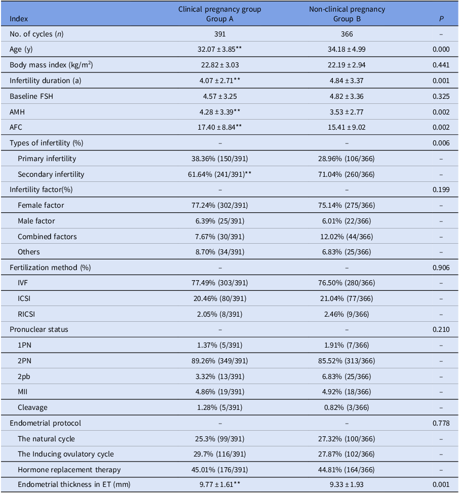
Frozen-thawed embryo transfer cycles were categorized into two groups based on pregnancy outcomes: clinical pregnancy group (Group A) and non-clinical pregnancy group (Group B). The results showed the following: Age (F = 19.204, P = 0.000) and infertility duration (F = 13.211, P = 0.001) were significantly higher in Group B compared to Group A. AMH levels (F = 8.754, P = 0.002), antral follicle count (F = 0.116, P = 0.002), and endometrial thickness on the day of transfer (F = 0.406, P = 0.001) were significantly lower in Group B compared to Group A (P < 0.01). Infertility type showed significant differences between the two groups (χ 2 = 7.466, P = 0.006 < 0.01), as shown in Table 1.
Comparison of basic data on pregnancy outcomes among patients with different high-quality blastocyst transfers in frozen-thawed embryo transfer cycles
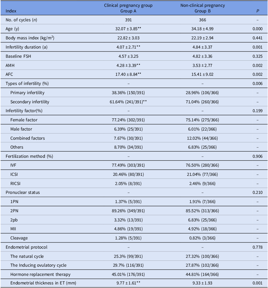
Table 1. Long description
The table compares clinical pregnancy group (Group A) and non-clinical pregnancy group (Group B) in frozen-thawed embryo transfer cycles. It includes data on age, body mass index, infertility duration, baseline follicle-stimulating hormone (FSH), anti-Müllerian hormone (AMH) levels, antral follicle count (AFC), types of infertility, infertility factors, fertilization methods, pronuclear status, cleavage, endometrial protocol, and endometrial thickness. Group A has 391 cycles, while Group B has 366 cycles. Age and infertility duration are significantly higher in Group B. AMH levels, AFC, and endometrial thickness are significantly lower in Group B. The table also shows percentages for primary and secondary infertility, female and male factors, combined factors, and others. Fertilization methods include IVF, ICSI, and RICSI. Pronuclear status includes 1PN, 2PN, 2pb, and MII. Endometrial protocols include the natural cycle, inducing ovulatory cycle, and hormone replacement therapy. The table highlights significant differences in various factors between the two groups.
Note: **indicates highly significant differences.
Comparison of pregnancy outcomes
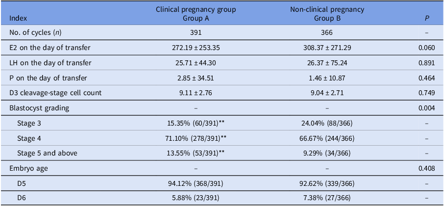
FET cycles were categorized into clinical pregnancy group (Group A) and non-clinical pregnancy group (Group B) based on pregnancy outcomes. The results revealed the following: There were statistically significant differences between Group A and Group B (χ 2 = 10.847, P = 0.004). Group A had a significantly higher proportion of blastocysts at stages 4 and 5 or above, while the proportion of stage 3 blastocysts was lower. No significant differences were observed between the two groups in terms of E2, LH, and P levels on the day of transfer, D3 cleavage-stage cell count, and embryo age (D5 vs. D6 blastocysts) (P > 0.05), as shown in Table 2.
Comparison of pregnancy related factors in different high-quality blastocyst transfer patients during freeze-thaw embryo transfer cycles
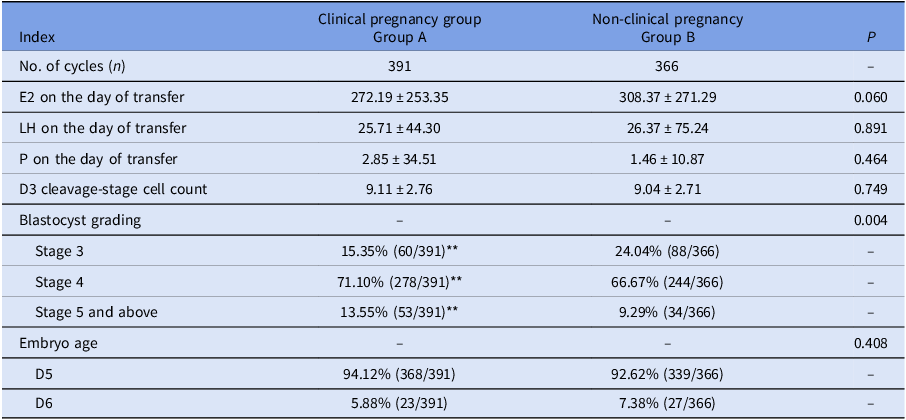
Table 2. Long description
The table compares pregnancy-related factors between clinical pregnancy group A and non-clinical pregnancy group B during freeze-thaw embryo transfer cycles. It includes data on the number of cycles, hormone levels on the day of transfer, D3 cleavage-stage cell count, blastocyst grading, and embryo age. Group A had 391 cycles, while Group B had 366 cycles. Estradiol (E2) levels were slightly higher in Group B, while luteinizing hormone (LH) and progesterone (P) levels showed no significant differences. The D3 cleavage-stage cell count was similar in both groups. Notably, Group A had a higher proportion of blastocysts at stages 4 and 5 or above, and a lower proportion at stage 3, indicating a statistically significant difference in blastocyst grading between the groups. Embryo age distribution (D5 vs. D6 blastocysts) did not differ significantly.
Note: **indicates highly significant differences.
Multivariate regression analysis
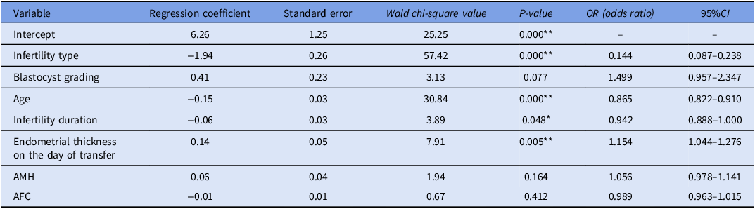
Multivariate logistic regression analysis of Group B indicated that age, infertility type, infertility duration, and endometrial thickness on the day of transfer were independent factors significantly influencing clinical pregnancy outcomes. Among these: Age was significantly negatively correlated with pregnancy success (OR 0.865 [95% CI: 0.822–0.910], P < 0.001), with younger patients demonstrating higher pregnancy success rates. For infertility type, secondary infertility was associated with significantly lower pregnancy rates compared to primary infertility (OR 0.144 [95% CI: 0.087–0.238], P < 0.001). Shorter infertility duration had a positive impact on pregnancy outcomes (OR 0.942 [95% CI: 0.888–1.000], P = 0.048). Increased endometrial thickness on the day of transfer significantly improved pregnancy success rates (OR 1.154 [95% CI: 1.044–1.276], P = 0.005). Although blastocyst grading and AMH did not reach statistical significance in this analysis, their odds ratio (OR) values suggest potential effects on pregnancy success, as shown in Table 3.
Multivariate logistic regression analysis of factors associated with clinical pregnancy outcome
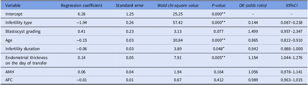
Table 3. Long description
The table presents a multivariate logistic regression analysis of factors associated with clinical pregnancy outcome. It includes eight variables: Intercept, Infertility type, Blastocyst grading, Age, Infertility duration, Endometrial thickness on the day of transfer, AMH, and AFC. Each variable is listed with its regression coefficient, standard error, Wald chi-square value, P-value, odds ratio (OR), and 95% confidence interval (CI). The table shows that age, infertility type, infertility duration, and endometrial thickness on the day of transfer are significant factors influencing clinical pregnancy outcomes. Age has a negative correlation with pregnancy success, with younger patients showing higher success rates. Secondary infertility is associated with lower pregnancy rates compared to primary infertility. Shorter infertility duration positively impacts pregnancy outcomes. Increased endometrial thickness improves pregnancy success rates. Blastocyst grading and AMH do not reach statistical significance but suggest potential effects on pregnancy success.
*Reference Category: Group B.
** indicates highly significant differences.
Discussion
In in vitro fertilization-embryo transfer technology, selecting the best single high-quality blastocyst for transfer is crucial for ensuring healthy offspring and maternal well-being. However, determining how to identify the blastocyst with the highest implantation potential remains a significant challenge in clinical practice. This study investigates the differences among high-quality blastocysts in terms of pregnancy outcomes and their influencing factors.
This study demonstrated that age and infertility duration were significantly higher in the non-clinical pregnancy group compared to the clinical pregnancy group, suggesting that clinical pregnancy rates decrease significantly with increasing age and longer infertility duration. Wang et al. (Wang et al., Reference Wang, Tian, Zhao, Lu, Dong and Zhang2024) reported a nonlinear relationship between female age and pregnancy outcomes in single embryo transfer patients, particularly noting a significant decline in pregnancy and sustained pregnancy rates in women over 34 years of age. For patients under 37 years, single embryo transfer should be prioritized, while selective single embryo transfer remains a reasonable strategy for patients over 38 years, which aligns with the findings of this study. Furthermore, Huang et al. (Huang et al., Reference Huang, Shi, Xing, Yan, Shen, Shan, Sun and Mei2024) noted that young women (<35 years) with infertility lasting more than five years had significantly lower clinical pregnancy and live birth rates. In contrast, Ozturk et al. (Ozturk et al., Reference Ozturk, Dilbaz, Ozelci and Dilbaz2024) suggested that infertility duration did not significantly differ between clinical and non-clinical pregnancy groups, which contradicts the findings of this study.
Additionally, this study found that AMH levels, antral follicle count, and endometrial thickness on the day of transfer were significantly lower in the non-clinical pregnancy group (P < 0.01). Ye et al. (Ye et al., Reference Ye, Du, Cao, Jiang, Qi, Sun, Zhou and Wang2023) indicated that lower AMH levels might be associated with poorer pregnancy outcomes, particularly in women under 35 years of age. Wang et al. (Wang et al., Reference Wang, Guan, Zhang, Jia, Wu, Yao, Zhang and Li2023) and Miyagi M et al. (Miyagi et al., Reference Miyagi, Mekaru, Nakamura, Oishi, Akamine, Heshiki and Aoki2021) identified AMH as a predictor of live birth rates in older women, but its impact on younger women was relatively limited. Ozturk et al. (Ozturk et al., Reference Ozturk, Dilbaz, Ozelci and Dilbaz2024) suggested a threshold AMH level of 3.34 ng/mL for predicting clinical pregnancy outcomes. Moreover, Wu et al. (Wu et al., Reference Wu, Sheng, Wu and Wu2023) found that endometrial thickness was significantly greater in the pregnancy group, consistent with this study’s results. Xu et al. (Xu et al., Reference Xu, Zhang, Jin, Mao, Shi, Huang, Han, Liang and Zhang2021) further pointed out that endometrial thickness was optimally associated with pregnancy outcomes when ≥12 mm, but excessive thickness (≥15 mm) might increase the risk of adverse outcomes. In this study, secondary infertility was more prevalent in the non-clinical pregnancy group, while endometrial thickness was higher in the clinical pregnancy group. It is hypothesized that uterine receptivity may be compromised due to endometrial damage in secondary infertility, underscoring the importance of endometrial thickness in pregnancy outcomes.
The relationship between blastocyst grading and pregnancy outcomes was also analysed in this study, showing that blastocysts at stage 4 or above were significantly more prevalent in the clinical pregnancy group, while stage 3 blastocysts were more common in the non-clinical pregnancy group. Pan et al. (Pan et al., Reference Pan, Zhou and Shen2024) reported no significant differences in birth outcomes and neonatal health between early and fully expanded blastocysts in fresh transfers. Ozgur et al. (Ozgur et al., Reference Ozgur, Berkkanoglu, Bulut, Donmez, Isikli and Coetzee2021) found that the implantation rates of fully expanded blastocysts with AA and BA scores were similar, while AB-scored blastocysts had lower implantation rates, which contradicts the findings of this study. Sciorio et al. (Sciorio et al., Reference Sciorio, Thong, Thong and Pickering2021) suggested a positive correlation between blastocyst size and clinical pregnancy rates, but further research is needed to confirm this relationship.
Multivariate regression analysis in this study identified age, infertility type, infertility duration, and endometrial thickness on the day of transfer as significant independent factors influencing pregnancy outcomes, whereas blastocyst grading, AMH levels, and antral follicle count were not statistically significant. This indicates that, provided high-quality blastocysts are available, pregnancy outcomes are more closely associated with patients’ baseline characteristics, such as age, infertility duration, and endometrial thickness. However, Jin et al. (Jin et al., Reference Jin, Shi, Lu, Bu, Huo and Zhang2021) suggested that changes in endometrial thickness during HRT cycles were unrelated to pregnancy outcomes, which differs from the results of this study. It is proposed that variations in endometrial thickness within the range suitable for transfer may not affect clinical pregnancy outcomes.
In the present study, we observed that the proportion of ≥Stage 4 blastocysts was significantly higher in the clinical pregnancy group, whereas Stage 3 blastocysts were more frequently observed in the non-clinical pregnancy group. These findings suggest that, even within the commonly defined category of ‘high-quality blastocysts’, the degree of blastocyst expansion may retain biological relevance. Previous evidence has indicated that earlier-stage blastocysts may exhibit relatively lower implantation potential compared with fully expanded blastocysts, which may partly explain the distribution observed in our cohort. However, the primary objective of this study was not to compare implantation potential across different expansion stages, but rather to determine whether patient-related background factors remain significant determinants of clinical pregnancy outcomes among embryos that already meet conventional high-quality criteria (stage ≥ 3 with ICM and TE grades ≥ B). Future studies may adopt a more stringent definition of high-quality blastocysts – such as restricting inclusion to stage ≥4 blastocysts with both ICM and TE grades ≥B – to further reduce biological heterogeneity and more precisely assess the independent contribution of blastocyst morphology to pregnancy outcomes.
The limitations of this study include the use of data from a single centre. Although the study was rigorously designed, it may not fully represent conditions in other regions or institutions. Variations in patient populations, treatment protocols, and laboratory conditions across centres could contribute to differences in pregnancy outcomes. Therefore, future multi-centre studies are needed to validate these findings and improve their generalizability.
In conclusion, this study demonstrates that the clinical pregnancy outcomes of high-quality blastocysts are primarily influenced by age, infertility type, infertility duration, and endometrial thickness on the day of transfer, while the impacts of blastocyst grading, AMH levels, and antral follicle count are relatively minor.
Data availability statement
The datasets used and/or analysed during the current study are available from the corresponding author upon reasonable request.
Competing interests
Authors declare no Conflict of Interest for this article.
Funding information
The authors declare that the publication of this research article was financially supported by the Science and Technology Research and Development Program of Yulin City, Guangxi (Project Number: Yushi Branch 202432038).
Consent to participate
All participants were informed about the study details, and their written informed consent was obtained before the study.
Consent for publication
Not applicable.