





Introduction
Neonatal pulmonary artery thromboembolism is a rare, life-threatening condition that presents with significant diagnostic challenges, often manifesting with non-specific signs such as respiratory distress or cyanosis. Reference Cowan, Emelue and Spyropoulos1 The incidence among patients admitted to the neonatal ICU is approximately 0.2%. Reference Robinson, Achey and Nag2 While known risk factors such as the presence of central venous catheters, severe dehydration, or genetic predisposition exist, a substantial proportion of cases—up to 55%—remain of undetermined aetiology. Reference Robinson, Achey and Nag2 This diagnostic uncertainty, compounded by the undetermined aetiology, ensures that early diagnosis and timely intervention remain critical challenges in the management of these critically ill neonates. Therefore, we present a rare case of idiopathic bilateral neonatal pulmonary artery thromboembolism, successfully treated with tissue plasminogen activator, to highlight a successful management approach in the absence of formal guidelines and to contribute to the limited evidence base for this challenging clinical scenario.
Case report
We present a term male newborn (38 + 5 weeks gestation, 3310g) delivered vaginally without complications. Following an unremarkable early course, he was discharged home on the second day of life. On postnatal day 9, he presented to an outside hospital with acute cardiorespiratory collapse.
Upon admission, the infant was in a pre-agonal state with severe hypoxaemia (SpO2 60%) and profound metabolic acidosis, requiring immediate intubation and inotropic support. A bedside echocardiogram revealed severe right ventricular dilation and dysfunction, with elevated right ventricular pressures suggestive of significant outflow obstruction. Crucially, a suspected thrombus was visualised in the left pulmonary artery (Figure 1), prompting urgent transfer to our Paediatric Heart Centre for further management.
Transthoracic echocardiogram (parasternal view) showing the proximal pulmonary arteries. A large occluding thrombus (long arrow) is seen in the right pulmonary artery, whereas a smaller thrombus (short arrow) is seen in the left pulmonary artery. Ao: Aorta, MPA: Main pulmonary artery.

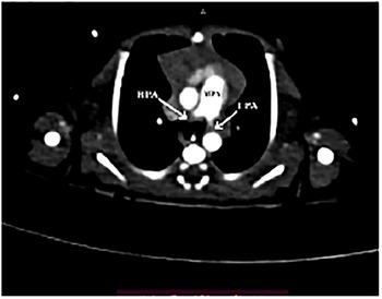
On arrival, a CT angiography confirmed massive bilateral pulmonary thromboembolism, with complete occlusion of the proximal left pulmonary artery and extension into the distal right pulmonary artery (Figure 2). A limb Doppler ultrasound was negative for peripheral thrombosis. Following a multidisciplinary consultation with paediatric cardiology, radiology, paediatric haematology, and paediatric cardiac surgery, a decision was made to initiate systemic thrombolysis with alteplase (tissue plasminogen activator) alongside therapeutic anticoagulation with unfractionated heparin. The tissue plasminogen activator infusion was initiated at 0.06 mg/kg/hour and gradually titrated up to a maximum of 0.4 mg/kg/hour based on serial echocardiographic assessments of thrombus size and right ventricular function. The patient was monitored closely for bleeding complications with regular cranial ultrasounds. aPTT, fibrinogen, and D-dimer levels were monitored regularly via blood tests every 4 hours. Bedside cranial ultrasounds and echocardiograms were repeated regularly every 6–8 hours during thrombolytic therapy. The patient was kept under intensive monitoring throughout thrombolysis, with close observation of neurological and hemodynamic parameters.
Computed tomography scan of the chest (axial view) showing a large thrombus (long arrow) in the right pulmonary artery and a small thrombus (short arrow) in the left pulmonaryartery artery.

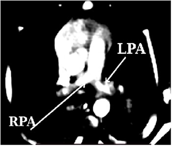
After 36 hours of therapy, a follow-up echocardiogram demonstrated significant thrombus resolution and normalisation of right ventricular function. The tissue plasminogen activator infusion was then tapered and discontinued. A subsequent control CT confirmed complete resolution of the thrombi (Figure 3) but revealed post-infarct cystic changes in the lung parenchyma, a recognised complication of the initial ischaemic event.
Computed tomography scan of the chest (axial view) showing complete resolution of pulmonary artery thrombi.

Post-thrombolysis, the patient was transitioned to subcutaneous low-molecular-weight heparin and was successfully extubated on the sixth day of hospitalisation. An extensive aetiological workup for thrombophilia was negative. The patient was discharged home on day 16 on low-molecular-weight heparin anticoagulation and remained asymptomatic with no evidence of recurrence at one-month follow-up.
Discussion
Our case involves a critically ill neonate with idiopathic neonatal pulmonary artery thromboembolism who was successfully managed with antithrombotic therapy. Isolated pulmonary artery thromboembolism in children is exceedingly rare, with a lack of reliable statistical data in the literature. While arterial thrombosis constitutes 24 to 34% of rare thrombosis cases in neonates, specific data regarding isolated pulmonary artery involvement is scarce. Reference Kenny3 One 30-year retrospective autopsy study found three infants (under 1 year of age) with massive pulmonary embolism, Reference Byard and Cutz4 while another single-centre study found an incidence of 0.7% in their neonatal ICU. Reference van Elteren, Veldt and Te Pas5 Pulmonary artery thrombi often mimic persistent pulmonary hypertension of the newborn and respiratory failure, and may consequently be underdiagnosed due to a low index of suspicion in this age group. Reference Werthammer, Pritt, Laura, Brown and Heydarian6
Known risk factors in neonates include central venous or arterial catheters, inflammation, disseminated intravascular coagulation, impaired liver function, fluctuations in cardiac output, and CHD. Reference Carpenter, Richardson and Hall7 The risk of PE increases with the number of risk factors, Reference Dijk, Curtin, Lord and Fitzgerald8 with the greatest risk posed by the presence of a CVL, severe dehydration, and maternal gestational diabetes. Reference van Elteren, Veldt and Te Pas5 None of these were aetiological factors in our patient. The primary challenge in such idiopathic cases is twofold: first, maintaining a high index of suspicion to consider this rare diagnosis in a neonate with non-specific cardiorespiratory collapse, and second, navigating complex therapeutic decisions in the absence of established protocols.
The diagnostic pathway for a neonate with unexplained acute respiratory distress and hypoxaemia must be rapid and systematic. As demonstrated in our case, a bedside echocardiogram is the crucial first step. It can identify indirect signs of PE, such as severe right ventricular dilation and dysfunction secondary to acute outflow obstruction, and may even visualise the thrombus, prompting definitive imaging with CT angiography. Reference Thacker and Lee9 To streamline this process, we propose a diagnostic and therapeutic algorithm for suspected neonatal PE (Figure 4). Reference Cowan, Emelue and Spyropoulos1,Reference Martinez Licha, McCurdy, Maldonado and Lee10
Proposed treatment pathway. ECHO, echocardiograph; ECMO, extracorporeal membranous oxygenation; iNO, inhaled nitric oxide.

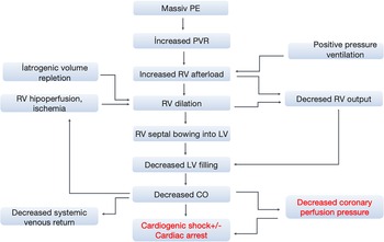
Managing the subsequent acute right ventricular failure is a delicate balance. The pathophysiology involves a vicious cycle wherein increased pulmonary vascular resistance leads to right ventricular dilation, which in turn impairs left ventricular filling and reduces cardiac output. This ultimately compromises coronary perfusion and exacerbates right ventricular ischaemia (Figure 5). Reference Cowan, Emelue and Spyropoulos1,Reference Martinez Licha, McCurdy, Maldonado and Lee10,Reference Konstantinides, Meyer and Becattini11 Therapeutic goals are to support cardiac output while avoiding interventions that could exacerbate right ventricular failure, such as excessive volume repletion or medications that cause systemic vasodilation. Reference Konstantinides, Meyer and Becattini11
Pathophysiology of clinical decompensation from pulmonary embolus. RV, right ventricle; LV, left ventricle; SVR, systemic vascular resistance; PVR, pulmonary vascular resistance. Circular elements represent iatrogenic contributors to pathophysiology.
Note: As both right ventricular pressure and left ventricular pressure increase, right ventricular pressure exceeds left ventricular pressure leading to bowing of the septum into the left ventricle and impaired left ventricular filling.

Once the diagnosis of neonatal pulmonary artery thromboembolism is confirmed, the therapeutic strategy must be tailored to the patient’s hemodynamic status and thrombus burden. Options range from anticoagulation alone to systemic thrombolysis and mechanical thrombectomy. Our institutional approach involves a dynamic risk–benefit assessment by a multidisciplinary team (paediatric cardiology, radiology, paediatric haematology, and paediatric cardiac surgery) to decide on the optimal course of action. The decision between systemic thrombolysis and surgical thrombectomy for massive neonatal pulmonary artery thromboembolism is a critical and complex one, with limited evidence to guide clinicians. In this case, the administration of tissue plasminogen activator through the strategically placed umbilical venous catheter was successful. Nevertheless, it is crucial to recognise that surgical intervention remains a vital alternative, particularly when rapid debulking of the thrombus is required or when contraindications to thrombolysis exist.
As highlighted by Kalanti et al., emergent surgical embolectomy can be life-saving in critically ill infants, particularly when thrombolysis is deemed too slow or carries an unacceptably high risk of haemorrhage. Reference Kalaniti, Lo Rito, Hickey and Sivarajan12 Similarly, Curry et al. argue that surgical thrombectomy offers the advantage of immediate and complete thrombus removal, which can rapidly restore hemodynamic stability and may circumvent the significant bleeding risks associated with tissue plasminogen activator in neonates. Reference DE, C and V13
In our case, the patient, while critically ill, demonstrated transient stabilisation following initial resuscitation, providing a narrow window to attempt a less invasive medical approach. Furthermore, the significant morbidity associated with emergent open-heart surgery in a neonate was carefully considered. Surgical removal of the thrombus is an invasive procedure and potentially carries a high mortality rate in this fragile population. Additionally, the anatomical location of the thrombus, with extension into the distal right pulmonary artery, made catheter-based embolectomy a particularly high-risk option due to the inherent dangers of distal embolisation, precipitous hemodynamic decompensation during the intervention, and the risk of stroke. Reference DE, C and V13 Despite having a comprehensive paediatric cardiac surgery programme capable of performing definitive surgical thrombectomy, our multidisciplinary team’s consensus was to utilise thrombolysis as the primary intervention. Surgical thrombectomy was held in reserve as a rescue therapy in the event of clinical failure.
This case, therefore, contributes to the literature by illustrating a scenario where a medical-first approach was successful, but it also underscores the necessity for institutions to have rapid access to paediatric cardiac surgical teams for cases where this strategy may fail.
Management is challenging in hemodynamically unstable infants, who have a heightened risk of bleeding complications, including intracranial haemorrhage, and require close monitoring. Reference Wang, Hays and Balasa14 Some patients may even require extracorporeal membrane oxygenation. Regarding medical management, tissue plasminogen activator is the thrombolytic agent of choice in children, but evidence-based guidelines regarding its use are limited. Reference Monagle, Chan and Goldenberg15 Our institutional approach involves a dynamic risk–benefit assessment by a multidisciplinary team (paediatric cardiology, haematology, intensive care and paediatric cardiac surgery).
Minimising the risk of serious bleeding is critical when administering tissue plasminogen activator. Reference Ballabh16 Although studies suggest a low-dose tissue plasminogen activator regimen (0.01–0.06 mg/kg/hour) is safer, we had to titrate the dose up to 0.4 mg/kg/hour based on the clinical response. Reference Monagle, Chan and Goldenberg15 The necessity for this dose titration in our patient demonstrates that a flexible, imaging-guided approach can be effective, but also underscores the urgent need for more research to establish optimal dosing regimens.
Given the risk of rapid hemodynamic collapse, even the suspicion of massive PE should prompt anticipatory contingency planning. This includes preparing for potential salvage therapies like extracorporeal membrane oxygenation well in advance, as standard resuscitation is unlikely to resolve the underlying mechanical obstruction. Reference Laher and Richards17
The optimal management strategy for a critically ill neonate with massive pulmonary embolism, particularly the choice between thrombolysis and extracorporeal membrane oxygenation, remains a significant clinical challenge. The decision is nuanced and requires a highly individualised, multidisciplinary approach. Factors such as the patient’s hemodynamic stability, the risks of haemorrhage with thrombolysis versus the procedural risks of extracorporeal membrane oxygenation, and the specific institutional expertise must be carefully weighed. The current literature provides limited guidance for this high-stakes scenario in the paediatric population, underscoring the necessity for a collaborative, case-by-case assessment by the clinical team to determine the most appropriate course of action.
Conclusion
In conclusion, idiopathic neonatal PE is a rare but critical diagnosis. While extrapolating from adult guidelines is often necessary, this case demonstrates a successful, imaging-guided approach to systemic thrombolysis in a neonate. It underscores that a structured pathway, seamless communication within a multidisciplinary team, Reference Tarango and Manco-Johnson18 and proactive planning are paramount to optimising the outcomes of life-preserving interventions for this vulnerable population. Further multicenter studies are desperately needed to develop standardised, evidence-based guidelines.
Data availability statement
The data that support the findings of this study are available from the corresponding author upon reasonable request.
Acknowledgements
The authors would like to thank the multidisciplinary team involved in the care of this patient, including the staff of the Neonatal İntensive Care Unit, Pediatric Cardiology, Radiology, Pediatric Hematology, and Pediatric Cardiac Surgery departments at Liv Bona Dea Hospital.
Author contribution
LS: Conceptualised and designed the case report, collected the data, drafted the initial manuscript, and reviewed and revised the manuscript.
BA, ShB: Critically reviewed the manuscript for important intellectual content.
GA, SA, SE: Contributed to data collection.
Financial support
This research did not receive any specific grant from funding agencies in the public, commercial, or not-for-profit sectors.
Competing interests
The authors declare that they have no competing interests.
Ethical standard
“Ethical approval was not required for this case report as it describes a single patient case retrospectively, and all patient data were fully anonymized. The study was conducted in accordance with the Declaration of Helsinki. Written informed consent to participate in this study was obtained from the patient’s parents/legal guardian.”
Consent for publication
Written informed consent was obtained from the patient’s parents/legal guardian for publication of this case report and any accompanying images.



 Open access

