





Introduction
CHD is the most common birth defect, and ventricular septal defects are the most common CHDs, occurring due to incomplete closure of the interventricular septum during fetal development. Reference Hoffman and Kaplan1 It affects approximately 0.3–0.4% of live births worldwide. Reference Reller, Strickland, Riehle-Colarusso, Mahle and Correa2,Reference Van Der Linde, Konings and Slager3 Ventricular septal defects occur in isolation or in combination with other CHDs, such as atrioventricular canal, Tetralogy of Fallot, and, occasionally, D-transposition of the great arteries.
Perimembranous ventricular septal defects, the most frequently observed type, are located in the membranous portion of the septum, adjacent to the tricuspid and aortic valves. Reference Pérez-Pomares and Kelly4 Perimembranous ventricular septal defects account for approximately 75–80% of all ventricular septal defect cases, making them the predominant type of ventricular septal defect observed in clinical practice. Reference Jacobs, Burke, Quintessenza and Mavroudis5 Clinical presentation and treatment approaches are influenced by the severity and hemodynamic relevance of these abnormalities. Restrictive perimembranous ventricular septal defects, characterised by a smaller defect size that limits the volume of left-to-right shunting due to higher resistance across the defect, represent a subset of perimembranous ventricular septal defects. These defects typically have minimal hemodynamic impact and, in many cases, close spontaneously over time. However, in some cases, they can still contribute to complications such as aortic valve prolapse or infective endocarditis. Reference Jacobs, Burke, Quintessenza and Mavroudis5, Reference Lang, Badano and Mor-Avi6
Understanding the embryological basis, pathophysiology, clinical implications, and management of perimembranous ventricular septal defects is essential for optimising patient outcomes. The aim of our study is to shed light on restrictive perimembranous ventricular septal defects whose management remains controversial.
Materials and methods
Children diagnosed with isolated perimembranous ventricular septal defect from 1991 to 2021 in the department of Paediatric Cardiology at Ankara University School of Medicine were enrolled in this retrospective study. The ethics committee at Ankara University approved this retrospective analysis. The medical records of patients diagnosed with isolated perimembranous ventricular septal defect were reviewed, and 448 patients diagnosed with isolated perimembranous ventricular septal defect were enrolled in this study. The children diagnosed with perimembranous ventricular septal defect and with other CHDs and missing data were excluded from the study. Also, patients with a follow-up period of less than 1 year and without at least 3 examinations were excluded from the study. The data of the patients at the time of admission, 3rd month, 6th month, 12th month, and annually after the first year, preoperative and postoperative period were collected. Echocardiographic and clinical findings, the requirement for treatment, intervention, and surgery were evaluated. The use of furosemide, ACE inhibitors, and/or digoxin was accepted as medical treatment. The long-term progression and rates of ventricular septal defects were categorised into three groups: remaining open, closing spontaneously, and undergoing surgical or interventional closure.
Echocardiography was performed by experienced paediatric cardiologists using Vivid 7 Pro and Vivid 9N Portable Ultrasound System (GE Medical Systems®, Vingmed Ultrasound, Horten, Norway), Philips ie33 (Philips Healthcare, Amsterdam, Netherlands), or Toshiba SSH 140-A scanner with a 3.5 MHz transducer (Toshiba, Tokyo, Japan). Standard echocardiographic measurements were obtained according to the American Society of Echocardiography guidelines. Reference Lang, Badano and Mor-Avi6 The patients were classified according to the size and location of the ventricular septal defect on echocardiography (Echo). The ventricular septal defect size was determined in comparison to the diameter of the aortic annulus (ventricular septal defect/aortic annulus diameter); as small (≤ 1/3 of the ratio), medium (between 1/3 and 2/3), and large (≥ 2/3 of the ratio). The location of the ventricular septal defect was determined according to the International Society for Nomenclature of Paediatric and Congenital Heart Disease Classification Scheme for Ventricular Septal Defect as Incorporated in the International Classification of Diseases-11th Iteration. Reference Lopez, Houyel and Colan7
Perimembranous ventricular septal defects were enrolled in this study and were classified as central, inlet, and outlet. The hemodynamic classification of restrictive perimembranous ventricular septal defects classifies abnormalities based on the pressure differential between the left and right ventricles. When the pressures of two ventricles equalise without pulmonary stenosis, this is referred to as a large or non-restrictive defect in isolated defects. A highly restrictive defect is one in which the pressure gradient between left ventricle and right ventricle /pulmonary artery pressure is more than 60 mmHg (ventricular septal defect peak velocity more than 3.87 m/s), and a moderately restrictive ventricular septal defect with a pressure difference of 25–60 mmHg (ventricular septal defect peak velocity 2.5–3.87 m/s). Reference Awasthy and Radhakrishnan8
Presence of aortic regurgitation, aortic valve prolapses, pulmonary hypertension, and left ventricular dilatation was also evaluated. Left ventricular dilatation was defined as a Z-score of >+2 of the left ventricular internal diameter end diastole. Reference Pettersen, Du and Skeens9 The left ventricular end-diastolic diameter z-scores at the time of diagnosis, middle of the treatment, and at the final follow-up were recorded. Pulmonary artery systolic pressure was measured by tricuspid regurgitation peak velocity, and a tricuspid regurgitation velocity above 2.8 m/s was defined as pulmonary hypertension.
The pulmonary artery pressures were also measured during right heart catheterisation for those undergoing catheterisation. Pulmonary hypertension was defined as an increase in mean pulmonary arterial pressure >20 mmHg at rest, pulmonary vascular resistance >2 Wood units, and pulmonary arterial wedge pressure ≤15 mmHg measured by right heart catheterisation. Reference Humbert, Kovacs and Hoeper10
Statistical analyses were performed by the SPSS 20.0 statistical package (IBM Corp., Armonk, NY, USA). Mean ± standard deviation, median (min-max), and percentage were used for descriptive statistics. The compatibility of the data to normal distribution was evaluated using the Shapiro–Wilk Test. Intergroup evaluation of categorical variables was made using the Pearson’s Chi-Square and Fisher’s Exact test. The Mann–Whitney U test was used to compare the continuous data. The level of statistical significance was determined as p < 0.05.
Results
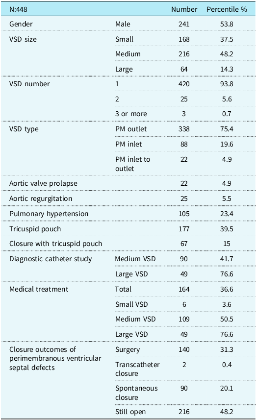
A total of 448 patients (53.8% male) were evaluated. The mean age at the time of diagnosis was 26.5 ± 47.6 months. The mean follow-up period was 47.8 ± 42.8 months (range: 1–29 years). 168(37.5%) of the patients had a small ventricular septal defect, 216 patients (48.2%) had a medium, and 64 patients (14.2%) had a large ventricular septal defect. The characteristics of a perimembranous ventricular septal defect are shown in Table 1.
General data of perimembranous ventricular septal defects

VSD = ventricular septal defect; N = number.
In the overall cohort (n = 448), 140 patients underwent surgical closure, two patients underwent transcatheter closure, 90 patients experienced spontaneous closure, and 216 patients’ ventricular septal defects remained open. (Table 1) The spontaneous closure rate for small, medium, and large ventricular septal defects were 29.2%, 17.6%, and 4.7%, respectively. The mean spontaneous closure age was 28 ± 32.5 months. The mean spontaneous closure age of the small ventricular septal defect was 29.9 ± 35.6 months, and the medium ventricular septal defect age was 24.5 ± 23.7 months. Surgical or interventional closure was performed in 41.6 % and 81.3% of the medium and large ventricular septal defects, respectively. Among patients with medium-sized perimembranous ventricular septal defects, 41.2% underwent surgery and 0.4% underwent transcatheter closure. The mean surgery age was 21.7 ± 29.8 months. The 70.8% of small ventricular septal defects remained open, whereas this ratio was 40.8% and 14.1% for medium and large ventricular septal defects, respectively. Although spontaneous closure did not occur, the ventricular septal defect size was reduced by the tricuspid pouch in six of the nine patients whose large ventricular septal defect did not close. Eisenmenger syndrome developed in the remaining three patients due to non-compliance with their follow-ups.
At baseline, 105 patients with perimembranous ventricular septal defects had pulmonary hypertension; however, only ten of these patients (9.5%) had persistent pulmonary hypertension at the last follow-up visit. Among them, five patients had large and five had medium-sized ventricular septal defects. For patients with large ventricular septal defects, two (one with Down syndrome) underwent surgical closure but continued to have pulmonary hypertension. The remaining three patients (one with Down syndrome, one with Edwards syndrome), who did not comply with follow-ups, are being followed up with Eisenmenger syndrome. Among patients with medium-sized ventricular septal defects, one with Down syndrome is being followed up with primary pulmonary hypertension. The other four patients (one with Down Syndrome) underwent surgical ventricular septal defect closure but continued to have pulmonary hypertension; three patients were diagnosed at a late stage, at 36, 72, and 132 months of age, respectively.
To evaluate temporal changes in management strategies and clinical outcomes, patients were additionally stratified according to diagnostic period. The cohort was divided into four diagnostic intervals (1991–2005, 2006–2010, 2011–2015, and 2016–2021). Due to the limited number of patients, the period between 1991 and 2005 was analysed as a single interval, whereas subsequent years were grouped into consecutive 5-year periods. Temporal trends in treatment strategies and outcomes are further illustrated in Figure 1. Overall, variations were observed in the rates of medical therapy, spontaneous closure, and surgical intervention across diagnostic periods, while transcatheter closure was observed only in the most recent periods.
Temporal trends in management and outcomes according to diagnostic period. Line graph showing temporal changes in medical therapy, spontaneous closure, surgical intervention, transcatheter closure, and persistence of ventricular septal defects across diagnostic periods.

There were 368(82.1%) patients with restrictive perimembranous ventricular septal defect. 45.1% of them had highly restrictive perimembranous ventricular septal defects, whereas 54.9% had moderately restrictive perimembranous ventricular septal defects. The size of the restrictive perimembranous ventricular septal defects were 45.6% small, 46.7% medium, and 7.6% large. 28.2% of those patients required medical treatment (ACE inhibitors and/or diuretic and/or digoxin), whereas 23.3% of the restrictive perimembranous ventricular septal defects’ patients had diagnostic catheterisation. 23.3% of the restrictive perimembranous ventricular septal defects closed spontaneously, whereas 22% of the restrictive perimembranous ventricular septal defects had surgical ventricular septal defect closure, and two had transcatheter ventricular septal defect closure. 54% of restrictive perimembranous ventricular septal defects remained open. Among patients with highly restrictive perimembranous ventricular septal defects (n = 166), 16.9% underwent surgery, 16.3% experienced spontaneous closure, and 66.9% remained open. In patients with moderately restrictive perimembranous ventricular septal defects (n = 202), 27.2% underwent surgery, 29.2% had spontaneous closure, and 43.6% remained open. Among non-restrictive perimembranous ventricular septal defects (n = 80), 73.8% underwent surgery, 5% experienced spontaneous closure, and 21.3% remained open.
Follow-up results of patients with restrictive perimembranous ventricular septal defects according to left ventricular end-diastolic diameter z-scores are shown in figure 2. The left ventricular end-diastolic diameter z-score of 68 patients with a left ventricular end-diastolic diameter z-score of >+2 did not have surgery, and during follow-up, the left ventricular end-diastolic diameter z-score declined <+2 in 62 (91.1%) (figure 3).
Follow-up of patients with restrictive perimembranous VSD and a LVEDD z-score of > +2. LVEDD = left ventricular end-diastolic diameter; VSD = ventricular septal defect; n = number.

Figure 2 Long description
The flowchart begins with restrictive perimembranous ventricular septal defects with a total of 368 cases. It branches into three categories: Small with 168 cases, Medium with 172 cases, and Large with 28 cases. Each category is further divided based on the z-score of left ventricular end-diastolic diameter greater than +2. The Small category has 21 cases, the Medium category has 81 cases, and the Large category has 24 cases. From these divisions, the flowchart shows the outcomes: Spontaneously close, Open, and Surgical Closure. For the Small category, 6 cases spontaneously close, and 15 cases remain open. For the Medium category, 8 cases spontaneously close, 35 cases remain open, and 38 cases undergo surgical closure. For the Large category, 1 case spontaneously closes, 1 case remains open, and 23 cases undergo surgical closure.
Changes in left ventricular end-diastolic diameter (LVEDD) Z-score of patients with restrictive perimembranous VSD who had a z-score > +2 and did not undergo surgery.

Discussion
Ventricular septal defects (perimembranous ventricular septal defects) are among the most prevalent congenital heart anomalies, and their management varies based on factors such as defect size, hemodynamic impact, and the patient’s clinical status. Reference Hoffman and Kaplan1 Across diverse regions and study designs, perimembranous ventricular septal defects consistently emerge as the most prevalent subtype of ventricular septal defect in children. In recent paediatric cohorts, between roughly 55% and 76% of ventricular septal defect cases are perimembranous. Reference Namuyonga, Lubega and Aliku11–Reference Bah, Sapian, Anuar and Alias13
Our long-term, single-centre study provides a comprehensive evaluation of the natural history and outcomes of perimembranous ventricular septal defects, with a particular focus on restrictiveperimembranous ventricular septal defects (rpVSD). Among 448 patients followed over a period of up to 29 years, the findings highlight important aspects regarding spontaneous closure rates, the impact of defect size and hemodynamics, and the necessity of surgical or medical intervention.
Pulmonary hypertension is a significant complication in patients with a ventricular septal defect and necessitates early diagnosis and treatment. In our cohort, pulmonary hypertension was observed in 23.4% of patients at baseline, aligning with existing literature suggesting that unrestrictive ventricular septal defects are associated with increased pulmonary blood flow and risk of pulmonary vascular disease. Reference Humbert, Kovacs and Hoeper10 Among these, only 9.5% exhibited persistent pulmonary hypertension at final follow-up. The presence of chromosomal syndromes in half of the persistent pulmonary hypertension cases highlights the role of genetic comorbidities in pulmonary hypertension prognosis. This underscores the importance of timely intervention to prevent irreversible pulmonary vascular disease, as noted in prior studies emphasising the necessity of early surgery in patients with unrestrictive ventricular septal defect and associated pulmonary hypertension. Reference Awasthy and Radhakrishnan8,Reference Humbert, Kovacs and Hoeper10 The fact that most patients with pulmonary hypertension showed improvement over time suggests that conservative management may be a viable option in selected cases, emphasising the importance of tailored follow-up strategies.
Spontaneous closure occurred in 29.2% of small, 17.6% of medium, and 4.7% of large ventricular septal defects. These findings are consistent with previous studies, which reported that small ventricular septal defects often close spontaneously, whereas large defects are less likely to do so and more often require surgical intervention. Reference Van Der Linde, Konings and Slager3 In infants diagnosed with small ventricular septal defects, long-term follow-up reveals excellent clinical outcomes without the need for medical or surgical intervention, supporting a conservative management approach in hemodynamically insignificant cases. Reference Gabriel, Heger and Innerhofer14 The role of tricuspid valvular tissue in perimembranous ventricular septal defect closure has been previously documented, particularly in the context of perimembranous defects where septal leaflet apposition or aneurysmal transformation may assist in reducing shunt flow. This structural contribution to spontaneous closure remains a clinically relevant observation. Reference Anderson, Lenox and Zuberbuhler15
In our study, medical therapy was used in 36.6% of patients, primarily those with symptomatic or moderate-to-large defects. The distribution of medical therapy, particularly in large ventricular septal defects (76.6%), reflects clinical efforts to stabilise hemodynamics and prevent volume overload in patients awaiting either spontaneous closure or surgical intervention, thereby allowing time for patient growth and potential spontaneous defect closure. In a cohort of 113 infants with ventricular septal defects, 59% had small defects and 41% had moderate or large defects. All infants with moderate or large ventricular septal defects required medical treatment (furosemide, ACE inhibitors, and digoxin) after four weeks of age due to symptoms of heart failure or pulmonary hypertension. By the end of the study, 52% of these patients underwent surgical closure. An additional 12% continued medical therapy despite defect size reduction, while 34% were able to discontinue treatment after outgrowing symptoms. Only 2% of moderate or large ventricular septal defects closed spontaneously.16 In a nationwide French study, 26 percent of the 212 patients with hemodynamically significant pressure-restrictive perimembranous ventricular septal defects who were older than a year had surgical or transcatheter ventricular septal defect closure. Clinical complaints in younger patients and physical defect factors in older patients affected the choices for treatment. Reference Sudaka, Thambo and Vaksmann17
In our study, surgical intervention was performed in 81.3% of patients with large perimembranous ventricular septal defects, and 80.7% of these patients received medical therapy. However, ten patients underwent surgical repair without prior medical treatment. Among them, four were operated due to the presence of aortic valve prolapse and progressive aortic regurgitation. One patient had undergone pulmonary artery banding. Another four patients had very large ventricular septal defects accompanied by pulmonary hypertension and were operated on at an early age. Additionally, one patient was diagnosed with a large ventricular septal defect at 144 months of age and subsequently underwent surgery. In these cases, surgical intervention was prioritised due to the severity of anatomical or hemodynamic findings, such as aortic valve prolapse, aortic regurgitation, or pulmonary hypertension, making medical therapy either insufficient or unnecessary as an initial approach.
A temporal analysis was performed to address the potential impact of evolving clinical practice over the long study period. When the cohort was stratified according to diagnostic periods, variations were observed in management strategies, including the use of medical therapy, diagnostic catheterisation, and interventional procedures. Notably, transcatheter closure was observed only in the most recent diagnostic periods, reflecting the gradual introduction of catheter-based techniques in the management of ventricular septal defects. The proportion of patients referred for surgical intervention was relatively higher during the 2011–2015 period; however, over the last decade, advances in diagnostic and monitoring technologies have enabled more patients to be managed with careful clinical follow-up, even if the defect does not close spontaneously or surgically. At the same time, spontaneous closure rates showed modest fluctuations across periods, suggesting that the natural history of restrictive perimembranous ventricular septal defect has remained relatively stable despite changes in therapeutic approaches. These findings highlight how advances in diagnostic technologies, increasing institutional experience, and evolving treatment strategies may influence clinical decision-making in the management of perimembranous ventricular septal defect over long observation periods.
Restrictive perimembranous ventricular septal defects constituted the majority (82.1%) of our cohort. Despite 54% of these defects remaining open, spontaneous closure occurred in 23.3% of cases. This study highlights that a significant proportion of perimembranous ventricular septal defects, 20.1%, may close spontaneously over time, and many restrictive ventricular septal defects remain asymptomatic in long-term follow-ups. However, these findings align with previous studies, which report that small and medium-sized ventricular septal defects often close spontaneously, while large defects frequently require surgical correction. Reference Reller, Strickland, Riehle-Colarusso, Mahle and Correa2,Reference Van Der Linde, Konings and Slager3 Although restrictive perimembranous ventricular septal defects have a better course compared to non-restrictive ventricular septal defects, a considerable proportion of restrictive perimembranous ventricular septal defects, 36.6%, received medical treatment, particularly those with symptomatic presentations.
The question of “how small is too small to close” remains central to restrictive ventricular septal defect management. Backer et al. emphasised that even small restrictive ventricular septal defects may be associated with risks such as aortic valve prolapse, bacterial endocarditis, or socio-economic implications, and thus may benefit from elective closure in some cases. Their results demonstrated excellent surgical outcomes with no mortality and minimal morbidity in over 140 patients undergoing elective closure. Reference Backer, Winters and Zales18 In contrast, data from Kleinman et al., as discussed by Daniels and Beekman, suggest that left ventricular dilation may regress spontaneously in asymptomatic children with pressure-restrictive ventricular septal defects, advocating for a conservative approach. These findings highlight the ongoing controversy regarding the timing and indications for closure, and the necessity of individualised, risk-stratified decision-making. Reference Daniels19
Our study also monitored left ventricular end-diastolic diameter Z-scores in patients with restrictive ventricular septal defects. Among those with initial left ventricular dilation (Z-score > +2), 91.1% demonstrated regression without intervention. This aligns with Soufflet et al., who observed improved systolic function and reduced left ventricular end-systolic dimension in 220 patients aged 16 years and above with restrictive perimembranous ventricular septal defects. Reference Soufflet, Van de Bruaene and Troost20 Similarly, Gabriel et al. reported that most small ventricular septal defects left unrepaired in childhood were associated with normal ventricular dimensions (89%) in adulthood. Reference Gabriel, Heger and Innerhofer14
Conclusion
Although we have definitive knowledge about perimembranous ventricular septal defect and restrictive perimembranous ventricular septal defect for several years, the management of these topics is one of the most controversial issues in paediatric cardiology, and there is still no clear consensus. The management of patients with perimembranous ventricular septal defect should consider defect size, hemodynamic effects, and clinical presentation. Early diagnosis of large ventricular septal defects and appropriate treatment are crucial in preventing complications such as pulmonary hypertension. Restrictive perimembranous ventricular septal defect poses clinical challenges due to its unique anatomical location and hemodynamic consequences. Although restrictive perimembranous ventricular septal defects do not usually close spontaneously (54%), their natural course is better than that of non-restrictive perimembranous ventricular septal defects, and in ventricular septal defects that do not close, spontaneous regression of left ventricular dilatation occurs over time. Therefore, clinicians should be more careful when deciding to intervene in restrictive perimembranous ventricular septal defects, keeping in mind that the hemodynamic effects of restrictive perimembranous ventricular septal defects generally regress over time. This could be crucial for optimising treatment management strategies and patient-specific risk stratification. Future multicenter prospective studies with longer follow-up are essential to refine indications for intervention and optimise long-term outcomes in this diverse patient population.
Limitation
This study has several limitations that should be acknowledged. First, its retrospective and single-centre design may limit the generalizability of the findings to broader or more diverse populations. Data collection was based on medical records spanning nearly three decades (1991–2021), during which diagnostic and treatment protocols may have evolved, potentially introducing heterogeneity in patient management and follow-up strategies. Although echocardiographic assessments were performed by experienced paediatric cardiologists, interobserver variability and the absence of standardised longitudinal imaging protocols across the entire study period may have influenced the consistency of measurements, particularly in the classification of ventricular septal defect size and left ventricular dimensions.
Data availability statement
The data that support the findings of this study are not openly available due to reasons of sensitivity and are available from the corresponding author upon reasonable request.
Author contributions
SK: Conceptualisation (supporting), methodology, investigation (lead), and writing original draft preparation (lead). MGR: Investigation (supporting), and writing original draft preparation (supporting). BA: Investigation (supporting). TU: Project administration, supervision (supporting). SA: Project administration, supervision (supporting). ET: Conceptualisation (lead), supervision (lead), and writing review and editing (lead).
Financial support
No specific funding was received from any bodies in the public, commercial, or not-for-profit sectors to carry out the work described in this article.
Competing interests
All authors declare that they have no conflict of interest.
Ethical standard
The present study was approved by the local ethics committee of the Ankara University Faculty of Medicine.


 Open access

