


1. Introduction
Oncological patients undergo radiotherapy as part of their cancer treatment, where high doses of radiation are used to destroy cancer cells and reduce tumors. There are two main techniques for applying radiotherapy: external beam therapy and brachytherapy. The selection between these techniques is based on the tumor’s location. For instance, in the treatment of deep-seated tumors, photon radiotherapy is recommended, as its highest dosage rate is achieved at a considerable depth within the patient’s body. In contrast, for the treatment of superficial lesions, such as skin cancer, electron therapy is suggested.
Regarding the cancer incidence rate in Guayaquil, Ecuador, data indicates an annual average of 4,078 new cases between 2010 and 2014 (Reference Tanca, Real, Jaramillo and QuintoTanca et al., 2019). Of these cases, over 80% will require radiotherapy at some point during their disease progression (Reference Algara LópezAlgara López, 2016), either as a curative treatment or for palliative purposes. Studies have demonstrated that the combination of surgery and radiotherapy increases patient survival rates, reaching between 50% and 58% at 15 years (Reference Abe, Abe, Enomoto, Kikuchi, Koyama, Masuda, Nomura, Sakai, Sugimachi, Tominaga, Uchino, Yoshida, Haybittle, Davies, Harvey, Holdaway, Kay, Mason, Forbes and CaffierAbe et al., 2005).
Radiotherapy treatment planning relies on imaging techniques such as computed tomography (CT), magnetic resonance imaging (MRI), or positron emission tomography (PET). The radiation therapist, in collaboration with the oncologist, is responsible for defining the planned target volume (PTV), which represents the area expected to receive the highest radiation dose during treatment. Additionally, organs at risk (OAR) and the planned risk volume (PRV) are identified, which correspond to the margin of surrounding healthy tissue that will receive radiation exposure without necessarily requiring it. The precise definition of PTV and PRV volumes, along with their respective margins, remains an area of research. The studies by Molinelli et al. (Reference Molinelli, de Pooter, Romero, Wunderink, Cattaneo, Calandrino and Heijmen2008) and Van Herk et al. (Reference Van Herk, Remeijer, Rasch and Lebesque2000) establish criteria for defining these margins based on treatment technique, dose fractionation protocols, and the imaging system used for medical imaging acquisition.
The accurate definition of these volumes is crucial, as it directly influences the complications that may arise when applying radiotherapy to healthy tissue. Statistics indicate that between 80% and 90% of patients undergoing radiotherapy develop some degree of radiation-induced dermatitis (Reference Salazar and Ocampo-CandianiGarza Salazar & Ocampo-Candiani, 2010), which, in some cases, can progress to Grade 4 radiodermatitis, leading to necrosis of the surrounding healthy tissue and affecting 25% of patients (Reference Hymes, Strom and FifeHymes et al., 2006).
To ensure that high doses of radiation precisely target the PTV, radiation therapists use accessories designed to immobilize the patient, establish a reference point for radiation application, protect surrounding tissues, and increase radiation dosage at the target volume. These accessories, known as boluses (Figure 1), provide superficial coverage (Reference BarbagelataBarbagelata, 2022). The materials used in bolus manufacturing aim to replicate human tissue properties in terms of radio-density, measured in Hounsfield Units (HU), which reflect a material’s ability to reduce or block radiation transmission. Traditionally, boluses are made from materials such as synthetic gel sheets, thermoplastic sheets, moldable waxes, and even gauze dressings moistened with water, all intended to conform to the patient’s body surface for treatment. While each material presents certain advantages, they all share common drawbacks, including limited adaptability to irregular surfaces, high variability in thickness (which complicates precise radiation dosing), air bubble formation, risk of fungal and bacterial infections, and non-reusability.
Bolus for radiotherapy (reproduced from Lu et al. (Reference Lu, Song, Yao, An, Shi and Huang2021) licensed under Creative Commons Attribution 4.0 license)

In this context, additive manufacturing (AM) emerges as an outstanding option for bolus fabrication, offering a more precise, rapid, and safer technology compared to traditional handcrafted manufacturing methods. Researchers such as Kong et al. (Reference Kong, Yan, Sun, Qian, Zhou, Cai and Tian2019) have studied photon radiotherapy dosimetry in boluses made from gels and hydrogels. Meanwhile, Lu et al. (Reference Lu, Song, Yao, An, Shi and Huang2021) expanded the application of additive manufacturing by analyzing a wide range of polymers commonly used in fused filament fabrication (FFF) and stereolithography (SLA). The results are promising, as AM-fabricated boluses perfectly conform to the patient’s anatomy, are reusable, prevent air bubble formation, and allow the isodose curve to adapt to the target volume (PTV), controlling radio-density based on bolus thickness or the printing parameters available for each technique, as illustrated in Figure 2(b).
Dose depth using two types of boluses: (a) with a handcrafted bolus, (b) with a bolus manufactured using AM

The design process for AM boluses begins with defining the mesh of the area of interest using Digital Imaging and Communications in Medicine (DICOM) files obtained from CT, MRI, or PET scans. Subsequently, the bolus is manually digitally sculpted onto the three-dimensional image of the treatment area, then exported to slicing software, and finally fabricated using a 3D printer.
However, a drawback of this process is that image acquisition requires exposing the patient to radiation sources or contrast agents, leading to costly procedures for patients that sometimes provide irrelevant information for planning certain radiotherapy cases. As an alternative, Dipasquale et al. (Reference Dipasquale, Poirier, Sprunger, Uiterwijk and Miralbell2018) propose the use of a HandyScan™ 700 surface scanner by Creaform, with a resolution of 0.05 mm, for image acquisition as a starting point for AM bolus design. However, this technique has not yet been explored locally due to the high cost of the equipment, which can exceed $30,000.
To implement the use of AM in electron radiotherapy, this study proposes a methodology for bolus design that begins with the acquisition of three-dimensional images using a Sense 2™ scanner by 3D Systems, applying the image acquisition protocol proposed by Helguero et al. (Reference Helguero, Cansing, Maldonado, Saldarriaga and Amaya-Rivas2024) for low-cost equipment. This approach includes a linear equation that correlates radio-density with the infill percentage of the fabricated piece, overcoming the limitation of the 150 HU available in locally manufactured paraffin boluses. This methodology provides a design tool that reduces material and imaging costs and can be applied by professionals outside the design field to obtain more efficient, reusable, personalized, and safer pieces that offer greater patient comfort during therapy compared to the handcrafted methods currently used in resource-limited and socially significant medical centers.
2. Methodology
The general steps for designing boluses for superficial radiotherapy consist of four main stages, as shown in Figure 3. The detailed methodology for the design and additive manufacturing (AM) of the bolus is broken down in Figure 6. One of the primary challenges in the design phase is ensuring that the bolus has the appropriate radio-density to achieve the maximum dose in the PTV. To control this variable, the radio-density is adjusted by modifying the infill percentage of the bolus while maintaining a constant thickness of 5 mm throughout the entire surface of the piece. This thickness was chosen because increasing it leads to the loss of surface details, which can promote the formation of air bubbles between the bolus and the patient’s surface. Additionally, tests confirmed that if the bolus needs to be sectioned due to its complex shape to incorporate coupling and fixation mechanisms, a thickness of 5 mm ensures secure grips that can withstand forces of up to 5 Newtons without catastrophic deformation of the piece.
Proposed methodology for designing boluses for electron radiotherapy

2.1. Image acquisition
The process is conducted during one of the patient’s medical appointments. The equipment used is a Sense 2™ scanner from 3D Systems, valued at approximately $700.00. No configuration changes or parameter modifications are required, as the software used for data collection is highly intuitive and provides guides and tips throughout the process. The scanning is performed by moving the scanner along the sagittal and transverse axes of the area of interest on the patient, ensuring a constant 40 cm distance from the patient and maintaining a 45° inclination relative to the horizontal plane (Figure 4). The procedure takes no more than 15 minutes and is easy for new operators to learn. The details of the 3D scanning process with low-cost equipment were previously explored by Helguero et al. (Reference Helguero, Cansing, Maldonado, Saldarriaga and Amaya-Rivas2024) and validated using a mathematical model.
Scanning process of the patient’s area of interest

The result is a digital file in .obj format, which is editable in most CAD (Computer-Aided Design) software or mesh modeling programs such as 3ds Max or Meshmixer.
2.2. Bolus design for AM
The resulting scan file is reviewed in collaboration with the radiation therapist to ensure that all areas of interest for therapy application are included. If confirmed, the 3D image is exported to mesh editing software.
Next, it is recommended to optimize the mesh to eliminate surface imperfections, such as folds or discontinuities, which are common in the image acquisition process (Figure 5(a)). After repairing the surface, the number of polygons is reduced, followed by applying a retopology function that modifies the shape of the polygons, making them uniform so that thickness can be applied without vertex intersections or overlapping faces. The result is a surface that completely covers the area of interest, has no sharp or straight edges, is continuous throughout, and consists of polygons with three or four vertices (Figure 5(b)).
Modification and optimization of the mesh in interest: (a) Unprocessed mesh with overlapping faces highlighted in green, (b) Optimized mesh with a smooth surface and no overlapping

The main criterion for removing irregular surfaces is to ensure that they are caused by image acquisition noise and not by the patient’s anatomical details. It is recommended that the designer use high-quality photographs of the area of interest. The scanner’s resolution allows detecting irregularities as small as 0.9 mm, ensuring product adaptability and preventing air bubbles. Retopology functions are easy to use since they only redefine the base polygon shapes without altering their distribution or quantity.
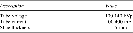
The bolus radio-density depends on the type of radiotherapy. Therefore, a preliminary process was conducted to characterize this parameter in polylactic acid (PLA) by fabricating several test specimens of 50×50 mm with a thickness of 5 mm, using infill percentages of 10%, 20%, and 50%. These specimens were analyzed using a CT scanner, using a commercial paraffin bolus as a reference. The general CT scanner parameters are detailed in Table 1.
General CT scanner parameters

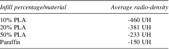
Radio-density of specimens with different infill percentages

The CT scan results are shown in Table 2, demonstrating a proportional relationship between the infill percentage and radio-density. From this information, Equation 1 was derived, establishing the relationship between these parameters, serving as the basis for determining the required infill percentage in slicing software to achieve the desired radio-density.
 $$y = 5.7x - 519.5$$
Where y represents the radio-density in Hounsfield Units (HU) and x represents the infill percentage using a linear pattern. Once the mesh is extruded to a thickness of 5 mm, it is exported as an STL file to slicing software for printing configuration. The orientation of the piece should be prioritized so that the extruder remains perpendicular to the radiation beam direction.
For complex tumor shapes such as melanomas on finger phalanges, the boluses are designed to be printed in sections and then assembled on the patient. Effective attachment methods include the use of hooks and perforations for fastening (Figure 6(a)).
Bolus design process from scanned images: (a) two-part bolus design for a hand, (b) validation of bolus design on the patient’s digital 3D scan

The final validation phase involves overlaying the designed bolus on the patient’s digital image to confirm its adaptability (Figure 6(b)). It is recommended to inspect the entire setup with a transparency level in the bolus to identify complex areas or those distant from the edges, ensuring proper surface contact to prevent air bubbles.
2.3. Additive manufacturing
Boluses are fabricated using extrusion-based additive manufacturing (FDM/FFF) due to its cost-effectiveness, availability of open-source equipment and software, and ability to control custom parameters to achieve the required radio-density. The average resolution of these printers is 0.3 mm, surpassing the scanner’s resolution and preserving fine details.
PLA was chosen for its surface quality, flexibility, and resistance. To minimize humidity, which can cause air bubbles in the extruded material and affect radiation dispersion Baltz et al. (Reference Baltz, Chi, Wong, Wang, Craft, Kry, Lin, Garden, Smith and Howell2019) with bubbles larger than 2 mm in diameter. Filament dryers and protective cabinets with humidity control are recommended. Printing parameters include an extruder temperature between 190-220 °C, a heated bed between 50-60 °C and a printing speed no greater than 60 m/min. To ensure adhesion, the first layer should have a maximum height of 0.3 mm and an initial speed of 40%, with organic supports for steep overhangs.
2.4. Validation and implementation
Once the bolus has been manufactured, validation is carried out to determine whether the target radio-density has been achieved with the established infill percentage. To do this, the piece is examined in a linear accelerator to obtain HU measurements across the entire surface of the bolus. With the radiologist’s approval, the adaptability of the piece to the area of interest on the patient is assessed. This process consists of a qualitative test in which the patient is positioned in the posture they will assume during treatment, ensuring that the skin and bolus surfaces maintain full contact around the contour. To validate internal areas, a methylene blue staining test can be performed, or alternatively, transparent PLA material can be used to visually verify the compatibility of the pieces, as it has been demonstrated that the filament color does not affect radio-density.
If the results of the radio-density and patient compatibility tests are satisfactory, the radiologist is notified to proceed with the radiotherapy application. The advantage of the bolus fabricated using additive manufacturing is its reusability throughout the entire treatment, which may extend up to 15 days.
If discrepancies are detected in the Hounsfield unit measurements or if it is determined that the piece does not properly fit the patient, it is recommended to repeat the process from the medical image acquisition stage using the 3D scanner.
The entire bolus design methodology is summarized in the flowchart shown in Figure 7, which groups each process along with its corresponding activities and parameters to consider.
Flowchart of the methodology for the design of boluses

3. Results and study case
The methodology was validated through the design and fabrication of a bolus for an 18-year-old male patient, weighing 110 kg and measuring 1.80 m in height, to undergo radiotherapy on the left genian, labial, and mentonian areas. The image acquisition was performed in 15 minutes during a medical appointment, generating three-dimensional images of the patient’s head (Figure 8(a)). The areas of interest were extracted (Figure 8(b)), and the polygon density was reduced to 10,000 units to facilitate manipulation, smoothing edges and filling empty spaces in the labial commissure (Figure 8(c)).
Summary of the case study in images: (a) Image acquisition, (b) limitation of the mesh to the area of interest, (c) polygon reduction, mesh smoothing, and optimization, (d) design for additive manufacturing, (e) extrusion-based manufacturing, (f) adaptability tests on the patient

The surface mesh was optimized and retopologized to convert triangles into rectangles, minimizing errors during extrusion (Figure 8(d)). To achieve the required -150 HU, a 65% infill was configured. The bolus was printed using an extrusion-based system and PLA filament, with organic supports to ensure proper orientation and a more compact initial layer (Figure 8(e)). The adaptability tests on the patient confirmed that the bolus fit correctly (Figure 8(f)). The radio-density measurement, performed with the same CT scanner, yielded an average value of -162.896 HU, without discontinuities in the results (Figure 9).
Radio-density versus position: the estimated average value is -162.896 HU, with fluctuations between -150 and -100 HU

The application of the proposed methodology for bolus design using AM results in a reduction in raw material costs. The fabrication of the bolus for this case study using AM cost $1.93, compared to $5.52 for manufacturing it with paraffin sheets available at the medical center, representing a 65% cost reduction.
Additionally, the current image acquisition method for bolus design entails a cost of $1,157.42 for the medical center. By using the 3D scanner, this cost was reduced to $219.90, including the image capture and mesh editing process up to the final printing file, leading to an 81% cost reduction in image acquisition and design.
4. Conclusions
The proposed methodology for bolus design using AM is an innovative, practical, and cost-effective alternative to traditional handcrafted bolus manufacturing methods, which are still used in clinics with limited financial resources. This approach is based on the premise of using low-cost hardware and open-source software. The effectiveness of the scanner as a source for medical images has already been demonstrated in studies by Sharma et al. (Reference Sharma, Sasaki, Rickey, Leylek, Harris, Johnson, Alpuche Aviles, McCurdy, Egtberts, Koul and Dubey2018) which found that 89% of the surface scanned using the Sense™ scanner from 3D Systems had a difference of less than 2 mm when compared to images obtained through traditional CT scans. An advantage of the Sense 2™ scanner used in this study over the Sense™ scanner used by Sharma et al. (Reference Sharma, Sasaki, Rickey, Leylek, Harris, Johnson, Alpuche Aviles, McCurdy, Egtberts, Koul and Dubey2018) is its ability to maintain a dynamic reference during scanning. This feature provides greater freedom of movement in extended scanning sessions without the need to immobilize the patient or use tripods to prevent reference loss, an issue for which Dipasquale et al. (Reference Dipasquale, Poirier, Sprunger, Uiterwijk and Miralbell2018) proposed a solution using an expensive scanner. On the other hand, the relationship between radio-density and infill percentage for a 5 mm thick piece, as expressed in Equation 1, showed a difference of 8.6% compared to the expected radio-density, which represents a clear improvement over the paraffin bolus it aims to replace, where the difference exceeds 20%. These results regarding radio-density and infill percentage remain valid as long as the infill pattern and constant thickness of 5 mm with PLA material are maintained, since variations in Hounsfield Units (HU) are more closely related to the size and spacing of air bubbles trapped in the infill pattern rather than the physical properties of the material. To explore the use of other polymers, it is recommended to refer to the studies by Pérez-Cualtán et al. (Reference Pérez-Cualtán, Abril and Suarez Venegas2024) which examine various materials and propose equations with 90% reliability in radio-density control.
The 65% cost savings in fabrication materials and the 81% reduction in image acquisition costs make this methodology an excellent alternative to conventional methods currently used in Ecuador. The proposed design methodology includes all the necessary criteria for additive manufacturing of boluses and organizes them in a way that facilitates implementation by radiation therapists with little or no experience in the design and fabrication of these medical devices, ultimately aiming to improve patients’ quality of life.




 Open access

