


Introduction
The family Anoplocephalidae Blanchard, 1891, is a tapeworm family of the order Cyclophyllidea van Beneden in Braun, 1900. The species of this family are characterized by a scolex without a rostellum (Georgiev et al., Reference Georgiev, Bray, Littlewood, Morand, Krasnov, Poulin and Degen2006). Anoplocephalidae includes four subfamilies, of which the subfamily Anoplocephalinae includes 59 genera of parasites of mainly mammals and birds (Mariaux et al., Reference Mariaux, Tkach, Vasileva, Waeschenbach, Beveridge, Dimitrova, Haukisalmi, Greiman, Littlewood, Makarikov, Phillips, Razafiarisolo, Widmer, Georgiev, Caira and Jensen2017). The diagnostic features of the subfamily include the presence of a sac-shaped or reticular uterus that persists even in gravid proglottides, eggs provided with a modified embryophore (known as pyriform apparatus), and well-developed internal and external seminal vesicles (Georgiev et al., Reference Georgiev, Bray, Littlewood, Morand, Krasnov, Poulin and Degen2006; Wickström et al., Reference Wickström, Haukisalmi, Varis, Hantula and Henttonen2005).
Wild mammals, particularly rodents, may play an important role as hosts in the diversity of the family Anoplocephalidae. One such rodent species is Lagidium viscacia (Molina), a member of the family Chinchillidae, broadly distributed from Southern Peru to Central Chile and Argentina. Locally known as ‘Chinchilla’ or, more specifically, ‘vizcacha’, these rodents typically inhabit rocky landscapes and feed primarily on grasses (Chebez et al. Reference Chebez, Pardiñas and Teta2014). Studies on anoplocephalid cestodes parasitizing vizcachas are relatively scarce, with only three published works to date. The first study, from Peru (Parra, Reference Parra1953), described Perutaenia threlkeldi Parra, Reference Parra1953 (currently Monoecocestus threlkeldi). The other two studies were from Argentina: Denegri et al. (Reference Denegri, Dopchiz, Elissondo and Beveridge2003) erected the genus Viscachataenia to accommodate Viscachataenia quadrata (von Linstow, 1904), and more recently, Martins et al. (Reference Martins, Robles, Navone and Callejn2023) investigated the diversity of Monoecocestus in rodents, recording M. threlkeldi from Formosa and also, three additional unidentified species of Monoecocestus from Buenos Aires and Chaco.
The aim of our study is to describe a new cestode species of the genus Monoecocestus parasitizing the vizcacha Lagidium viscacia, based on specimens collected in the area of Puno, Peru. The species description includes morphological traits as well as nuclear and mitochondrial DNA sequences.
Materials and methods
A total of 20 specimens of L. viscacia were examined for the presence of parasitic worms. Three specimens were collected in the province of Moho (15°21′39″S, 69°29′59″W), eight from the province of Melgar (14°52′55″S, 70°35′24″W), and nine from the province of Lampa (15°25′00″S, 70°35′00″W), in the region of Puno, Peru. All specimens were sampled from November to December, 2024. Host specimens were collected using the method described by Bautista et al. (Reference Bautista, Palacios, González, Selem-Salas, Sosa-Escalane and Hernandez-Betancourt2011), using a Tomahawk live trap. Specimens were euthanized according to the protocols of the American Veterinary Medical Association (AVMA, 2013) and the Peruvian regulations. Collection of all the specimens was conducted under the permit D000144-2024-MIDAGRI-SERFOR-ATFFS-PUNO issued to Henry Apaza-Añamuro by the Servicio Nacional Forestal y de Fauna Silvestre (SERFOR), Peru. All the parasitic worms were recovered and stored in 90% ethanol.
Morphological analysis
Five cestode specimens were stained with Mayer’s paracarmine, and 24 with Gömöri’s trichrome. Specimens were mounted in permanent slides in Canada balsam following the procedure described by Lamothe-Argumedo (Reference Lamothe-Argumedo1997). For the observation of eggs, 2–3 gravid proglottides of seven specimens were digested with proteinase K in temporal preparations, following Coronado-Morones et al. (Reference Coronado-Morones, Alonso Panti-May, Torres-Carrera and Garcia-Prieto2025). Measurements are reported in micrometres unless other units are indicated. Drawings were made using a drawing tube attached to a Zeiss Standard 25 optical microscope and were digitized with Adobe Illustrator v. 10. Type specimens were deposited in the Colección Helmintológica del Departamento de Protozoología, Helmintología e Invertebrados Afines (MUSM-HEL), Universidad Nacional Mayor de San Marcos (UNMSM); other paratypes are in the National Helminth Collection (CNHE) at the Institute of Biology, UNAM (IB-UNAM), Mexico City, Mexico. A single specimen was processed for scanning electron microscopy (SEM) as follows: the specimen was dehydrated through a graded series of EtOH, critical point dried with CO2, mounted on metal stubs with conductive carbon tape, sputter-coated with gold, and observed in a Hitachi Stereoscan Model SU1510 SEM at 10 kV (Hitachi Ltd., Tokyo, Japan) at the Laboratorio Nacional de la Biodiversidad (LANABIO), IB-UNAM, Mexico City, Mexico.
For comparative purposes, microphotographs of type specimens of M. threlkeldi (NMNH No. 37,380) were requested from the U.S. National Parasite Collection.
Molecular analysis
Total DNA was extracted from 3–5 proglottides from two specimens, using the Molecular Biology kit, BIOBASIC, following the manufacturer’s instructions. A fragment of the mitochondrial cytochrome Oxidase subunit 1 (cox1) was amplified with primers JB3 (5′-TTTTTTGGGCATCCTGAGGTTTAT 3′) and COIR-trema (5′ CAACAAATCATGATGCAAAAGG 3′) (Bowles et al., Reference Bowles, Hope, Tiu, Liu and McManus1993; Miura et al., Reference Miura, Kuris, Torchin, Hechinger, Dunham and Chiba2005). Partial fragments of the nuclear small ribosomal subunit (18S rDNA) and large ribosomal subunit (28S rDNA), were amplified with primers A (5′-AACCTGGTTGATCCTGCCAGT 3′) L (5′-CCAACTACGAGCTTTTTAACTG 3′) and C (5′-CGGTAATTCCAGCTCCAATAG3′) Y (5′-CAGACAAATCGCTCCACCAAC 3′) (Apakupakul, Reference Apakupakul, Siddall and Burreson1999), for 18S rDNA and 391 (5′-AGCGGAGGAAAAGAAACTA 3′) (Smythe & Nadler Reference Smythe and Nadler2006) 536 (5′-CAGCTATCCTGAGGGAAAC 3′) (Garcia-Varela & Nadler, Reference García-Varela and Nadler2005), for 28S rDNA. Finally, a fragment of the internal transcribed spacer 1 (ITS-1), 5.8S rDNA, and Internal transcribed spacer 2 (ITS-2) were amplified with primers BD1 (5′ GTCGTAACAAGGTTTCCGTA 3′) and BD2 (5′ TATGCTTAAATTCAGCGGGT 3′) (Bowles et al., Reference Bowles, Hope, Tiu, Liu and McManus1993). Products of the PCR reactions were visualized by electrophoresis in a 1.6% agarose gel. Successful amplifications were purified using CentriSep 96 filter plates (ThermoFisher Scientific) with Sephadex G-50 columns (Cytiva, Marlborough, Massachusetts, USA). Sequencing reactions were composed of 0.4 μL BigDye Terminator v.3.1 (Applied Biosystems, Waltham, Massachusetts, USA), 2 μL 5 × buffer, 4 μL ddH2O, 1 μL 10 μM primer, and 3 μL purified PCR product (total volume 10 μL). Samples were purified using Sephadex G-50 columns, then 25 μL 0.5 mM EDTA was added, and the samples were finally sequenced in an ABI-PRISM 3100 sequencer (Applied Biosystems) at the LANABIO, IB-UNAM. Finally, sequences were compared with those available in GenBank, using the Blast tool (Madden, Reference Madden2013). Due to the limited availability and the fragmentary nature of Monoecocestus DNA sequences deposited in GenBank, a robust phylogenetic analysis with a meaningful interpretation was precluded in the present study. The DNA sequences generated herein are publicly accessible and are intended to promote the acquisition of more data on related taxa in the near future.
Results
Systematics
Anoplocephalinae Blanchard, 1891
Monoecocestus Beddard, 1914
Monoecocestus viscaciae Zapata, Coronado-Morones, and Torres-Carrera, n. sp.
Taxonomic summary
Type host: Lagidium viscacia (Molina) (Rodentia: Chinchillidae).
Type locality: Moho, Puno, Peru (15°21′39″S 69°29′59″W 3932 m.a.s.l.).
Site of infection: Intestine.
Number of infected hosts: three of the eight hosts analysed P = 37.5% (Melgar); two of three hosts analysed P = 66.6% (Moho); and two of nine hosts analysed P = 22.2% (Lampa).
Other localities: Melgar, Puno (14°52′55″S 70°35′24″W), Lampa, Puno (15°25′00″S 70°35′00″W).
Type material: MUSM-HEL (from Moho): Holotype: 5675; 8 paratypes: 5676; CNHE 12554 (1 paratype); Other paratypes: from Melgar, MUSM-HEL: 5677 6 specimens; CNHE 12555: 3 specimens; from Lampa, 8 paratypes MUSM-HEL: 5678 and 3 specimens CNHE 12556.
GenBank Accession Numbers: cox1 (PZ190066, PZ190067); 18S rDNA (PZ195148); ITS (PZ195147) and 28S rDNA (PZ195145, PZ195146).
Zoobank registration:
Etymology: The specific name is a noun in genitive case, which refers to the Monoecocestus of the vizcacha Lagidium viscacia.
Description
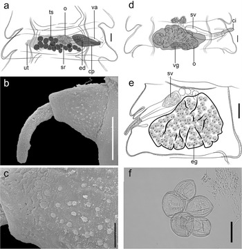
Description based on 30 specimens; with characteristics of Monoecocestus (see Beveridge, Reference Beveridge, Khalil, Jones and Bray1994). Total length, 3.79 mm (2.07–4.55, n = 10) (Figure 1a, b). Scolex 262 (207–315, n = 21) long and 320 (216–360; n = 21) wide; circular suckers directed anterolaterally, 140 (117–180; n = 21) in diameter, sucker aperture 85 (71–102, n = 9) diameter (Figure 1b,c). Neck short, 73 (59–111, n = 9) long by 317 (288–351, n = 9) wide. Strobila 4680 (2999–8678) long; maximum width 898 (720–1114). Proglottides 26 (21–35; n = 21) in number (Figure 1a), craspedote. Immature proglottides 13 (11–16; n = 21) in number, 83 (53–113; n = 21) long and 377 (270–468; n = 21) wide. Mature proglottides 4 (3–5) in number, wider than long, 83 (53–113; n = 21) long and 377 (270–468; n = 21) wide, respectively. Gravid proglottides 14 (10–16) in number, 458 (342–639; n = 21) long and 782 (459–1028; n = 21) wide. Genital pores in the posterior portion of proglottid in immature and mature proglottides, more posterior in gravid proglottides; alternating regularly (Figure 1d). The genital atrium may protrude, forming a well-expressed genital papilla bearing bud-shaped sensory receptors, each with 4 in diameter (Figure 2b), which are distributed regularly. Body surface covered with lineate spinitriches (sensu Chervy, Reference Chervy2009) (Figure 1e).
Monoecocestus viscaciae n. sp. (a) Drawing of a whole specimen. (b) SEM microphotographs of whole specimen; (c) scolex, apical view; (d) lateral view showing position of genital pores; (e) lineate spinitriches. Scale bars a, 500 μm; b, 1 mm; c, 100 μm; d, 250 μm; e, 5 um.
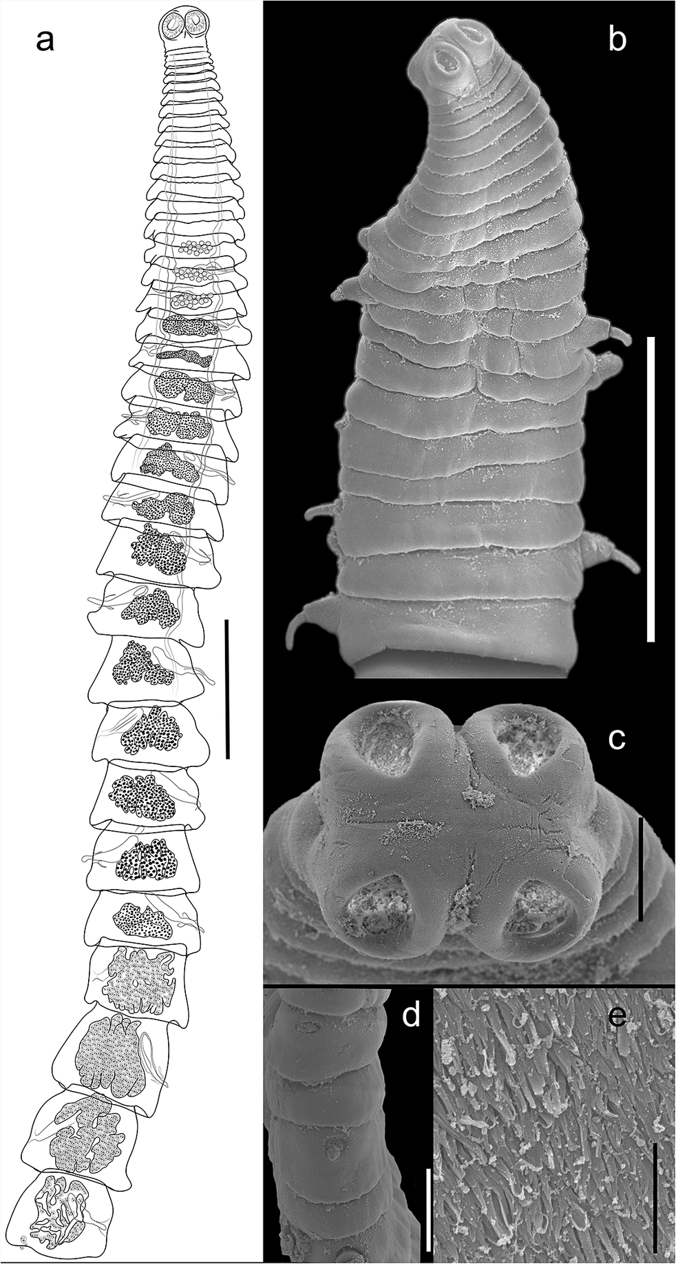
Figure 1. Long description
Panel a at top-left is a line drawing of the entire Monoecocestus viscaciae specimen, showing segmented body structure and internal clusters. Panel b at top-right is an S E M microphotograph of the whole specimen, highlighting external segmentation and surface texture. Panel c below b presents an apical S E M view of the scolex, revealing four rounded suckers and surface details. Panel d at lower left shows a lateral S E M view, indicating the position of genital pores along the body margin. Panel e at lower right displays a close-up S E M of lineate spinitriches, with densely packed, elongated surface projections. Scale bars are labeled as follows: a, 500 micrometers; b, 1 millimeter; c, 100 micrometers; d, 250 micrometers; e, 5 micrometers.
Monoecocestus viscaciae n. sp. (a) Drawing of mature ploglotis; (b) Cirrus everted; (c) genital papilla; (d) drawing of mature ploglotis with developed seminal vesicle inside cirrus pouch; (e) gravid proglotis; (f) microphotograph of eggs. Scale bars a, d, f, 50 μm; b, 100 um; c, 25 μm.
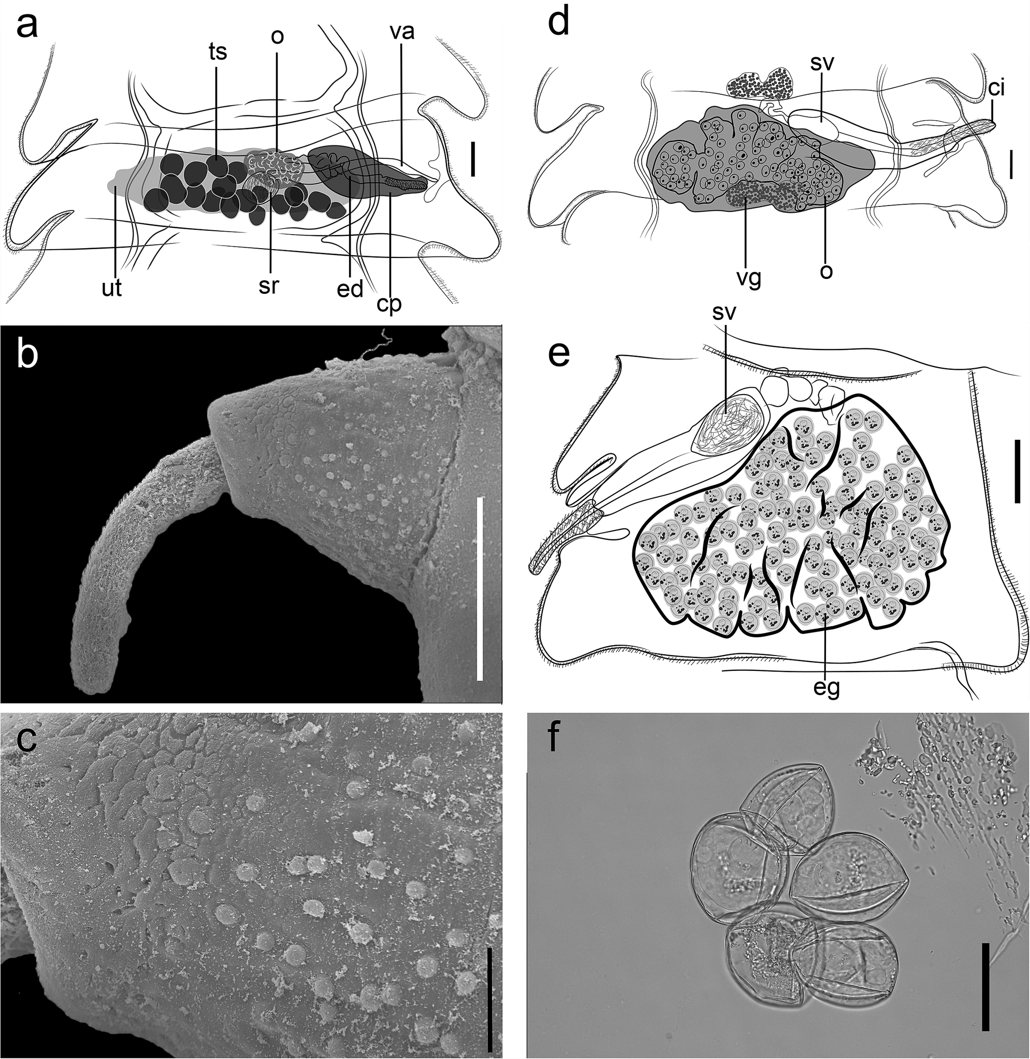
Figure 2. Long description
Panel a, top-left, is a labeled anatomical drawing of a mature proglottid, with structures including ts, o, va, ut, sr, ed, and cp, and a scale bar of 50 micrometers. Panel b, top-center, is a micrograph showing an everted cirrus with a scale bar of 100 micrometers. Panel c, top-right, is a close-up micrograph of the genital papilla, scale bar 25 micrometers. Panel d, bottom-left, is a drawing of a mature proglottid with a developed seminal vesicle inside the cirrus pouch, labeled sv, ci, vg, o, and a 50 micrometer scale bar. Panel e, bottom-center, is a drawing of a gravid proglottid with sv, o, and eg labeled, scale bar 50 micrometers. Panel f, bottom-right, is a micrograph of four eggs, scale bar 50 micrometers. All panels are oriented with internal structures centered and labeled for anatomical reference.
Testes regularly spherical, forming a single cluster in the median field of proglottis; some testes overlapping ovary; testes 17 (15–19; n = 8) per proglottis, with a diameter of 33 (31–37; n = 8) (Figure 2a). Cirrus-sac 292 (215–360; n = 21) long and 87 (54–108; n = 21) wide, containing an armed cirrus (Figure 2b, c), that may or may not be everted, 193 (177–208, n = 10) long by 22 (19–26, n = 11) wide. Internal seminal vesicle present in postmature proglottids, 89 (46–135; n = 21) diameter (Figure 2d).
Ovary multilobed, 259 (219–366; n = 8), wide. Vitellarium bilobed, posterior and central to the ovary, 88 (71–102; n = 8) wide. Uterus reticulated, with several transversely arranged lobes (Figure 2e). Vaginal opening anterior to the cirrus. Vagina distinct in a few mature proglottides, 246 (228–259; n = 4) long and 34 (26–44; n = 4) wide (Figure 2a). Seminal receptacle almost spherical, partially posterior to the medial region of cirrus sac, 43 (38–47; n = 6) long and 33 (22–40; n = 6) wide. Eggs 35 (28–42; n = 21) in diameter, with an embryophore forming a pyriform apparatus. Egg outer shell not spinose (glabrous), 48 (44–64, n = 10) in diameter; oncosphere 17 (13–19, n = 8) in diameter (Figure 2e, f).
Remarks
Among the 26 valid species of Monoecocestus, only 18 are distributed in South America, with M. threlkeldi being the unique species known to parasitize rodents of the genus Lagidium (Parra, Reference Parra1953; Caira et al., Reference Caira, Jensen and Barbeau2022). From all of the South American species, Monoecocestus viscaciae n. sp. can be differentiated by total body length, being considerably smaller. In addition to the differences of the body length, only three South American species of Monoecocestus share the same number of testes with the new species, ranging 15–19: Monoecocestus machadoi (Rego, 1964) Beveridge, Reference Beveridge, Khalil, Jones and Bray1994 from Proechimys guyannesis Geoffroy Saint-Hilaire, from Brazil, Monoecocestus petiso Haverkost & Gardner, Reference Haverkost and Gardner2010 from Galea musteloides Meyen, from Bolivia, and M. threlkeldi from Lagidium peruanum Meyen, from Peru. For the remaining 15 species, the number of testes may be used to clearly differentiate the new species.
Monoecocestus viscaciae n. sp. is distinguished from M. machadoi by having a smaller scolex (207–315 by 216–360 vs. 1,120 by 1,050) and sucker diameter (117–180 vs. 300–330). In addition, the ovary is wider in M. viscaciae n. sp. (219–366 vs. 112), and cirrus length is smaller (177–208 vs. 300). Monoecocestus petiso can be differentiated from the new species by having a higher number of proglottids (49–55 vs. 21–35), a larger seminal receptacle (92 by 68 vs. 38–47 by 22–40), and a larger neck (155–219 vs. 59–111 length).
Monoecocestus threlkeldi is clearly the most similar species to Monoecocestus viscaciae n. sp.; both species are distributed in Peru, with M. threlkeldi originally described from Jauja, Junin, while the new species is known only from Puno, Peru. Both species parasitize rodents (L. peruanum or Northern vizcacha, and L. viscacia or Southern vizcacha, respectively) and also have similar numbers of testes. However, these species can be differentiated from one another based on the size of the scolex (330–470 by 440–530 in M. threlkeldi) being larger than in the new species (207–315 by 216–360). Furthermore, the cirrus pouch is smaller in the new species (215–360 by 54–108 vs. 360–440 by 130–160). Likewise, the egg diameter of M. viscaciae n. sp. is smaller than in M. threlkeldi (44–46 vs. 60–66). Finally, the new species possesses an internal seminal vesicle while M. threlkeldi possesses both internal and external vesicles (Parra, Reference Parra1953).
Molecular results
DNA sequences generated here have a total length of 282 bp (cox1); 781 bp (ITS); 1554 bp (28S rDNA), and 1486 (18S rDNA). Blast query of these sequences confirms the overall similarity of M. viscaciae n. sp. with samples of specimens of the genus Monoecocestus. Sequence of then 28S resulted in a similarity of 95% with Monoecocestus americanus (Stiles, 1895) Fuhrmann, 1932 ex. Erethizon dorsatum (Linnaeus) from Alaska, USA (AY569772) (Wickström et al. Reference Wickström, Haukisalmi, Varis, Hantula and Henttonen2005), regarding cox1 and ITS, resulted in a maximum similarity of 87.2% (AY568184) and 83.7% (AY752652), respectively, with the same Monoecocestus species. Finally, a Blast query of the 18S rDNA sequence resulted in a similarity of 95.6% with Moniezia benedeni (Moniez, 1879) Blanchard, 1891 (AB862304) ex Bos taurus Linnaeus from Senegal (Diop et al. Reference Diop, Yanagida, Hailemariam, Menkir, Nakao, Sako, Tidiane-Ba and Ito2015).
Discussion
Peruanotaenia threlkeldi was described by Parra (Reference Parra1953) as a new genus and species of anoplocephalid cestode based on specimens collected from L. peruanum (Rodentia: Chinchillidae) in Jauja Province, Central Peru. The original description provides a detailed characterization of the specimens and a general justification for the erection of a new genus. However, some inaccuracies seem to be present in the original description. The ovary was described as ‘small and bilobed’ by Parra (Reference Parra1953). However, based on our study of the type material, we conclude that this structure is, in fact, the vitellarium. Furthermore, the position of the testes was reported as ‘mainly anterior to the female glands’ by Parra (Reference Parra1953). Based on this, Haverkost and Gardner (Reference Haverkost and Gardner2009) suggested that the original description was based on immature proglottides. However, based on our observations of the type specimens, we conclude that the distribution of the testes and the female glands actually overlap, indicating that the original description included mature specimens (USNM 37380).
Beveridge (Reference Beveridge, Khalil, Jones and Bray1994) synonymized the genus Peruanotaenia with Monoecocestus, based on the similarity in the position of the vaginal opening, which is anterior to the cirrus sac within the genital atrium. However, his conclusions were not completely supported due to the poor condition of the type specimens, which he described as ‘extremely macerated’. For this reason, he urged the need to study new specimens to confirm its inclusion in Monoecocestus. Haverkost and Gardner (Reference Haverkost and Gardner2009) provided a redescription of M. threlkeldi based on specimens collected from a different host, Holochilus brasiliensis Desmarest (Cricetidae), in Beni, Bolivia. According to the original description, Haverkost and Gardner (Reference Haverkost and Gardner2009) mentioned that type specimens of M. thelkeldi lacked gravid proglottids, and therefore emphasized the need for the collection of mature specimens. These authors highlighted the differences in size and position of the testes; however, no comparisons regarding additional morphological traits were provided, such as the smaller scolex and the fact that the neck is shorter or absent in the type specimens (Table 1). In contrast, photographs of the type specimens of M. threlkeldi analysed in the present study, include terminal proglottids containing eggs (Figure 3). Consequently, the measurements reported by Parra, such as the body size, were based on gravid specimens and are comparable with the measurements of species described herein.
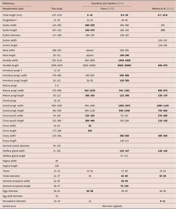
Comparative morphometric characteristics between Monoecocestus viscaciae n. sp. and Monoecocestus threlkeldi from Peru, Bolivia, and Argentina. Differences among M. threlkeldi and the new species are highlighted in bold
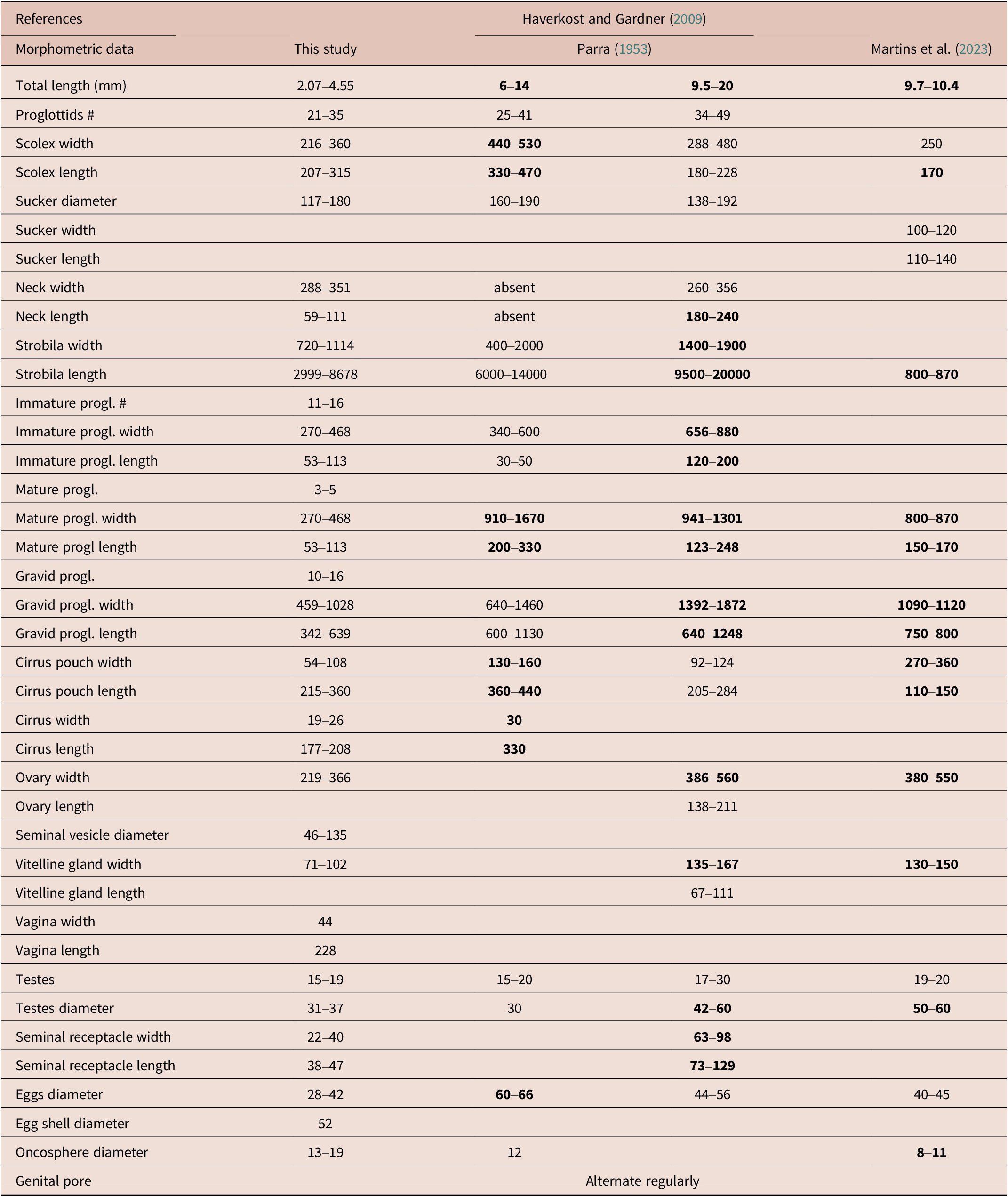
Table 1. Long description
The table consists of 36 rows and 5 columns. The first column lists morphometric traits such as total length, proglottid number, scolex width and length, sucker and neck dimensions, strobila measurements, proglottid counts and sizes, reproductive organ dimensions, and egg characteristics. The next four columns present values for each trait from ‘This study’, Parra 1953, Haverkost and Gardner 2009, and Martins et al. 2023. For each trait, values are given as ranges or single numbers, with some cells marked as ‘absent’ or left blank if data are unavailable. Bolded values in the Parra, Haverkost and Gardner, and Martins columns indicate measurements that differ from the new species. For example, total length in ‘This study’ is 2.07–4.55 mm, while in Parra it is 6–14 mm (bold), in Haverkost and Gardner 9.5–20 mm (bold), and in Martins 9.7–10.4 mm (bold). The number of proglottids ranges from 21–35 in ‘This study’, 25–41 in Parra, and 34–49 in Haverkost and Gardner. Scolex width in ‘This study’ is 216–360, compared to 440–530 (bold) in Parra, 288–480 in Haverkost and Gardner, and 250 in Martins. Several rows, such as sucker width and length, have missing data in some columns. Neck is absent in Parra, but present in other studies. Strobila width and length, proglottid measurements, and reproductive organ sizes are detailed for each study, with bold values marking differences. The genital pore is noted as alternating regularly across all columns. The table highlights that Monoecocestus viscaciae n. sp. is generally smaller in most measurements compared to M. threlkeldi, with the most pronounced differences in total length, scolex width, and strobila length.
Microphotograph of Monoecocestus threlkeldi specimen from the type series NMNH. No. 37,380. Scale bar 1 mm.
Source: Y. Villacampa NMNH, New York.

More recently, Martins et al. (Reference Martins, Robles, Navone and Callejn2023) recorded M. threlkeldi parasitizing Holochilus chacarius Thomas (Cricetidae) from Formosa, Argentina, providing a brief morphological description alongside a 311 bp sequence of the nuclear ITS1 region. The Argentinean specimens show a clear similarity with the specimens identified as M. threlkeldi from Bolivia by Haverkost and Gardner (Reference Haverkost and Gardner2009) (Table 1). In fact, Martins et al. (Reference Martins, Robles, Navone and Callejn2023) identification of M. threlkeldi was based exclusively on Haverkost and Gardner (Reference Haverkost and Gardner2009) redescription, and included only a limited set of characters, such as the number and position of the testes relative to female organs and to osmoregulatory canals. It is important to note that some differences in morphological traits of taxonomic relevance, such as the size of suckers, in the cirrus pouch, and in the scolex (Table 1), were not discussed in the characterization of the specimens identified as M. threlkeldi, parasitizing Holochilus species.
Morphological comparison between Monoecocestus viscaciae n. sp. with the type specimens and original description of M. threlkeldi, as well as the comparison with the redescription by Haverkost and Gardner (Reference Haverkost and Gardner2009) and the record of Martins et al. (Reference Martins, Robles, Navone and Callejn2023), provides a clear distinction between the new species described herein. These morphological differences are summarized in Table 1, and discussed below.
Specimens of M. threlkeldi from Bolivia differ from the new species primarily in the length of the neck, which is shorter in M. viscaciae n. sp. (59–111 vs. 180–240). The overall size of the strobila is also smaller in M. viscaciae n. sp. than in M. threlkeldi, being 720–1,114 by 2,999–8,678 in the new species, and 1,400–1,900 by 9,500–20,000 in the specimens from Bolivia. Furthermore, the ovary is narrower in the new species (219–366 vs. 386–560), and the seminal receptacle is smaller in the new species (22–40 by 38–47 vs. 63–98 by 73–129) in comparison with the specimens of M. threlkeldi from Bolivia.
Samples identified as M. threlkeldi from Argentina mainly differ from M. viscaciae n. sp. by having a shorter scolex (170 vs. 207–315). The cirrus sac is narrower in the new species (54–108 vs. 270–360) but larger than M. threlkeldi (214–360 vs. 110–150). Ovarian width is smaller in M. viscaciae n. sp. (219–366 vs. 380–550) as well as testes diameter (31–37 vs. 50–60).
The collection of additional specimens of M. threlkeldi from their type host and locality, with a thorough description of mature specimens as well as the generation of DNA sequences of nuclear and mitochondrial loci, is needed in order to resolve whether the samples from Bolivia and Argentina are conspecific. However, based on the morphological evidence provided here, and notwithstanding the limitations of the original description of M. threlkeldi and the poorly preserved type specimens, M. viscaciae n. sp. clearly represents a new species. Here, we described the 27th species of Monoecocestus and the 19th one for the Neotropics.
Acknowledgements
Thanks to members of LANABIO, UNAM, México: Laura Márquez Valdelamar, Nelly López Ortíz, and A. Jiménez for their assistance in generating DNA sequences; Berenit Mendoza Garfias for preparing specimens for SEM. To Georgina Ortega-Leite for providing important bibliography references. Special thanks to Lidia Sánchez Pérez, curator of the parasite collection, Museo de Historia Natural, Lima Perú providing catalogue numbers and as well as to Anna Phillips and Yolanda Villacampa, Curator of Clitellata, parasitic worms, and meiofauna and Research assistant, respectively, from Smithsonian Institution, National Museum of Natural History, US, for providing us high-resolution digital photos of the type specimens of P. threlkeldi. We thank Jorge Cárdenas and José Iannacone for their assistance with the logistical aspects of this study. To two anonymous reviewers to considerably improve the original version of the manuscript.
Financial support
Programa de Apoyo a Proyectos de Investigación e Innovación Tecnológica (PAPIIT-UNAM IN215722 and IN226525), granted to AO-F. C-Z acknowledges the Universidad del Altiplano-Puno, Perú, for financial support to carry out a research stay in the Laboratorio de Helmintología, UNAM, from August to December, 2025. To the Secretaría de Ciencia, Humanidades, Tecnología e Innovación (SECIHTI), Mexico, for providing a scholarship to DC-M (CVU 2095706).
Competing interests
The author(s) declare none.
Ethical standard
The authors assert that all procedures contributing to this work comply with the ethical standards of the relevant national and institutional guides on the care and use of animals.


 Open access

