


Introduction
Eggs are widely favored by global consumers as an affordable source of available animal protein (Mosayyeb Zadeh et al. Reference Mosayyeb Zadeh, Mirghelenj and Hasanlou2023). With the growing demand for eggs, ensuring the healthy breeding of laying hens has become increasingly crucial. The egg production cycle of commercial laying hens can be categorized into three distinct phases: the early laying period, the peak laying period and the late laying period (Liu et al. Reference Liu, Fu and Zhou2023a). During the peak laying stage, laying hens exhibit optimal laying performance, characterized by the robust activation of hepatic lipid synthesis and metabolism to supply sufficient lipid for egg yolk formation (Wang et al. Reference Wang, Yue and Liu2019). Whereas, as a result of high-intensity egg production, laying hens are vulnerable to a range of metabolic and immune diseases especially during the late laying period. Previous studies have demonstrated that fatty liver haemorrhage syndrome (FLSH) mainly occurs in caged laying hens during the peak laying period (Zhang et al. Reference Zhang, Meng and Li2022), while oxidative stress is also a prevalent phenomenon for high-yield commercial laying hens especially in the late laying period (Ding et al. Reference Ding, Du and Zhang2020).
Likewise, during the late laying period, laying hens are susceptible to experiencing imbalances in lipid oxidation and a decrease in anti-oxidant capacity, which are often accompanied by FLSH (Liu et al. Reference Liu, Wang and Wang2023b), oxidative stress (Ding et al. Reference Ding, Du and Zhang2020), salpingitis (Li et al. Reference Li, Xu and Liu2023b) and avian osteoporosis (Chen et al. Reference Chen, Turner and Applegate2020). Consequently, an increasing number of researchers are concentrating on how to improve production performance of laying hens in the late laying stage. Previously, it was reported that dietary quercetin, genistein (Xiaodi et al. Reference Xiaodi, Shao and Li2023), naringin (Li et al. Reference Li, Xu and Liu2023b) and xylo-oligosaccharides (Wen et al. Reference Wen, Wang and Li2022) have the effect on alleviating oxidative stress or regulating lipid metabolism of laying hens during the late laying period. Nevertheless, the disease formation is a process of slow progression along with laying eggs, and those metabolic diseases are usually irreversible in the late laying period. Then, it is pivotal to reveal physiological changes when reaching the peak laying stage in hens and explore the nutritional strategies to prevent the progression of diseases in advance.
Hence, the current study was carried out to compare the physiological metabolism difference between the early laying (around 30% laying rate) and peak laying stages (more than 95% laying rate) in laying hens, aiming to reveal what we should focus on in this process and establish a theoretical basis for exploring nutritional regulation strategies during the peak laying stage.
Materials and methods
Animals and experimental design
The experimental design and procedures in this study were approved by the Animal Care and Use Committee of the College of Animal Science and Technology of the Northwest A&F University (Shaanxi, China). A total of 360 Hy-Line Brown laying hens were randomly allotted into two groups with nine replicates of 20 birds each. With the same diet, blood samples were collected from the wing vein of hens when the same flock laying rate reached around 30% or more than 95%, respectively. The serum sample were obtained by centrifugation at 3,000 × g for 10 min after blood samples were left at 37°C water bath. After the blood sampling, one bird from each replicate was randomly selected and killed by cervical dislocation and dissected for tissue collection. Then, about 1 cm3 piece of liver were immediately frozen in liquid nitrogen, and fresh caecal contents were also collected after the cecum intestines were opened. Lastly, all samples were stored at −80°C for further analysis. The basal diet used in the study was typical of the diets commonly used in the northwest part of China, and the ingredients and nutrient levels of the diets are shown in Supplementary Table 1.
Egg quality detection
Egg production was recorded daily in the layer flock. When the flock reached 30% or more than 95% laying rate, three eggs were selected randomly per replicate to measure egg quality parameters. In brief, eggshell thickness and eggshell strength were measured respectively using a dial pipe gauge (ETG-1061; Robotmation, Co., Ltd., Tokyo, Japan) and a texture analyzer (EFG-0503; Robotmation, Co., Ltd., Tokyo, Japan). Then multifunction egg quality analyzer (EMT-5200; Robotmation, Co., Ltd., Tokyo, Japan) was employed to evaluate yolk color, albumen height and haugh units. Finally, the eggshell and yolk were weighed, respectively.
Serum biochemical parameters analyses
The concentration of total cholesterol (TC), triglyceride (TG), total bilirubin (TBIL), total bile acid (TBA), low-density lipoprotein cholesterol (LDL-c) and glutamate (GLU) as well as the activities of aspartate aminotransferase (AST) and alkaline phosphatase (ALP) in the serum were detected by Yangling Demonstration Zone Hospital (Yangling, China). Total anti-oxidant capacity (T-AOC), malondialdehyde (MDA), S-adenosyl methionine (SAM), S-adenosyl homocysteine (SAH) were determined using commercial kits based on manufacturer’s guidelines (Jiancheng Bioengineering Institute, Nanjing, Jiangsu, China).
Hepatic transcriptomics analysis
Total RNA was extracted from the chicken liver tissues using TRIzol reagents according to the manufacturer’s instructions. After the construction of RNA-seq library, sequencing was performed by Shanghai Personal Biotechnology Co., Ltd using Illumina NovaSeq platform. The quality of sequencing reads data was initially examined. Then, HISAT2 software (http://ccb.jhu.edu/software/hisat2/index.shtml) was utilized to correspond the filtered reads with the reference genome (GRCg7b, https://www.ncbi.nlm.nih.gov/genome/111). Thereafter, the differential expression genes (DEGs) which have been normalized were pinpointed through DESeq2 with the criteria of log2 fold change >1 and P-value <0.05. Subsequently, the heatmap packages were implemented to perform bivariate clustering analysis. Eventually, based on P-value <0.05, all identified DEGs were used for gene ontology and Kyoto Encyclopedia of Genes and Genomes (KEGG) functional-enrichment applying Clusterprofiler.
Western blotting
A piece of frozen liver tissue was homogenized in cold RIPA Lysis Buffer supplemented with protease inhibitors and phosphatase inhibitors (DEEYEEBio, Shanghai, China). The protein concentration was preliminarily calibrated by bicinchoninic acid assay kit (Xi’an AccuRef Scientific Co., Ltd, Xi’an, China). The detailed procedures of western blotting analysis refer to the previous method (Zheng et al. Reference Zheng, Zhao and Yang2023). The primary antibodies were diluted at 1:1000, and their detail information was as follows: β-actin (PTMBIO, PTM-5028), PI3K85 (Abways, CY6915), AKT (Abways, CY5561), P-AKT (Abways, CY6569), mTOR (Abways, CY5306), P-mTOR (Abways, CY6571), FoxO1 (Abways, CY7234), STAT3 (Abways, CY5165), P-STAT3 (Abways, CY6566), BCL2 (Abways, CY6717), P38 MAPK (Abways, CY5262), ERK (Abways, CY5487), P-ERK (Abways, CY5277), Caspase-3 (Abways, CY5051), P53 (Abways, CY5131), PPARγ (Abways, CY6675), SREBP1 (Wanleibio, WL02093) and C/EBPα (Abways, CY5723). The goat anti rabbit secondary antibody (DIYIBio, Shanghai, China) was diluted at a ratio of 1:2000. β-Actin was used as internal control to standardize target proteins. Then ImageJ software (National Institutes of Health, Bethesda, MD, USA) was employed to quantify the western blots.
Metabolomics analysis of serum
The serum sample treatment was based on the previous study (Guo et al. Reference Guo, Kuang and Zhuang2021). Metabolomics was performed using the Vanquish UHPLC system (Thermo Fisher, Waltham, MA, USA) accompanied by the Orbitrap Q Exactive HF-X mass spectrometer (Thermo Fisher, Waltham, MA, USA). The detailed mass spectrometer conditions and parameters can according to the previous study (Liu et al. Reference Liu, Zeng and Qu2021). Afterwards, the methodology outlined by Dunn (Dunn et al. Reference Dunn, Broadhurst and Begley2011) was employed for matching peaks, taking peaks and quantifying each metabolite from the raw GC-TOF-MS data. Based on the orthogonal partial least-squares discriminant analysis (OPLS-DA), the variable importance in projection (VIP) values (VIP > 1) coupled with the Student’s t-test (P < 0.05) were employed to identify significantly differential metabolites. Ultimately, the pathway enrichment analysis was performed, and all differential metabolites were annotated to MetaboAnalyst to carry out pathway enrichment analysis.
Caecal microbiome
Microbial genomic DNA extraction was performed following the manufacturer’s instructions using the TIANGEN DNA Kit (TIANGEN, Beijing, China). Then, DNA purity and concentration was determined by NanoDrop 2000 UV–Vis spectrophotometer (Thermo Scientific, Wilmington, DE, USA) and agarose gel electrophoresis, respectively. The purified DNA was amplified using the primer targeting the V3-V4 region of bacterial 16S rDNA (F: ACTCCTACGGGAGGCAGCA, R: GGACTACHVGGGTWTCTAAT). After PCR product purification, paired-end sequencing libraries construction was conducted by the Illumina platform, which were carried out by Personalbio Technology Co. Ltd. (Shanghai, China).
Statistic analysis
In the study, statistical software SPSS 23.0 (SPSS162 Inc., Chicago, IL, USA) and GraphPad Prism 9.4.0 (673) (GraphPad Software, San Diego, CA, USA) were used for our statistical analysis and plotting. All data were expressed as the mean with standard error, and the P value <0.05 was considered statistically significant.
Results
Egg quality
Egg quality parameters from the flock with 30% or 95% laying rate are shown in Fig. 1A–H. The result showed that the egg weight and yolk weight were significantly higher in peak laying hens when compared with the early laying hens (P < 0.05), whereas there was no significant difference in other parameters including albumen weight, haugh unit, albumen height, shell strength and shell thickness (P > 0.05).
The egg quality parameters for eggshell thickness, eggshell strength, eggshell weight, haugh units, albumen height, albumen weight, yolk weight and egg yolk color. Serum biochemical parameters including TC, LDL-c, TG, GLU, SAH, SAM, ALP, AST, T-AOC, MDA, TBA and TBIL. Data are expressed as mean ± SEM. *P < 0.05, **P < 0.01 represent that the comparison between the early and peak laying periods is statistically significant.

Blood biochemical parameters
As displayed in Fig. 1I–T, the contents of serum MDA and TBA were heightened in peak laying hens, while the serum T-AOC, TBIL and LDL-c concentrations were markedly reduced (P < 0.05). No significant difference was observed in serum TC, TG, GLU, SAH, ALP and AST levels between these two different physiological periods (P > 0.05).
DEGs identification and KEGG pathway analysis
As exhibited in Fig. 2A and B, principal component analysis and heat map showed obvious difference in hepatic genes expression pattern between the early laying and peak laying stages. A total of 809 of DEGs were discovered including 540 up-regulated and 269 down-regulated genes (Fig. 2C). Among them, FASN and ACACA related to lipogenesis were up-regulated, while the expression of fatty acid oxidant gene CPT1 was reduced (Supplementary Table 2). KEGG enrichment analysis showed that total DEGs were enriched into fatty acid degradation, pyruvate metabolism, peroxisome, PPAR signalling pathway, p53 signalling pathway and many amino acid metabolism pathways (Fig. 2D). Additionally, the up-regulated pathways, including fatty acid biosynthesis, insulin signalling pathway, pyruvate metabolism, were observed in the peak laying period (Fig. 2E). Conversely, fatty acid degradation, PPAR signalling pathway, peroxisome and p53 signalling pathway were attenuated in the peak laying stage (Fig. 2F).
Hepatic DEGs identification and KEGG pathway enrichment analysis. (A) Principal component analysis. (B) Cluster analysis heat map. Red represents up-regulated genes and blue represents down-regulated genes. (C) The number of DEGs. (D) KEGG pathways enriched form all DEG. (E) KEGG pathways enriched form up-regulation genes. (F) KEGG pathways enriched form down-regulation genes. Larger circles indicate that more genes are involved in the enrichment pathway, and colours indicate that pathway impact values range from brown to green. Abbreviations: DEG, differential expression genes; KEGG, Kyoto Encyclopedia of Genes and Genomes.

Serum metabolomic analysis
As shown in the Fig. 3A, OPLS-DA showed clear separation between the early and peak laying stages. The heat map (Fig. 3B) also indicated distinct discrimination between these two phases. A total of 151 metabolites were identified including 74 up-regulated differential metabolites and 77 down-regulated differential metabolites (Fig. 3C). Particularly, a number of metabolites possessing anti-oxidant function such as alpha-tocopherol were reduced in the peak laying period (Supplementary Table 3). Functional KEGG pathway enrichment analysis indicated that insulin signalling pathway, FoxO signalling pathway, phenylalanine, tyrosine and tryptophan biosynthesis, citrate cycle (tricarboxylic acid [TCA] cycle), mTOR signalling pathway, primary bile acid biosynthesis and tryptophan metabolism were significantly enriched (Fig. 3D). Notably, as depicted in Fig. 3E, there is a close relationship among the metabolites, where one metabolite can transform into others through specific reactions.
Serum metabolomics analysis. (A) OPLS-DA score map of serum metabolites combining positive and negative ions. (B) Clustered heatmap of differential metabolites. (C) The number of differential metabolites. (D) Pathway enrichment based on differential metabolites. (E) The network diagram of differential metabolites. Data were expressed as mean ± SEM. *P < 0.05, **P < 0.01 represent that the comparison between the early and peak laying periods is statistically significant.

Protein expression
As shown in Fig. 4A, the protein expression related to lipid metabolism or apoptosis was examined. At the peak laying stage, there was a notable increase in the protein levels of PI3K85, whereas the protein expressions of the phosphorylation of AKT and mTOR showed no significant changes (P > 0.05) (Fig. 4B–D). Concurrently, the results of Fig. 4E–K revealed that there was a significant decrease in the abundance of inflammation-related proteins, including FOXO1 and p38, as well as antiapoptosis-related proteins such as p53 and bcl2 (P < 0.05); no significant difference was obtained in the phosphorylation of STAT3 and ERK as well as the expression level of caspase-3 (P > 0.05). In addition, the CEBPα protein expression levels, which are associated with lipid synthesis, exhibited a distinct increase, while the level of PPAR decreased (P < 0.05) (Fig. 4L–N).
The protein expression of certain genes. (A) The western blotting bands of related genes. (B-N) Quantitative analysis of western blotting bands. Data are expressed as mean ± SEM. The asterisk indicates statistically significant differences (two-tailed unpaired t-test, *P < 0.05, **P < 0.01).

Caecal microbiome and SCFAs
As exhibited in Fig. 5A, the result showed that Chao1 index was decreased trendily in peak laying hens (P = 0.054), while there were no significant differences in the Shannon or Simpson indices. The beta diversity of caecal microbiota also indicated distinct discrimination between these two phases (Fig. 5B). Further, LEfSe analysis was conducted to identify the specific microbiota between two periods. As shown in Fig. 5C, Ruminococcus, Oxalobacter, Paracoccus, Subdoligranulum, Coprobacillus, Sphaerochaeta, Clostridium, Caulobacter, Prevotella and Thermus were identified as the dominant microflora in peak laying hens. The dominant microflora at the genus level in early laying hens were Nautella, Bifidobacterium, Treponema, Demequina, Allobaculum and Dorea. Functional prediction analysis showed that fatty acid metabolism, peroxisome, lipoic acid metabolism, folate biosynthesis, Valine, leucine and isoleucine degradation, geraniol degradation, citrate cycle (TCA cycle) were attenuated in the peak laying period (Fig. 5D). Conversely, starch and sucrose metabolism, ABC transporters, RNA polymerase, cysteine and methionine metabolism, pentose phosphate pathway, limonene and pinene degradation were enhanced during the peak laying period.
Caecal microbiome analysis. (A) The alpha diversity of caecal microbiota including Chao1, Shannon, Simpson. (B) The beta diversity of caecal microbiota. (C) Differential microbiota analysis based on the LEfSe method. The default parameters were LDA score >2 and P < 0.05). (D) Functional enrichment analysis of differential caecal microorganisms on the level 3 of the metabolism pathway. Data are expressed as mean ± SEM. The asterisk indicates statistically significant differences (two-tailed unpaired t-test, *P < 0.05, **P < 0.01).

Discussion
With the increase of egg production, the hepatic lipid synthesis of laying hens was enhanced to meet the yolk formation during the peak laying period. It has been previously shown that the laying hens in the peak laying period are vulnerable to the occurrence of inflammatory reactions, fat deposition, autophagy and hepatic oxidative stress (Zhu et al. Reference Zhu, Zhang and Du2022). Aiming to investigate the physiological difference between the early and peak laying stages, an integrated analysis of the transcriptome, microbiome and metabolomics was conducted in the study. A previous study has established that T-AOC is commonly employed to assess the anti-oxidant capacity of the host (Liu et al. Reference Liu, Fu and Zhou2023a). Meanwhile, MDA levels, as a by-product of lipid peroxidation, are typically used to gauge the extent of oxidative stress (Biazus et al. Reference Biazus, Da Silva and Bottari2017). In this study, decrease of T-AOC and increase of MDA levels were observed in the serum of laying hens during the peak laying stage, indicating a decrease in the anti-oxidant function of birds during this stage. Normally, there is a balance between the accumulation of reactive oxygen species (ROS) in the organism and the scavenging of ROS by the anti-oxidant defense system. Nevertheless, with the arrival of the peak laying period, a large amount of ROS produced during the peak ovulation stage might disrupt the ROS balance, potentially leading to oxidative stress, resulting in a decrease in T-AOC and an increase in the MDA levels of laying hens during the peak laying period.
To explore the gene expression differences between the early and peak laying stages, transcriptome analysis was employed. RNA-Seq results revealed that FASN, ACACA and PRKAA2 related to de novo fatty acid synthesis were up-regulated, which was similar to the previous research that lipid synthesis is activated strongly in chicken liver during the egg laying period (Li et al. Reference Li, Wang and Xu2015). It has also been evidenced that ACACA and FASN are a rate-limiting enzyme and a key enzyme respectively in the de novo biosynthesis of fatty acid (Chen et al. Reference Chen, Chen and Ding2023; Lou et al. Reference Lou, Zhou and Li2021). Meanwhile, the result of western blotting showed that the expression level of CEBPα was also increased in the peak laying period. Earlier evidence has indicated that CEBPα is known as an important adipogenic factor which can regulate the activity of acetoacetyl-CoA synthetase and fatty acid binding protein 4 (Yang et al. Reference Yang, Zhang and Yang2023). Furthermore, according to KEGG enrichment analysis, it was observed that the pyruvate metabolism, peroxisome and p53 signalling pathway were down-regulated during the peak laying stage. Earlier researches have revealed that pyruvate metabolism and peroxisome play a crucial role in oxidative stress (Tian et al. Reference Tian, Wang and Zhang2022). As a well-known transcription factor, p53 also has a potent effect on alleviating oxidative stress including activating the transcription of anti-oxidant genes, inhibiting the accumulation of ROS and regulating anti-oxidant-related signalling pathways (Budanov Reference Budanov2011; Chipuk et al. Reference Chipuk, Kuwana and Bouchier-Hayes2004). Oxidative stress is usually recognized as an imbalance between cellular anti-oxidant capacity and ROS. Meanwhile, the necrosis and apoptosis of hepatocytes are outcomes of oxidative stress (Kim et al. Reference Kim, Lee and Noh2023). Therefore, we further detected the protein expression level of apoptosis-related genes. Bcl2 is usually considered as an anti-apoptotic factor (Chen et al. Reference Chen, Chen and Ding2023), whose expression level was decreased during the peak laying period in this trail. Those results suggested that lipid synthesis was enhanced while lipid oxidation was decreased in the peak laying period, which may be accompanied with oxidative stress.
As the final products of life activities, metabolites can better reflect the disparity in the physiological status of host. Therefore, the serum metabolomics analysis was employed to explore the physiological metabolism difference between two periods. Among the differential metabolites, a large number of metabolites possess anti-oxidant function such as hesperetin, apigenin, SAM, zingerone and alpha-tocopherol, but they were down-regulated in peak laying hens, which further confirmed a decrease in the anti-oxidant capacity of peak laying hens. As common flavonoids are abundantly present in a variety of fruits and leafy vegetables, hesperetin and apigenin have been demonstrated to trigger the Nrf2 signalling pathway, resulting in alleviation of oxidative stress and inhibiting subsequent inflammation reduced by ROS overproduction (Feng et al. Reference Feng, Yu and Li2017; Li et al. Reference Li, Wang and Liu2021b). It has also been proven that SAM can regulate the level of GSH for its remarkable anti-oxidant feature (Valdés et al. Reference Valdés, Paredes and García Carreras2023). A previous study revealed that zingerone which have a powerful anti-oxidant can activate the AMPK to mitigate hepatic oxidative stress, inflammation and apoptosis (Gungor et al. Reference Gungor, Ekici and Onder Karayigit2020). Alpha-tocopherol, a crucial component of the anti-oxidant defense system, has the capability to prevent lipid peroxidation induced by the elevation of free radicals (Wallert et al. Reference Wallert, Ziegler and Wang2019). Functional KEGG pathway enrichment analysis indicated that FoxO signalling pathway, citrate cycle (TCA cycle), mTOR signalling pathway and primary bile acid biosynthesis pathways were significantly enriched. Consistently, the western blotting result showed the protein expression level of FOXO1 was decreased in the study. FOXO1 has been proven that have a role in up-regulating the anti-oxidant genes in oxidative stress, further alleviating DNA damage and apoptosis (Zhao et al. Reference Zhao, Qin and Wang2023). Taken together, the decline of anti-oxidant metabolites and anti-inflammatory factors suggesting the anti-oxidant capacity of host was impaired.
The TCA cycle is usually considered a hub in glucose, lipid and amino acid metabolism. Hence, the result of liver transcriptomic and serum metabolomic was carried out in a further analysis centring around the TCA cycle (Fig. 6). A series of genes related to de novo synthesis of fatty acids, such as FASN, ACACA and PRKAA2, were elevated, while genes linked to fatty acid degradation, including HADHA, ACADL, CPT1A, ACAA1, ECI2 and HADHB, were decreased. Among these genes, HADHA, a fatty acid β-oxidation enzyme, has been proven that have a role in alleviating oxidative stress and liver damage in mice with non-alcoholic fatty liver disease (Liu et al. Reference Liu, Yang and Liu2023c). Therefore, the down-regulated gene – HADHA – in the study indicates the hens may be experiencing oxidative stress. Consistently, the important metabolic intermediates such as fumaric acid and oxaloacetic acid were seen down-regulated in the peak laying period, which have been proven as strong anti-oxidants to alleviate the oxidative stress and inflammation of the host (Kaur et al. Reference Kaur, Shivanandappa and Kumar2020; Li et al. Reference Li, Li and Chen2022). The results presented here suggested that the TCA cycle is also affected by oxidative stress of the host. On the other hand, the level of TBA in the serum was increased during the peak laying stage. Earlier study has revealed that the bile acid is closely related to liver and intestinal diseases, and excessive bile acid levels may cause fatty liver and inflammatory bowel diseases (Zhang et al. Reference Zhang, Lyu and Wang2023b). These findings indicated that with the decline of anti-oxidant capacity of laying hens, the metabolism of the host was affected profoundly.
Network diagram of genes and metabolites related to the TCA cycle. Red means up-regulated and blue means down-regulated.

Increasing pieces of evidence indicated that the gut microbiota is vital for its function on the physiological and pathological states of the host by regulating the energy metabolism, inflammatory factors and intestinal barrier function (Fan et al. Reference Fan, Xia and Wang2023). The composition of gut microbiota is variable and closely related to the age, diet and metabolism of host. The result of Chao1 index showed a decreasing trend in the peak laying period which indicated the diversity of gut microbes was reduced. The alpha diversity result was in accordance with that of the previous study revealing that the diversity of bacteria was commonly higher in the early laying period than that in the peak laying period (Wang et al. Reference Wang, Wang and Li2023b). Accordingly, the LEfSe analysis revealed an increase in the abundance of Clostridium and a decrease in Bifidobacterium during the peak laying stage. It has been proposed that Clostridium species is importantly related to obesity and metabolic diseases (Zhang et al. Reference Zhang, Lyu and Wang2023b). Bifidobacterium, an important component of probiotics, has been revealed to have anti-oxidant capacity and plays an important role in maintaining the homeostasis of intestinal flora, improving immune level and alleviating inflammatory (Wang et al. Reference Wang, Wang and Li2023b). Altogether, the reduction of intestinal microbial diversity and the decrease of beneficial bacteria abundance indicated that intestinal homeostasis of peak laying hens was affected by the oxidative stress of host. Therefore, it is essential to take measures to maintain the healthy intestinal flora and improve the intestinal homeostasis. Additionally, a recent study had shown that Bifidobacterium V9 is used to regulate oxidative stress and lipid metabolism (Lv et al. Reference Lv, Tao and Peng2023). Further studies should be carried to verify the function of probiotics in laying hens especially before the peak laying period.
Conclusion
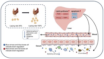
The current study has revealed that the lipid synthesis was enhanced in the peak laying stage, which companied with a decline in the anti-oxidant capacity of hens. In addition, the physiological metabolic changes also lead to the reduction in intestinal microbial diversity and beneficial microbes during the peak laying period (Fig. 7). These results all supported the topic that it is vital to pay attention to enhancing the anti-oxidant capacity in the early laying stage. Additionally, the findings of plentiful anti-oxidants and probiotics in this study may provide a theoretical basis for the nutrient regulation strategy of laying hens during the peak laying period.
The difference of physiological status in laying hens between the early and peak laying periods.

Supplementary Material
The supplementary material for this article can be found at https://doi.org/10.1017/anr.2024.21.
Acknowledgements
This work was funded by the National Science Foundation of China (32372910); National Key Research & Development Program of China (2023YFD1301400 and 2023YFF1001900); the Program for Shaanxi Science & Technology (2022GD-TSLD-46-0302, 2023KXJ-243, 2023GXJS-02-01 and L2022-QCYZX-NY-004) as well as Innovation and entrepreneurship training program for college students (202410712240 and X202410712257). We also express our sincerely thanks to HPC of NWAFU for data analysis.
Author Contributions
Yumeng Wang: Data curation; formal analysis; writing – original draft; writing – review & editing. Xi Sun: Writing – review & editing, Formal analysis. Xiaoying Liu: Writing – review & editing, Data curation. Chaohui Wang: Writing – review & editing, Methodology. Zhouzheng Ren: Writing – review & editing. Xin Yang: Writing – review & editing, Supervision. Xiaojun Yang: Writing – review & editing, Resources. Yanli Liu: Writing – review & editing, Supervision, Project administration, Formal analysis, Conceptualization, Funding acquisition.
Conflicts of Interest
The authors declare that they have no competing interests.
Ethical Standards
The use of all the birds and experimental protocols in this study were approved by the Institution Animal Care and Use Committee of the Northwest A&F University (Yangling, China, protocol number NWAFAC1008).


 Open access