





Introduction
Tetralogy of Fallot is the most common cyanotic CHD, accounting for approximately 7–10% of all CHDs. Reference Zimmerman, Smith and Sable1–Reference Šamánek, Slavík, Zbořilová, Hroboňová, Voříšková and Škovránek3 In many low- and middle-income regions, tetralogy of Fallot accounts for an even higher share of congenital heart cases, representing 7–15% of case series, compared with the 3–6% prevalence reported in population-based studies from high-income countries. Reference Jivanji, Qureshi, Reel and Lubega4–Reference Zimmerman and Sable6 While complete repair in infancy is standard practice in high-income settings, presentation is frequently delayed in low- and middle-income countries due to delayed diagnosis, fragmented referral pathways, and limited surgical capacity. Reference Jivanji, Qureshi, Reel and Lubega4,Reference Hoffman and Hoffman7
Children undergoing late repair present with a distinct and complex physiologic burden. Chronic cyanosis results in increased blood viscosity, microcirculatory hypoxia, and varying degrees of pre-operative organ dysfunction. Reference Hoffman and Hoffman7–Reference Corno, Milano, Samaja, Tozzi and Von Segesser12 Concurrently, long-standing right ventricular pressure overload drives myocardial hypertrophy, interstitial fibrosis, and impaired relaxation, culminating in restrictive diastolic physiology. Reference Chaturvedi, Shore and Lincoln9,Reference Munkhammar, Cullen, Jögi, De Leval, Elliott and Norgård13–Reference Ganni, Yen Ho and Reddy15 These changes are often compounded by elevated pulmonary vascular resistance due to hypoxic vasoconstriction, endothelial dysfunction, and structural remodelling, as well as by the development of substantial aortopulmonary collateral circulation. Reference Cordina and Celermajer10,Reference Oechslin, Kiowski, Schindler, Bernheim, Julius and Brunner-La Rocca16–Reference Edraki, Naghshzan, Amoozgar, Keshavarz, Mehdizadegan and Mohammadi19
Following surgical correction, these pathophysiologic features converge to create a fragile post-operative state. A stiff, hypertrophied right ventricle with limited preload reserve must abruptly accommodate increased pulmonary blood flow into a reactive vascular bed, often in the presence of residual collateral runoff. This imbalance disrupts right ventricular–pulmonary arterial coupling and reduces cardiovascular reserve, increasing the risk of post-operative right ventricular failure, low cardiac output, and prolonged post-operative recovery. Reference García and Santos20–Reference Naeije and Badagliacca22
Pharmacologic support in this setting must therefore address impaired right ventricular relaxation and elevated pulmonary afterload while preserving effective ventriculo-arterial coupling. Milrinone, with its combined inotropic, lusitropic, and vasodilatory properties, is particularly attractive in this context. Reference Colucci23–Reference Farah and Frangakis25 Its use has become widespread in congenital cardiac surgery, including in patients undergoing tetralogy of Fallot repair. However, current practice is largely informed by extrapolation from studies conducted in infants and heterogeneous paediatric cardiac populations, rather than from trials specifically focused on older, cyanotic children undergoing late repair. Reference Smith, Owen, Borgman, Fish and Kannankeril26–Reference Hoffman, Wemovsky and Atz29
While early randomised data suggested a reduction in low cardiac output syndrome, more recent comparative studies and systematic reviews have questioned the consistency of these benefits, reporting no clear improvement in major post-operative outcomes and, in some cohorts, increased requirements for fluid or vasoactive support. Reference Burkhardt, Hummel, Rücker and Stiller27–Reference Cavigelli-Brunner, Hug and Dave30 This uncertainty is particularly consequential in low- and middle-income country settings, where delayed repair is common and post-operative management is effectively guided by integrated clinical and echocardiographic assessment.
This review synthesises the mechanistic basis for milrinone use, critically appraises the existing clinical data, and delineates a pragmatic, assessment-guided approach to its application in low- and middle-income country settings where the burden of late repair is high. In doing so, it aims to inform judicious clinical practice and to define critical priorities for lesion-specific research.
Pathophysiologic basis for haemodynamic vulnerability after late tetralogy of Fallot repair
Post-operative vulnerability following late tetralogy of Fallot repair arises from maladaptation of the right ventricular–pulmonary circuit, shaped by two fundamental and interrelated processes: structural right ventricular remodelling from chronic pressure overload and pulmonary vascular maladaptation from chronic low flow and cyanosis (Figure 1).
Pathophysiologic cascade in late-presenting tetralogy of Fallot (TOF) and mechanistic targets of milrinone. The left panel illustrates the maladaptive pathophysiology of late-presenting TOF. Chronic right ventricular (RV) pressure overload leads to hypertrophy, interstitial fibrosis, and restrictive physiology, while prolonged cyanosis drives erythrocytosis, endothelial dysfunction, and pulmonary vascular hyperreactivity. These processes converge to create a fragile RV–pulmonary arterial (PA) unit characterised by impaired ventriculo-arterial coupling and limited physiologic reserve. The right panel depicts milrinone’s cellular mechanism as a phosphodiesterase-3 (PDE3) inhibitor, increasing intracellular cyclic adenosine monophosphate (cAMP) in cardiomyocytes and vascular smooth muscle, thereby producing coordinated lusitropy, pulmonary and systemic vasodilation, and modest inotropy. Collectively, these effects target the dominant haemodynamic vulnerabilities after late TOF repair and aim to stabilise the high-risk post-operative state. PVR = pulmonary vascular resistance; SERCA2a = sarco/endoplasmic reticulum calcium ATPase 2a.

Figure 1 Long description
A diagram of the pathophysiologic cascade in late-presenting tetralogy of Fallot (TOF) and the mechanistic targets of milrinone. The left panel illustrates the maladaptive pathophysiology of late-presenting TOF. Chronic right ventricular (RV) pressure overload leads to hypertrophy, interstitial fibrosis, and restrictive physiology. Prolonged cyanosis drives erythrocytosis, endothelial dysfunction, and pulmonary vascular hyperreactivity. These processes converge to create a fragile RV-pulmonary arterial (PA) unit characterized by impaired ventriculo-arterial coupling and limited physiologic reserve. The right panel depicts milrinone's cellular mechanism as a phosphodiesterase-3 (PDE3) inhibitor, increasing intracellular cyclic adenosine monophosphate (cAMP) in cardiomyocytes and vascular smooth muscle. This produces coordinated lusitropy, pulmonary and systemic vasodilation, and modest inotropy. Collectively, these effects target the dominant hemodynamic vulnerabilities after late TOF repair and aim to stabilize the high-risk post-operative state.
The restrictive, preload-sensitive right ventricle
Chronic pressure overload in unrepaired tetralogy of Fallot drives concentric right ventricular hypertrophy, interstitial fibrosis, and progressive increases in myocardial stiffness. This structural remodelling impairs active relaxation and reduces diastolic compliance, creating a restrictive physiologic phenotype. Reference Hoffman and Hoffman7,Reference Jost, Connolly and Burkhart8,Reference Munkhammar, Carlsson, Arheden and Pesonen14,Reference Ganni, Yen Ho and Reddy15,Reference Heinisch, Laetitia and Damian31 Consequently, the right ventricle becomes highly preload-dependent and operates along the steep portion of its diastolic pressure–volume relationship, rendering cardiac output exquisitely sensitive to even modest changes in loading conditions. Reference Ganni, Yen Ho and Reddy15,Reference García and Santos20,Reference Naeije and Badagliacca22
Clinically, restrictive right ventricular physiology is characterised by impaired relaxation, elevated right ventricular end-diastolic pressures, and antegrade diastolic pulmonary flow. 32,Reference Van den Eynde, Derdeyn and Schuermans33 Although this restrictive pattern may limit post-operative right ventricular dilation, it does so at the cost of elevated systemic venous pressures, reduced preload reserve, and heightened sensitivity to afterload changes. Perioperatively, this substrate predisposes patients to low cardiac output syndrome and prolonged ventilatory support, especially if pulmonary vascular resistance remains elevated. Importantly, right ventricular diastolic dysfunction persists long after surgical repair. Reference Van den Eynde, Derdeyn and Schuermans33–Reference Sachdev, Bhagyavathy, Varghese, Coelho and Kumar35
Pulmonary maladaptation
The pulmonary vasculature in late-presenting tetralogy of Fallot is shaped by two key insults: chronically reduced pulmonary blood flow (anatomic) and prolonged hypoxaemia (cyanosis). Together, these factors result in a vasculature that is underdeveloped, structurally remodelled, and functionally hyperreactive.
-
• Chronic Low Flow: Inherent right ventricular outflow tract obstruction limits pulmonary arterial flow, which may lead to vascular hypoplasia and reduced effective cross-sectional area of the pulmonary vascular bed.
-
• Chronic Hypoxaemia: Cyanosis induces secondary erythrocytosis, increasing blood viscosity and impairing microcirculatory flow. Reference Corno, Milano, Samaja, Tozzi and Von Segesser12 Concurrently, it promotes endothelial dysfunction, reducing nitric oxide bioavailability and increasing basal and reactive pulmonary vascular tone. Reference Zabala and Guzzetta11
Collectively, these processes produce a pulmonary vascular bed characterised by elevated and labile resistance.
Following surgical correction, the abrupt increase in pulmonary blood flow confronts a vasculature that is poorly adapted to higher flow states and prone to exaggerated vasoconstrictive responses. This mismatch markedly increases right ventricular afterload, placing the already compromised right ventricle at high risk of failure during the early post-operative period.
Systemic vascular dysfunction and microcirculatory impairment
Beyond the pulmonary circulation, chronic cyanosis exerts widespread systemic effects. Reference Zabala and Guzzetta11,Reference Corno, Milano, Samaja, Tozzi and Von Segesser12,Reference Oechslin, Kiowski, Schindler, Bernheim, Julius and Brunner-La Rocca16 Endothelial dysfunction impairs vasoregulation and organ perfusion, while increased blood viscosity contributes to microcirculatory sludging and end-organ hypoxia. These systemic alterations further reduce physiologic reserve, heighten sensitivity to haemodynamic perturbations, and contribute to the overall fragility of the post-operative state.
The fragile post-operative state: impaired coupling and ventricular interdependence
The interaction between a hypertrophied, restrictive right ventricle and a dysfunctional pulmonary vascular bed creates a precarious post-operative state. Two closely interrelated phenomena are central to this vulnerability:
-
1. Compromised Ventriculo-arterial Coupling: The pressure-loaded right ventricle operates with limited contractile and diastolic reserve. Its performance is poorly matched to a reactive pulmonary vasculature, resulting in inefficient right ventricular–PA coupling. In this setting, even modest increases in pulmonary vascular resistance or decreases in right ventricular compliance can precipitate disproportionate declines in forward flow and cardiac output. Reference Redington, Penny, Rigby and Hayes36
-
2. Ventricular Interdependence: Elevated right ventricular pressures and impaired right ventricular compliance adversely affect right ventricular performance. Right ventricular dilation or septal shift can distort LV geometry, impair diastolic filling, and reduce systemic output, further limiting cardiovascular reserve in the early post-operative period. Reference Naeije and Badagliacca22,Reference Santamore and Dell’Italia37
The burden of aortopulmonary collaterals
Aortopulmonary collateral arteries are prevalent in older cyanotic children and introduce distinct perioperative challenges. Reference Fang, Xiong and Wang18,Reference Edraki, Naghshzan, Amoozgar, Keshavarz, Mehdizadegan and Mohammadi19 While they may augment pulmonary blood flow pre-operatively, collaterals impose a significant volume load on the left heart, increase pulmonary venous return, and complicate intraoperative haemostasis and post-operative fluid management. Following repair, the abrupt increase in pulmonary blood flow through both the reconstructed right ventricular outflow tract and persistent collaterals can overwhelm the non-compliant right ventricle, precipitating acute haemodynamic instability if loading conditions are not meticulously controlled.
Integrated right ventricular–pulmonary circulatory vulnerability
Taken together, a stiff hypertrophied right ventricle, a reactive pulmonary vascular bed, significant collateral burden, and adverse ventricular interdependence create a narrow physiologic window for maintaining adequate cardiac output. The right ventricular–pulmonary unit in late-presenting tetralogy of Fallot is functionally fragile and highly sensitive to perturbations in preload, afterload, and ventricular compliance. This integrated pathophysiologic state provides the foundational rationale for targeted inodilatory therapy, such as milrinone, which aims to simultaneously improve lusitropy, reduce pulmonary vascular resistance, and optimise ventriculo-arterial coupling during the high-risk post-operative period.
Milrinone use in late tetralogy of Fallot repair: pharmacologic rationale
Milrinone’s pharmacologic profile offers a targeted strategy to address the predominant haemodynamic vulnerabilities encountered after late tetralogy of Fallot repair. As a selective phosphodiesterase-3 inhibitor, it increases intracellular cyclic adenosine monophosphate concentration in cardiomyocytes and vascular smooth muscle, producing coordinated lusitropy, vasodilation, and modest inotropic support. Reference Farah and Frangakis25 This integrated action aligns directly with the pathophysiologic substrate of the restrictive, pressure-loaded right ventricle and the reactive pulmonary vascular bed observed in late-presenting tetralogy of Fallot (Figure 1).
Targeting diastolic dysfunction: enhancement of right ventricular lusitropy
Augmentation of lusitropy is arguably milrinone’s most physiologically relevant effect in this setting. By increasing intracellular cyclic adenosine monophosphate, milrinone enhances phosphorylation of phospholamban, accelerates sarcoplasmic reticulum calcium reuptake via SERCA2a, and reduces diastolic calcium sensitivity, all of which promote earlier and more complete myocardial relaxation. Reference Farah and Frangakis25 These cellular effects counteract the delayed relaxation and reduced compliance characteristic of the hypertrophied, fibrotic right ventricle in late tetralogy of Fallot. Clinically, improved lusitrophy translates into lower right ventricular filling pressures, improved early diastolic filling, and greater preload reserve, which are critical for maintaining forward flow in a preload-sensitive ventricle.
Reducing right ventricular afterload and improving right ventricular–PA arterial coupling
Milrinone reduces right ventricular afterload by promoting pulmonary vasodilation, addressing the elevated and labile pulmonary vascular resistance commonly present in delayed tetralogy of Fallot repair. Pulmonary vasodilation via phosphodiesterase-3 inhibition is largely independent of the nitric oxide pathway, which may be impaired in chronic cyanosis, a practical advantage in settings where inhaled nitric oxide is unavailable. Reference Allbritton-King and García-Cardeña17,Reference Wessel38–Reference Bailey, Miller, Lu, Tosone, Kanter and Tam40
By lowering pulmonary vascular impedance and improving ventricular relaxation, milrinone favourably shifts right ventricular–PA coupling towards a more efficient operating point. Reference García and Santos20,Reference Kozlik-Feldmann, Hansmann, Bonnet, Schranz, Apitz and Michel-Behnke41 The combined effect enhances stroke volume at a lower metabolic cost, which is critical for the stiff, pressure-loaded right ventricle in the early post-operative period.
Inotropic support and arrhythmogenic potential
Milrinone provides mild inotropic support via improved intracellular calcium handling without β-adrenergic stimulation. Reference Bailey, Miller, Lu, Tosone, Kanter and Tam40 This mechanism avoids the pronounced tachycardia and increased myocardial oxygen demand associated with catecholamines. However, milrinone is not without electrophysiologic effects; phosphodiesterase inhibition may predispose to arrhythmias, Reference King, Thompson and Foote42,Reference Öztürk, Kafalı and Tanıdır43 particularly in the presence of electrolyte imbalance, atrial dilation, or surgical scarring. Some observational data suggest an association between post-operative milrinone use and increased tachyarrhythmia risk in congenital heart surgery cohorts, although causality is difficult to establish and many confounders exist. Reference Burkhardt, Hummel, Rücker and Stiller27,Reference Saengsin, Sperotto and Lu28 However, in the specific context of late tetralogy of Fallot—where the substrate is dominated by diastolic dysfunction, elevated afterload, and catecholamine sensitivity—the net electrophysiologic profile of milrinone may still be favourable compared to pure β-agonists. Careful monitoring and maintenance of normal electrolyte concentrations remain essential.
Potential effects on microcirculatory and endothelial dysfunction
Beyond its macrohaemodynamic actions, milrinone may influence endothelial and microcirculatory function. Experimental and clinical evidence in other paediatric contexts suggests that phosphodiesterase-3 inhibition can preserve microvascular perfusion, maintain capillary density, and mitigate endothelial glycocalyx degradation, potentially facilitating tissue oxygen delivery and capillary recruitment. Reference Sarta-Mantilla, Fernández-Sarmiento and Acevedo44 While direct evidence in late tetralogy of Fallot is limited, these effects are physiologically plausible and may be particularly relevant in erythrocytotic children, in whom increased blood viscosity and microcirculatory sluggishness contribute to post-operative organ dysfunction.
Potential influence on post-operative effusions
While no studies have directly evaluated milrinone’s effect on post-operative effusions, its capacity to reduce pulmonary vascular resistance and improve right ventricular diastolic function may lower right-sided filling pressures and systemic venous congestion, providing a plausible mechanistic link to altered pleural fluid formation and clearance. This potential effect remains speculative and warrants focused investigation. Reference Gagliardi, Ceccherelli, Lovato and Gagliardi45
Pharmacokinetic considerations in older cyanotic children
Children undergoing late tetralogy of Fallot repair exhibit altered pharmacokinetics due to polycythaemia, expanded plasma volume, chronic hypoxaemia, and variable renal perfusion, all of which influence milrinone clearance and volume of distribution. Reference Bailey, Miller, Lu, Tosone, Kanter and Tam40,Reference Bailey, Hoffman and Wessel46 Dosing paradigms derived from infant cohorts may be unreliable in this population, and comorbid renal dysfunction may further prolong drug elimination. Consequently, milrinone exposure is often unpredictable, reinforcing the importance of careful clinical titration guided by perfusion indices, lactate trends, and serial echocardiography rather than fixed dosing alone.
Clinical evidence for milrinone use after tetralogy of fallot repair
Evidence from landmark trials and heterogeneous cohorts
The haemodynamic rationale for phosphodiesterase-3 inhibition in paediatric cardiac surgery was established through early physiologic studies conducted before the availability of randomised outcome trials. Reference Hammett and Griksaitis47 Seminal investigations by Wernovsky, Chang, and colleagues demonstrated that milrinone increases cardiac output while reducing systemic and pulmonary vascular resistance, without a proportional rise in myocardial oxygen consumption or significant tachycardia in children following cardiopulmonary bypass. These studies highlighted the importance of enhancing lusitropy and reducing afterload in the post-operative congenital heart population and provided the mechanistic foundation for subsequent clinical trials.
The most influential clinical evidence supporting milrinone use derives from the landmark PRIMACORP trial, a multicentre, randomised, placebo-controlled study evaluating prophylactic milrinone after paediatric congenital heart surgery. Reference Hoffman, Wemovsky and Atz29,Reference Hoffman, Wernovsky and Atz48 PRIMACORP demonstrated a significant reduction in the incidence of low cardiac output syndrome and shorter ICU stay in patients receiving milrinone compared with placebo. However, the trial population consisted predominantly of infants with mixed cardiac diagnoses, with minimal representation of older, cyanotic children or patients with chronically pressure-loaded right ventricles.
Following PRIMACORP, multiple observational studies and single-centre series evaluated milrinone in heterogeneous paediatric cardiac surgery cohorts, generally reporting associations with improved cardiac index, reduced catecholamine requirements, and shorter ICU duration. Reference Burkhardt, Hummel, Rücker and Stiller27,Reference Cavigelli-Brunner, Hug and Dave30,Reference Feneck, Sherry, Withington and Oduro-Dominah49,Reference Bailey, Miller, Lu, Tosone, Kanter and Tam50 Subsequent systematic reviews and network meta-analyses have confirmed that milrinone reduces the incidence of low cardiac output syndrome compared with placebo or no inotrope, although effects on mortality remain inconsistent and highly context-dependent. Reference Burkhardt, Hummel, Rücker and Stiller27,Reference Lechner, Hofer and Leitner-Peneder51 Collectively, these data support milrinone’s physiologic efficacy in paediatric cardiac surgery but offer limited insight into its effectiveness in specific lesion phenotypes such as late-presenting tetralogy of Fallot.
Lesion-specific evidence, current gaps, and evolving efficacy debate
Robust, lesion-specific evidence for milrinone in repaired tetralogy of Fallot is scarce. Existing data are largely derived from small observational series, retrospective analyses, and subgroup analyses. Reference Hammett and Griksaitis47,Reference Gatzoulis, Clark, Cullen, Newman and Redington52,Reference Therrien, Marx and Gatzoulis53 These reports suggest potential benefits, including improved right ventricular diastolic indices, reduced filling pressures, and earlier extubation, particularly in patients with elevated pulmonary vascular resistance or significant restrictive physiology. Importantly, no randomised controlled trials have been designed specifically to evaluate milrinone in older children undergoing late tetralogy of Fallot repair, representing a critical evidence gap.
The limitations of existing evidence are most consequential in low- and middle-income countries, where delayed tetralogy of Fallot repair remains common and post-operative physiology differs substantially from early-repair cohorts in high-income settings. Reference Jivanji, Qureshi, Reel and Lubega4,Reference Zimmerman and Sable6 In these environments, milrinone is frequently adopted based on biologic plausibility, extrapolation from global practice norms, and the absence of alternative pulmonary vasodilators such as inhaled nitric oxide. Recent analyses have associated milrinone use with increased fluid requirements, higher catecholamine exposure, and a lack of clear mortality benefit in broader paediatric cardiac surgery populations. Reference Burkhardt, Hummel, Rücker and Stiller27,Reference Saengsin, Sperotto and Lu28 While hypotension is a well-recognised adverse effect, data on arrhythmogenicity are inconsistent and appear context-dependent. Reference Smith, Owen, Borgman, Fish and Kannankeril26–Reference Saengsin, Sperotto and Lu28,Reference Karamlou, Silber and Lao54 This debate underscores that milrinone’s effects, both beneficial and adverse, are likely mediated by underlying physiology.
Summary of the evidence landscape
In summary, the evidence supporting milrinone in late tetralogy of Fallot repair is indirect and derived from physiologically distinct populations (Figure 2). While its pharmacologic profile is mechanistically attractive, high-quality, lesion-specific data are absent. This evidence gap is most acutely felt in clinical environments where the burden of late repair is high, and therapeutic decisions must balance physiologic rationale against uncertain comparative effectiveness.
The milrinone evidence landscape in repaired tetralogy of Fallot (TOF): existing data and critical gaps. The schematic illustrates a hierarchy of evidence supporting the use of milrinone after congenital heart surgery. The broad upper tier represents robust data from landmark randomised controlled trials (e.g., PRIMACORP) conducted in heterogeneous paediatric cardiac surgery populations, predominantly infants. The intermediate tier reflects limited observational data from CHD cohorts that include some TOF patients, often across mixed age groups. The narrow lower tier highlights the critical evidence gap: the absence of dedicated, high-quality studies focusing specifically on older, cyanotic children undergoing late TOF repair—particularly in low- and middle-income country (LMIC) settings, where delayed presentation is common and post-operative physiology differs fundamentally from early-repair cohorts. This gap underscores the need for future pragmatic, lesion-specific research to define optimal therapeutic use. RCT = randomised controlled trial.

Towards a pragmatic, physiology-guided approach in resource-variable settings
The absence of definitive, lesion-specific evidence for milrinone use after late tetralogy of Fallot repair should not be interpreted as a barrier to rational post-operative management. Rather, it highlights the need for a deliberate, physiology-guided strategy—one that aligns therapeutic choice with dominant haemodynamic mechanisms and is particularly suited to resource-variable settings, where invasive monitoring is uncommon and therapeutic options are limited.
Within this framework, milrinone should not be considered a routine or prophylactic intervention. Its use is best reserved for patients whose post-operative physiology reflects the vulnerabilities described in preceding sections and in whom the drug’s pharmacologic profile directly addresses the dominant disturbance.
Milrinone selection should therefore be guided by concordant clinical and echocardiographic evidence of right ventricular–pulmonary arterial instability. Clinical indicators suggestive of low cardiac output in a preload-sensitive right ventricle include cool extremities with delayed capillary refill, rising or persistently elevated serum lactate, oliguria despite apparently adequate intravascular volume, and escalating catecholamine requirements to maintain perfusion in the absence of overt systolic dysfunction.
Echocardiography plays a central role in identifying restrictive right ventricular physiology. Features supporting this diagnosis include impaired early-diastolic right ventricular filling with an abbreviated filling time, antegrade diastolic pulmonary artery flow reflecting elevated end-diastolic pressure and reduced compliance, right atrial enlargement indicative of chronically elevated filling pressures, and reduced respiratory variation in trans-tricuspid inflow, consistent with limited preload reserve.
Markers of elevated right ventricular afterload and pulmonary vascular reactivity further refine patient selection. These include interventricular septal flattening or leftward septal shift, elevated tricuspid regurgitation-derived right ventricular systolic pressure estimates, shortened pulmonary artery acceleration time, and dynamic changes in right ventricular size or septal geometry with ventilation or agitation, suggesting a labile pulmonary vascular bed.
Taken together, these findings define a physiologic state characterised by impaired right ventricular relaxation, increased effective afterload, and compromised ventriculo-arterial coupling. Serial reassessment of the same clinical and echocardiographic parameters—rather than reliance on invasive haemodynamics—allows clinicians to judge whether milrinone is achieving its intended effects: improved diastolic filling, reduced right ventricular afterload, enhanced forward flow, and stabilisation of systemic perfusion. This selective approach prioritises patients in whom diastolic dysfunction or pulmonary vascular reactivity is the principal driver of post-operative instability, while avoiding unnecessary exposure in those with preserved ventricular reserve or primarily volume-responsive physiology.
Equally important is recognising clinical scenarios in which milrinone is unlikely to confer benefit and may contribute to haemodynamic instability. In patients without evidence of restrictive right ventricular physiology or elevated pulmonary vascular resistance, particularly those with preserved diastolic function and primarily volume-responsive or distributive physiology, the vasodilatory effects of milrinone may lead to hypotension without meaningful improvement in forward flow. Similarly, in the presence of significant vasoplegia or low systemic vascular resistance, further vasodilation may necessitate escalating vasopressor support and increase overall haemodynamic complexity. In such contexts, milrinone does not address the dominant physiologic disturbance and may be detrimental if used indiscriminately. These considerations further underscore the importance of aligning therapy with underlying physiology rather than routinely administering milrinone in post-operative care.
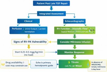
Figure 3 illustrates a pragmatic, physiology-guided approach to post-operative haemodynamic assessment and selective milrinone use after late tetralogy of Fallot repair.
Proposed physiology-guided algorithm for post-operative management after late tetralogy of Fallot (TOF) repair in resource-limited settings. This algorithm emphasises integrated clinical and echocardiographic assessment rather than invasive haemodynamic monitoring. Key decision points focus on identifying physiologic markers of RV–PA vulnerability, including echocardiographic evidence of restrictive RV filling, elevated RV afterload, or clinical signs of low cardiac output. In selected patients, milrinone is initiated cautiously—typically without a loading bolus—and titrated according to clinical and echocardiographic response. Boxes highlighted in yellow denote considerations particularly relevant to low- and middle-income country (LMIC) contexts, including drug availability, cost constraints, and the role of milrinone as a primary pulmonary vasodilator in the absence of inhaled nitric oxide or advanced mechanical support. The algorithm reinforces a pragmatic, patient-tailored strategy rather than a universal protocol. RV = right ventricle; LCOS = low cardiac output syndrome; iNO = inhaled nitric oxide.

From principles to practice
Synthesising these principles into bedside decision-making clarifies how physiology, rather than protocol, should guide post-operative care after late tetralogy of Fallot repair. Such a pragmatic approach moves away from rigid algorithms towards context-sensitive clinical reasoning. It requires identification of the dominant physiologic disturbance, whether restrictive right ventricular failure, elevated pulmonary afterload, or their interaction, selection of a therapy whose mechanism directly addresses that disturbance, and focused monitoring of the relevant physiologic response. In this setting, where a stiff, hypertrophied right ventricle confronts a reactive pulmonary vascular bed, milrinone, used selectively, dosed cautiously, and guided by integrated clinical and echocardiographic assessment, represents a rational application of pharmacologic principles within the realities of resource-variable care.
Conclusion and future directions
Milrinone should not be considered a routine component of post-operative care following late tetralogy of Fallot repair. Its role is inherently conditional, determined by the presence and severity of restrictive right ventricular physiology, elevated pulmonary vascular load, and impaired ventriculo-arterial coupling. In this context, milrinone functions not as a generic inotrope but as a targeted inodilator whose principal value lies in improving right ventricular relaxation and reducing effective afterload.
Ultimately, the relevant question is not whether milrinone “works” after tetralogy of Fallot repair, but in whom, when, and why it should be used. Addressing this question requires a shift away from extrapolation from heterogeneous populations towards lesion-specific investigation grounded in post-operative physiology.
Future research should prioritise pragmatic study designs and prospective registries that enrol late-presenting tetralogy of Fallot patients and link milrinone exposure to clearly defined physiologic phenotypes, echocardiographic response patterns, and clinically meaningful outcomes. Such efforts, particularly if embedded within routine care in resource-variable settings, would allow evaluation of milrinone as a targeted intervention rather than a protocolised default. Until such data are available, best practice after late tetralogy of Fallot repair should be defined by careful patient selection, cautious dosing, and serial reassessment of physiologic response using integrated clinical and echocardiographic markers.
Data availability statement
Not applicable.
Acknowledgements
Not Applicable.
Author contributions
JLN conceptualised, drafted, reviewed, and edited the manuscript.
Financial support
This study received no external funding.
Competing interests
The authors have no conflicts of interest to disclose.
Ethical standard
Not Applicable.
Consent for publication
Not applicable.
Use of artificial intelligence tools
Artificial intelligence-assisted tools were used during the preparation of this manuscript to generate figures based on author-defined scientific content and physiologic frameworks; artificial intelligence tools were used to assist with layout and visual structuring only. Final figure content, labelling, and scientific interpretation were reviewed, modified as needed, and approved by the authors. ChatGPT (GPT-5.2, OpenAI), accessed via the web-based interface (https://chat.openai.com), was used in January 2026. The tool was used in its standard configuration, without modification, fine-tuning, or augmentation with additional data by the authors. The authors declare no competing interests or conflicts of interest arising from the use of these artificial intelligence tools.



 Open access

