





Introduction
Bipolar disorder (BD), a chronic psychiatric condition affecting over 60 million people globally, is characterized by debilitating oscillations between mania and depression (Anderson, Haddad, & Scott, Reference Anderson, Haddad and Scott2012; Grande, Berk, Birmaher, & Vieta, Reference Grande, Berk, Birmaher and Vieta2016), with >70% of disease burden attributable to depressive episodes (Miller, Dell’Osso, & Ketter, Reference Miller, Dell’Osso and Ketter2014; Nierenberg et al., Reference Nierenberg, Agustini, Köhler-Forsberg, Cusin, Katz, Sylvia and Berk2023) and a staggering 20- to 30-fold increased suicide risk (Miller & Black, Reference Miller and Black2020; Plans et al., Reference Plans, Barrot, Nieto, Rios, Schulze, Papiol and Benabarre2019). Despite decades of pharmacological development, first-line therapies (e.g. lithium, antipsychotics) face critical limitations: up to 33% of patients exhibit treatment resistance, relapse rates approach 50% within 2 years, and metabolic side effects from second-generation antipsychotics exacerbate comorbidities (Elsayed, Ercis, Pahwa, & Singh, Reference Elsayed, Ercis, Pahwa and Singh2022; Hirsch et al., Reference Hirsch, Yang, Bresee, Jette, Patten and Pringsheim2017; Perlis et al., Reference Perlis, Ostacher, Patel, Marangell, Zhang, Wisniewski and Thase2006). This therapeutic crisis stems from a fundamental disconnect – while genome-wide association studies (GWASs) have identified >30 risk loci for BD (Cichon et al., Reference Cichon, Mühleisen, Degenhardt, Mattheisen, Miró, Strohmaier and Nöthen2011; Ferreira et al., Reference Ferreira, O’Donovan, Meng, Jones, Ruderfer and Jones2008; Hou et al., Reference Hou, Bergen, Akula, Song, Hultman, Landén and McMahon2016; Mühleisen et al., Reference Mühleisen, Leber, Schulze, Strohmaier, Degenhardt, Treutlein and Cichon2014; Stahl et al., Reference Stahl, Breen, Forstner, McQuillin, Ripke and Trubetskoy2019), translating these genetic signals into actionable targets has been hindered by incomplete proteomic validation and poor mechanistic mapping to neural circuits.
Recent multi-omics convergence provides a breakthrough path. Proteome-wide association studies (PWAS), by integrating brain-specific protein quantitative trait loci (pQTLs) with GWAS summary statistics, directly implicate disease-associated protein abundance shifts, overcoming the interpretative limitations of genomic associations alone (Brandes, Linial, & Linial, Reference Brandes, Linial and Linial2020). For instance, 2025 multi-omics analyses of energy metabolism pathways revealed mitochondrial complex I genes (e.g. NDUFS2) as cross-disorder risk factors in BD and schizophrenia (Zou et al., Reference Zou, Mendes-Silva, Dos Santos, Ebrahimi, Kennedy and Goncalves2025). Similarly, graph convolutional networks fusing brain imaging and gut microbiome data demonstrated 84% accuracy in classifying psychiatric disorders (H. Wang et al., Reference Wang, Peng, Huang, Liang, Wang, Zhu and Wu2025), underscoring the power of multimodal integration. These advances highlight a paradigm shift: bridging molecular cascades from gene→protein→circuit is essential for target discovery in BD’s heterogeneous landscape.
Critically, synaptic dysfunction and bioenergetic deficits constitute converging pathological axes in BD (Cikankova et al., Reference Cikankova, Sigitova, Zverova, Fisar, Raboch and Hroudova2017; Schloesser, Huang, Klein, & Manji, Reference Schloesser, Huang, Klein and Manji2008). Postmortem studies consistently show synaptic dysfunction in the dorsolateral prefrontal cortex (DLPFC) and hippocampus – key regions regulating emotional processing (Das et al., Reference Das, Hjelm, Rollins, Sequeira, Morgan, Omidsalar and Vawter2022; Vawter et al., Reference Vawter, Thatcher, Usen, Hyde, Kleinman and Freed2002). Mitochondrial impairments disrupt ATP-dependent neurotransmission (Ly & Verstreken, Reference Ly and Verstreken2006; Machado et al., Reference Machado, Pan, da Silva, Duong and Andreazza2016), while dysregulated Ca2⁺ signaling (e.g. via calcium sensor proteins) perturbs vesicle exocytosis and plasticity (Bello et al., Reference Bello, Jouannot, Chaudhuri, Stroeva, Coleman, Volynski and Krishnakumar2018; Naoki, Sakumura, & Ishii, Reference Naoki, Sakumura and Ishii2005). The DOC2 family, particularly DOC2A, regulates spontaneous or asynchronous neurotransmitter release, integrating into presynaptic machinery to fine-tune release dynamics (Pang et al., Reference Pang, Bacaj, Yang, Zhou, Xu and Südhof2011; Yao, Gaffaney, Kwon, & Chapman, Reference Yao, Gaffaney, Kwon and Chapman2011). However, prior genetic studies overlooked DOC2A’s protein-level dysregulation and its system-level impacts on BD-relevant pathways.
Here, we deploy a brain-anchored multi-omics framework to explore the molecular mechanisms underlying BD (Figure 1). We first perform PWAS leveraging pQTLs from DLPFC to prioritize protein-coding risk genes. Bayesian colocalization and summary-data-based Mendelian randomization (SMR) refine causality, followed by differential expression analysis in cell-type-specific contexts (neurons/astrocytes) and disease-vulnerable brain regions. Weighted gene co-expression network analysis (WGCNA) deciphers DOC2A-associated co-expression modules, while molecular docking evaluates its potential druggability. Our study nominates DOC2A as a synaptic-associated candidate gene in BD pathogenesis, offering a potential novel target for mechanism-based therapeutics.
Integrated multi-omics workflow for identifying druggable synaptic targets in BD. Our study implemented a sequential causal inference framework grounded in brain proteomics. First, PWAS were performed by integrating GWAS summary statistics with brain-specific pQTL data from two independent cohorts (ROSMAP and Banner). Second, Bayesian colocalization and SMR were applied to prioritize putatively causal genes. Third, transcriptional dysregulation of the identified genes was evaluated in induced pluripotent stem cell (iPSC)-derived neurons and astrocytes, as well as in prefrontal cortex (PFC) and hippocampus tissues. Fourth, WGCNA was used to identify DOC2A-associated modules, followed by functional enrichment analyses (GO, KEGG) to elucidate biological relevance. Finally, molecular docking was performed to assess the binding affinity of DOC2A with FDA-approved and neuroprotective compounds relevant to BD.

Figure 1. Long description
From the left, BD G W A S data (158,036 BD cases, 2,796,499 controls) and brain p Q T L data (R O S M A P: 376 d l P F C samples, Banner: 152 d l P F C samples) feed into proteomic putative causal gene prioritization. P W A S identifies overlapping genes in R O S M A P and Banner: D O C 2 A, W I P F 3, L A R S, P A C S I N 2, L Y R M 9, C C D C 92, P D F, D H R S 11. Colocalization analysis highlights genes with shared causal variants: D O C 2 A and P A C S I N 2. Causal inference refinement uses S M R (D O C 2 A) and H E I D I test (D O C 2 A, no significant horizontal pleiotropy). Transcriptomic dysregulation is assessed by differential expression, showing downregulation of D O C 2 A in BD brain regions and cell types, with cross-diagnostic comparison (S C Z, A S D). Brain transcriptional data includes hippocampus (18 BD, 18 controls) and frontal cortex (17 BD/19 controls, 28 S C Z/23 controls, 16 A S D/16 controls). Cellular transcriptional data covers i P S C-derived neurons (36 BD/52 controls, 6 BD/8 donors) and astrocytes (19 BD/14 controls, 5 BD/4 donors). Functional pathway elucidation involves co-expression network construction via W G C N A, identifying D O C 2 A as a potential regulator, and functional enrichment of co-expression module genes (BD-relevant synaptic pathways in neuron and astrocyte). Druggable target validation uses molecular docking, revealing high-affinity binding with BD therapeutics and neuroprotective compounds, conserved binding anchor (K130/K142), and a potential synaptic ligand hub and candidate therapeutic target for BD.
Materials and methods
This article integrates multi-omics data from several public resources, as summarized in Supplementary Table S1.
GWAS data
Summary statistics from the largest BD GWAS (meta-analysis of 79 cohorts; 158,036 cases and 2,796,499 controls as effective samples) were analyzed, spanning European, East Asian, African American, and Latino populations (O’Connell et al., Reference O’Connell, Koromina, van der Veen, Boltz, David, Yang and Andreassen2025). Downstream analyses in this study used these quality-controlled GWAS summary statistics without additional sample-level filtering performed. Quality control protocols and data preprocessing workflows are described in the original publication (O’Connell et al., Reference O’Connell, Koromina, van der Veen, Boltz, David, Yang and Andreassen2025).
Human brain pQTL data
Human brain proteomes from DLPFC were integrated from two cohorts: ROSMAP (n = 376) and Banner (n = 152) (Beach et al., Reference Beach, Adler, Sue, Serrano, Shill, Walker and Sabbagh2015; Bennett et al., Reference Bennett, Buchman, Boyle, Barnes, Wilson and Schneider2018). Cis-regulated pQTL data for 1,475 (ROSMAP) and 1,139 (Banner) proteins with significant pQTL associations were obtained from Wingo et al., and used as reference weights for PWAS. Complete processing protocols are available in the source study (Wingo et al., Reference Wingo, Liu, Gerasimov, Gockley, Logsdon, Duong and Wingo2021).
Proteome-wide association studies
Using the FUSION computational framework, we carried out PWAS to elucidate the role of genetically regulated protein abundance in BD pathogenesis (Gusev et al., Reference Gusev, Ko, Shi, Bhatia, Chung, Penninx and Pasaniuc2016). Analyses adopted multiple predictive models (top1, blup, lasso, enet, and bslmm), under which the core PWAS statistic was computed by FUSION as the linear sum of the product of z‐score×weight for the independent SNPs at the locus. False discovery rate (FDR) correction was performed via the Benjamini–Hochberg (BH) procedure, with statistical significance defined at P FDR < 0.05, ensuring robust associations between proteins and BD.
Bayesian colocalization for shared causal variants
To evaluate whether protein-disease associations arise from shared causal mechanisms, we conducted Bayesian colocalization analysis integrating GWAS signals with pQTL data from the ROSMAP cohort. Using the ‘coloc’ R package (Giambartolomei et al., Reference Giambartolomei, Vukcevic, Schadt, Franke, Hingorani, Wallace and Plagnol2014), we computed posterior probabilities for five alternative hypotheses (H 0–H 4): no association with either trait (H 0); association with GWAS only (H 1); association with pQTL only (H 2); two distinct causal variants for each trait (H 3); or a single shared causal variant (H 4), and focused on Hypothesis four (H 4) which denotes a single causal variant driving both GWAS and pQTL associations. A threshold (H 4 > 0.8) was adopted, ensuring empirical probability of true colocalization (Zheng et al., Reference Zheng, Haberland, Baird, Walker, Haycock, Hurle and Gaunt2020).
Summary-based Mendelian randomization
To validate the results from PWAS and colocalization analysis, SMR was employed (Zhu et al., Reference Zhu, Zhang, Hu, Bakshi, Robinson, Powell and Yang2016). This approach performed linear regression on large-scale GWAS loci and pQTLs from ROSMAP and Banner cohorts, estimating the causal relationship between genetic loci and disease with increased statistical power. To detect potential pleiotropy, we applied the heterogeneity in dependent instruments (HEIDI) test, with a P HEIDI value of above 0.01 indicating no significant horizontal pleiotropy or linkage effects, thereby supporting the reliability of causal association inference (Chauquet et al., Reference Chauquet, Zhu, O’Donovan, Walters, Wray and Shah2021).
External validation of risk gene expression in iPSC-derived neuronal and astrocytic transcriptomes
To independently validate transcriptional dysregulation of BD risk genes, we analyzed bulk RNA-sequencing data from two independent datasets of iPSC-derived neural cell types. The neuronal dataset included dentate gyrus-like neuronal transcriptomes from six lithium-nonresponsive BD type I patients and eight healthy controls, yielding 36 BD and 52 control neuronal cultures (Santos et al., Reference Santos, Linker, Stern, Mendes, Shokhirev, Erikson and Gage2021). The astrocytic dataset included transcriptomes from five BD type I patients and four controls, yielding 19 BD and 14 control astrocyte cultures (Vadodaria et al., Reference Vadodaria, Mendes, Mei, Racha, Erikson, Shokhirev and Gage2021). All samples met predefined quality criteria in the original studies, and were used in the present analyses.
Linear mixed models (LMMs) were implemented via the R package ‘lmerTest’ (v3.1–3) (Kuznetsova, Brockhoff, & Christensen, Reference Kuznetsova, Brockhoff and Christensen2017), with target gene expression modeled as a function of the fixed effect Disease and random intercept Donor, which accounts for donors as the biological replication unit. Model assumptions (normality, homoscedasticity) were validated, with astrocyte gene expression log-transformed to meet distributional requirements. Both datasets were normalized per original study protocols (Santos et al., Reference Santos, Linker, Stern, Mendes, Shokhirev, Erikson and Gage2021; Vadodaria et al., Reference Vadodaria, Mendes, Mei, Racha, Erikson, Shokhirev and Gage2021) to minimize technical variation.
Differential expression analysis in postmortem brain tissues
Postmortem hippocampal and PFC tissues from 19 BD patients and 19 unaffected controls were obtained through the University of Pittsburgh Brain Tissue Donation Program (Glausier, Kimoto, Fish, & Lewis, Reference Glausier, Kimoto, Fish and Lewis2015). Key demographic and tissue quality metrics (mean age, postmortem interval, pH, and RNA integrity number) showed no significant intergroup differences. Normalized microarray data, provided by Lanz et al., included 17 BD samples and 19 matched controls for the prefrontal cortex, as well as 18 BD samples and 18 matched controls for the hippocampus, which were utilized in our analysis (Glausier, Kimoto, Fish, & Lewis, Reference Glausier, Kimoto, Fish and Lewis2015; Lanz et al., Reference Lanz, Reinhart, Sheehan, Rizzo, Bove, James and Kleiman2019). To evaluate the cross-diagnostic relevance of the identified risk genes, target gene expression was evaluated in independent postmortem brain tissue cohorts for SCZ (SCZ: n = 28; control: n = 23) (Maycox et al., Reference Maycox, Kelly, Taylor, Bates, Reid, Logendra and de Belleroche2009) and ASD (ASD: n = 16; control: n = 16) (Voineagu et al., Reference Voineagu, Wang, Johnston, Lowe, Tian, Horvath and Geschwind2011). Data were processed per original protocols, with identical tests applied as for BD samples. Effect sizes were quantified using Cohen’s d to estimate the magnitude of dysregulation independent of sample size.
Differential expression analysis was performed via two-sample, two-tailed t tests. For genes mapped to multiple probes, expression values were averaged prior to testing. Given the targeted evaluation of a prespecified candidate gene within independent validation datasets, genes with nominal P < 0.05 and consistent directional effects across iPSC-derived neural cell types and brain regions were considered biologically relevant.
Identification of risk gene-associated modules in iPSC-derived neurons and astrocytes
To systematically identify risk gene-associated co-expression modules, we performed WGCNA on the expression profiles of iPSC-derived neurons and astrocytes using the ‘WGCNA’ R package (v1.73), which is designed for describing gene association patterns between different samples (Langfelder & Horvath, Reference Langfelder and Horvath2008). High-variability genes were selected by retaining transcripts above the first trough in the kernel density distribution of median absolute deviation values. To ensure sample quality control, quantitative outlier detection was employed based on hierarchical clustering, with outliers defined using Tukey’s fences method (Tukey, Reference Tukey1977). All samples were retained, and a soft-thresholding power was determined to achieve scale-free topology (R 2 > 0.8) while minimizing mean connectivity. An unsigned topological overlap matrix was constructed and subjected to dynamic hybrid tree-cutting with parameters: minModuleSize = 30, mergeCutHeight = 0.25, deepSplit = 2. To validate module robustness, network stability analysis was performed via the sampledBlockwiseModules framework with 10 independent resampling runs and 80% sample retention (Langfelder & Horvath, Reference Langfelder and Horvath2012). Module stability was quantified using the Jaccard index (0.5 as the threshold for preliminary stability) (Hennig, Reference Hennig2007) and a stability score (Monti, Tamayo, Mesirov, & Golub, Reference Monti, Tamayo, Mesirov and Golub2003), defined as the average proportion of genes consistently assigned to the reference module across resampling runs. For target genes-containing modules, we calculated module membership (MM, correlation of gene expression with module eigengene) and gene significance (GS, correlation with BD phenotype). Hub genes were defined as the top 10 genes ranked by intramodular connectivity, followed by validation of their correlations with target genes. All correlations were FDR-corrected (BH method), with significance defined at P FDR < 0.05, ensuring robust association signals.
Functional characterization of risk-associated modules
Building on the WGCNA-derived co-expression modules, we extracted risk gene-containing ones for functional enrichment analysis to further elucidate the molecular mechanisms underlying BD. Specifically, GO (Gene Ontology Consortium, 2021) and KEGG (Kanehisa et al., Reference Kanehisa, Furumichi, Sato, Kawashima and Ishiguro-Watanabe2023) pathway analyses were implemented via the ‘Clusterprofiler’ R package (v4.14.6) (Wu et al., Reference Wu, Hu, Xu, Chen, Guo, Dai and Yu2021). As no custom background gene set was defined, default gene backgrounds comprising all annotated genes in the corresponding GO and KEGG databases were used. Similarly, significance was adjusted using the BH method, with terms retained at a threshold of P FDR < 0.05.
Molecular docking-based druggability screening for top-ranked risk proteins
To further interrogate the feasibility of risk genes and their encoded proteins as therapeutic targets to complement conventional drug-target association databases, we selected a set of representative small-molecule compounds including transcriptomically prioritized candidates, FDA-approved BD reference therapeutics, and pharmacological negative/comparative controls targeting non-BD neurological conditions, based on prior literature describing transcriptional and neurobiological relevance to the prioritized risk gene. Chemical structure files of risk proteins and small-molecule ligands were retrieved from the PDB and PubChem databases, and we executed cavity-detection guided blind docking using the CB-Dock2 tool (Liu et al., Reference Liu, Yang, Gan, Chen, Xiao and Cao2022). Briefly, a curvature-based cavity detection algorithm was applied to identify the binding pockets of target proteins (Cao & Li, Reference Cao and Li2014). Protein–ligand blind docking was performed using AutoDock Vina, yielding binding energy values and interaction sites. A threshold of binding energy <−4.0 kcal/mol was adopted to better capture potential pharmacological affinities (Kalasariya et al., Reference Kalasariya, Patel, Gacem, Alsufyani, Reece, Yadav and Jeon2022; Maiti, Banerjee, & Kanwar, Reference Maiti, Banerjee and Kanwar2022; Wei et al., Reference Wei, Zhang, Zuo, Dang, Chen, Liu and Yu2025). Binding interfaces were visualized using Discovery Studio Visualizer and PyMOL. Structural and sequence data from UniProt were used to compare binding sites and define key interaction domains.
Results
Cross-dataset convergence identifies high-confidence BD risk genes
Integrative PWAS analysis of ROSMAP and Banner cohorts (P FDR < 0.05) identified 73 and 59 brain cis-protein abundance associations with BD, respectively (Supplementary Table S2). Intersection of top-scoring genes revealed eight high-confidence risk genes conserved across both datasets: DOC2A, WIPF3, LARS, PACSIN2, LYRM9, CCDC92, PDF, and DHRS11 (Supplementary Table S3). The consistent identification of these genes across independent cohorts supports their reproducible association with BD-related molecular signals despite cohort-specific differences.
Colocalization analysis confirms shared causal variants for priority risk genes
To quantify the likelihood of shared causal variants underpinning both GWAS and pQTL signals in BD, we performed Bayesian colocalization analysis with the ROSMAP dataset. Applying a threshold of H 4 > 0.8, we identified two prioritized genes – DOC2A (H 4 = 0.927) and PACSIN2 (H 4 = 0.870) – which also exhibited overlapping support across both ROSMAP and Banner datasets in our PWAS framework. This dual-line evidence strongly reinforced their potential critical roles as BD risk genes.
Integrative Mendelian randomization unveils DOC2A as a key causal gene for BD
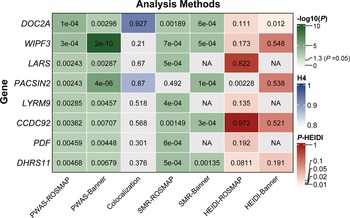
We conducted SMR to filter overlapping genes across two datasets under a stringent significance threshold (P SMR < 0.05), which were further cross-referenced with identified results derived from PWAS and colocalization analysis. Of particular note, DOC2A emerged with consistently supportive evidence across all analytical frameworks (SMR: P ROSMAP = 1.89 × 10⁻³, P Banner = 6.26 × 10⁻⁴), with no significant horizontal pleiotropy detected (HEIDI test: P ROSMAP = 1.11 × 10⁻¹, P Banner = 1.20 × 10⁻²), collectively highlighting its pivotal contribution to the pathogenesis of BD (Figure 2).
Prioritization of BD risk genes through integrated proteomic and genomic analyses. DOC2A emerges as a top-ranked risk gene with robust evidence across PWAS, Bayesian colocalization, SMR, and HEIDI refinement. ‘NA’ indicates nonapplicability of the gene/SNP to the specific analytical pipeline.

Figure 2. Long description
Starting at the top-left, the matrix lists genes vertically: D O C 2 A, W I P F 3, L A R S, P A C S I N 2, L Y R M 9, C C D C 92, P D F, D H R S 11. Analysis methods are labeled horizontally: P W A S dash R O S M A P, P W A S dash Banner, Colocalization, S M R dash R O S M A P, S M R dash Banner, H E I D I dash R O S M A P, H E I D I dash Banner. Each cell contains a numerical value, colored by three scales: green for negative log ten of P, blue for H four, red for P dash H E I D I. D O C 2 A shows strong evidence across all methods, with values: 1 e minus 4, 0.00298, 0.927, 0.00189, 6 e minus 4, 0.111, 0.012. W I P F 3 has a standout value of 2 e minus 10 in P W A S dash Banner. L A R S and P A C S I N 2 have N A in some columns. Color legends at the right indicate green for high negative log ten of P, blue for high H four, and red for high P dash H E I D I. N A denotes nonapplicability for certain gene-method combinations.
Cell- and region-specific dysregulation of DOC2A in BD pathophysiology
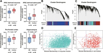
As neurons and astrocytes play pivotal roles in nervous system physiology, we integrated gene transcriptomic data across both cell types to evaluate the pathogenic contributions of risk genes in BD etiology. Using LMMs accounting for donor structure, DOC2A downregulation was detected in neurons (P = 4.26 × 10⁻²) and astrocytes (log-transformed P = 2.09 × 10⁻²) (Figure 3A; Supplementary Table S6). As supporting evidence, a similar downregulation pattern was observed in hippocampus (P = 9.80 × 10⁻³, t = −2.738, d = −0.91) and PFC (P = 1.44 × 10⁻², t = −2.580, d = −0.86), suggesting transcriptional suppression of DOC2A as a putative critical contributor to BD through reduced protein abundance. In contrast, DOC2A showed no significant dysregulation in SCZ (P = 1.49 × 10⁻¹, t = −1.467, d = −0.41) and ASD (P = 4.38 × 10⁻¹, t = −0.786, d = −0.28) cohorts, highlighting that the magnitude of its downregulation is more pronounced in BD.
Disease-associated dysregulation and co-expression networks anchored by DOC2A. (A) Significant downregulation of DOC2A in BD-relevant cell types (iPSC-derived neurons/astrocytes) and brain regions (PFC/hippocampus). For iPSC data, statistical significance was determined using Linear Mixed Models. (B, C) Gene co-expression modules derived from neuronal (B) and astrocytic (C) transcriptomes. (D, E) DOC2A localizes within synaptic modules showing strongest disease/module association (D: neuronal; E: astrocytic).

Figure 3. Long description
Panel A contains four box plots comparing normalized expression levels of D O C 2 A between B D and control groups in iPSC-derived neurons, iPSC-derived astrocytes, pre-frontal cortex, and hippocampus. Each plot includes sample sizes and p-values: neurons (n=36 B D, n=52 control, p=4.26 x 10 super -2), astrocytes (n=19 B D, n=14 control, p=2.09 x 10 super -2), pre-frontal cortex (n=17 B D, n=19 control, p=1.44 x 10 super -2), hippocampus (n=18 B D, n=18 control, p=9.80 x 10 super -3). Panel B shows a cluster dendrogram for neuronal transcriptomes with module colors below, mostly blue and turquoise. Panel C displays a cluster dendrogram for astrocytic transcriptomes with module colors, including blue, turquoise, and red. Panel D is a scatter plot of gene significance versus module membership in the turquoise module, with most points clustered below 0.5 gene significance and 0.8 module membership. Panel E is a scatter plot of gene significance versus module membership in the red module, with points distributed up to 0.8 gene significance and 0.8 module membership. D O C 2 A is localized within synaptic modules showing strong disease association.
Linking DOC2A-associated co-expression networks to synaptic pathways
To identify co-expression modules linked to risk genes, we performed WGCNA. Based on the average link hierarchical clustering and optimized threshold power (Supplementary Figure S4), we identified 47 modules (Figure 3B) in neuronal and 66 modules (Figure 3C) in astrocytic datasets, with DOC2A, respectively, located in the turquoise and red modules. In both contexts, DOC2A exhibited strong associations with the module and the BD phenotype (neuron: MM = 0.71, P = 1.90 × 10⁻²³; GS = −0.40, P = 2.19 × 10⁻⁵; astrocyte: MM = 0.46, P = 2.90 × 10⁻²; GS = −0.57, P = 8.41 × 10⁻³) (Figure 3D, E). Moreover, DOC2A showed robust correlations with all hub genes (defined as the top 10 genes ranked by intramodular connectivity) in neuron (e.g. STXBP1) and five hub genes in astrocyte (e.g. CD81) (Supplementary Table S8), implying its potential regulatory relevance to core functional hubs within these modules. Stability analysis confirmed the DOC2A-associated Turquoise module in iPSC-derived neurons had relatively high stability (Jaccard Index = 0.549 > 0.5; Stability Score = 0.668), validating the reliability of core findings. By contrast, the Red module in astrocytes exhibited markedly lower stability (Jaccard Index = 0.254; Stability Score = 0.320), indicative of a less robust co-expression architecture.
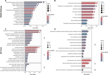
KEGG and GO analyses further delineate molecular mechanisms underlying these modules, prioritizing functional terms closely relevant to neuropsychiatric pathophysiology (Supplementary Tables S9–S12). In terms of biological pathways, KEGG analysis highlighted pathways including neuroactive ligand–receptor interaction, glutamatergic synapse, calcium signaling pathway, dopaminergic synapse, and neurodegeneration pathways (Figure 4A, B). For functional attributes, GO analysis similarly identified neuropsychiatric-relevant biological processes, notably synaptic transmission, glutamatergic synapse, regulation of synapse organization, neurotransmitter secretion, postsynaptic actin cytoskeleton organization, and mitochondrial gene expression (Figure 4C, D). These results collectively advanced our understanding of the roles of DOC2A and its co-expressed genes in the pathophysiology of BD.
Functional enrichment of DOC2A-associated co-expression modules. KEGG pathways and Gene Ontology (GO) terms enriched for neuronal (A, C) and astrocytic (B, D) modules converge on synaptic function regulation. BP: Biological Processes; CC: Cellular Components; MF: Molecular Functions.

Figure 4. Long description
Starting at the top-left, panel A displays KEGG pathway enrichment for neuronal modules, with horizontal bars for pathways such as neuroactive ligand-receptor interaction, calcium signaling, and c A M P signaling. The x-axis is gene ratio, ranging from zero percent to seven point five percent. Bar color intensity reflects negative log p adjust values, from four to twelve, and circle size indicates gene count, from forty to one hundred. Panel B, top-right, shows astrocytic module KEGG pathway enrichment, with bars for amyotrophic lateral sclerosis, pathways of neurodegeneration, Alzheimer disease, Huntington disease, prion disease, Parkinson disease, spinocerebellar ataxia, and retrograde endocannabinoid signaling. Gene ratio ranges from zero percent to fifteen percent, negative log p adjust from four to eight, and gene count from fifteen to forty. Panel C, bottom-left, presents Gene Ontology term enrichment for neuronal modules, divided into biological processes, cellular components, and molecular functions. Biological processes include modulation of chemical synaptic transmission, synapse organization, regulation of synapse structure or activity, vesicle-mediated transport in synapse, synapse assembly, regulation of synapse organization, neurotransmitter transport, neurotransmitter secretion, synaptic transmission, glutamatergic, and glutamate receptor signaling pathway. Cellular components feature synaptic membrane, neuron to neuron synapse, postsynaptic specialization, glutamatergic synapse, G A B A-ergic synapse, presynaptic active zone, excitatory synapse, Schaffer collateral C A one synapse, neurotransmitter receptor complex, ionotropic glutamate receptor complex. Molecular functions include neurotransmitter receptor activity, glutamate receptor activity, neurotransmitter transmembrane transporter activity, L-glutamate transmembrane transporter activity, and G protein-coupled neurotransmitter receptor activity. Gene ratio ranges from zero to zero point zero six, negative log p adjust from ten to thirty, and gene count from fifty to one hundred fifty. Panel D, bottom-right, shows astrocytic module Gene Ontology enrichment, with biological processes such as proteasome-mediated ubiquitin-dependent protein catabolic process, regulation of protein stability, A T P metabolic process, mitochondrial gene expression, oxidative phosphorylation, mitochondrial translation, mitochondrial respiratory chain complex assembly, proton motive force-driven mitochondrial A T P synthesis, glutathione catabolic process, postsynaptic actin cytoskeleton organization. Cellular components include mitochondrial inner membrane, mitochondrial protein-containing complex, mitochondrial ribosome. Molecular function is structural constituent of postsynaptic actin cytoskeleton. Gene ratio ranges from zero to zero point zero eight, negative log p adjust from two to seven, and gene count from ten to fifty. Color gradients and circle sizes consistently represent negative log p adjust and gene count across all panels.
DOC2A as a synaptic ligand hub links to BD pathology
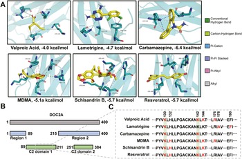
Molecular docking revealed high-affinity binding of FDA-approved BD therapeutics – valproic acid, lamotrigine, and carbamazepine (Nierenberg et al., Reference Nierenberg, Agustini, Köhler-Forsberg, Cusin, Katz, Sylvia and Berk2023; Ramadan et al., Reference Ramadan, Tay, Kaur, Parrish, Abdelmoteleb, Totlani and IsHak2025) – to DOC2A (domain 1) (Figure 5A), suggesting that DOC2A may function as a potential ligand-interacting node within BD-relevant synaptic networks. This finding gains further support from the similarly strong binding of three psychoactive compounds – N-methyl-3,4-methylenedioxyamphetamine (MDMA), schisandrin B, and resveratrol – suggesting that DOC2A may represent a convergence point for diverse therapeutic classes with shared neuroprotective mechanisms. Notably, several of these agents (valproic acid, MDMA, schisandrin B, and resveratrol) have previously been shown to modulate DOC2A expression levels in neuronal systems (Eun et al., Reference Eun, Kwack, Noh, Jung, Kim, Bae and Nam2010; Schulpen, Pennings, & Piersma, Reference Schulpen, Pennings and Piersma2015; Tomé-Carneiro et al., Reference Tomé-Carneiro, Larrosa, Yáñez-Gascón, Dávalos, Gil-Zamorano, Gonzálvez and García-Conesa2013; Zhang et al., Reference Zhang, Chen, Dahan, Xue, Wei, Tan and Zhang2019), reinforcing its putative functional relevance in BD pathology.
Binding site profiling further demonstrated striking spatial overlap: all ligands interacted with highly similar amino acid residues within DOC2A’s C2 domain 1 (Figure 5B, C), a critical functional module of interaction. In particular, lysine residues K130 and K142 emerged as conserved binding anchors across multiple ligand–receptor complexes. These residues may constitute regulatory switches of DOC2A activity, providing mechanistic insight into how pharmacological modulation of DOC2A might influence psychiatric disorders.
Molecular docking reveals potential binding affinity between DOC2A and multiple neuroactive compounds. (A) High-affinity binding of BD therapeutics and neuroprotective compounds to DOC2A, with the binding energy (kcal/mol) for each pair shown below the image. (B) Domain architecture of DOC2A: Main chain (gray), protein–protein interaction interfaces (blue), C2 domains (green). (C) Sequence alignment of interactive amino acid residues, with binding sites marked in red, conserved sites in green.

Figure 5. Long description
Panel A contains six molecular docking visualizations arranged in two rows and three columns. Each subpanel shows a neuroactive compound (Valproic Acid, Lamotrigine, Carbamazepine, M D M A, Schisandrin B, Resveratrol) docked to DOC2A, with the ligand in yellow and interacting amino acids labeled (e.g., L Y S 130, L Y S 142, H I S 132, A R G 176). Interaction types are color-coded: green for conventional hydrogen bonds, yellow for carbon-hydrogen bonds, blue for pi-cation, purple for pi-alkyl, gray for alkyl, and light blue for pi-pi stacked. Binding energies are listed below each compound: Valproic Acid negative 4.0 kcal per mol, Lamotrigine negative 4.7 kcal per mol, Carbamazepine negative 6.4 kcal per mol, M D M A negative 5.1 kcal per mol, Schisandrin B negative 5.7 kcal per mol, Resveratrol negative 5.7 kcal per mol. Panel B shows the linear domain architecture of DOC2A from residue 1 to 400, with the main chain in gray, two protein-protein interaction regions in blue (Region 1: 1 to 89, Region 2: 215 to 400), and two C2 domains in green (C2 domain 1: 89 to 211, C2 domain 2: 251 to 384). Panel C presents a sequence alignment of interactive residues for each compound, highlighting binding sites in red and conserved sites in green (K at positions 130, 142). The alignment shows that all compounds interact with the same core motif P Y V K L H L L P G A C K A N K L K T.
In contrast, the negative control (antiepileptic drug) levetiracetam showed negligible binding affinity to DOC2A in the same pocket (binding energy = −3.7 kcal/mol), while the comparative control topiramate (antiepileptic drug) (binding energy = −4.5 kcal/mol) bound to distinct residues (Y128, K142, and K144), which further identifies K130 as a conserved binding anchor (Supplementary Table S13).
Discussion
Our study nominates DOC2A as a candidate risk gene for BD, supported by convergent genetic, proteomic and network-level associations. PWAS, Bayesian colocalization, and SMR analyses collectively provide statistical genetic evidence supporting the association between DOC2A and BD. Transcriptomic profiling revealed consistent DOC2A downregulation across iPSC-derived neurons/astrocytes and postmortem PFC/hippocampus, indicating cell-type and regional dysregulation. DOC2A is significantly associated with synaptic co-expression modules enriched for neurotransmitter release, postsynaptic organization, and mitochondrial metabolism – processes putatively central to BD. Critically, molecular docking reveals high-affinity binding of DOC2A to BD-related therapeutics and neuroprotective compounds (e.g. valproic acid), nominating it as a mechanistically grounded, druggable candidate target.
DOC2A, a brain-specific calcium sensor belonging to the dual C2-domain protein family, is enriched at synaptic vesicles (Orita et al., Reference Orita, Sasaki, Naito, Komuro, Ohtsuka, Maeda and Takai1995; Verhage et al., Reference Verhage, de Vries, Røshol, Burbach, Gispen and Südhof1997; Yao, Gaffaney, Kwon, & Chapman, Reference Yao, Gaffaney, Kwon and Chapman2011) and plays a central role in regulating neurotransmitter release, synaptic efficacy, and plasticity (Ramirez et al., Reference Ramirez, Crawford, Chanaday, Trauterman, Monteggia and Kavalali2017; Sakaguchi et al., Reference Sakaguchi, Manabe, Kobayashi, Orita, Sasaki, Naito and Takai1999; Q.-W. Wang et al., Reference Wang, Qin, Chen, Tu, Xing, Wang and Yao2023; Xiao et al., Reference Xiao, Meng, Chen, Wang, Wu, Chen and Li2022) – processes tightly linked to BD pathophysiology (Elvsåshagen et al., Reference Elvsåshagen, Moberget, Bøen, Boye, Englin, Pedersen and Andersson2012; Valstad et al., Reference Valstad, Roelfs, Slapø, Timpe, Rai, Matziorinis and Elvsåshagen2021). Although previously implicated in suicidality, epilepsy and other disorders (Hu, Tang, Lan, & Mi, Reference Hu, Tang, Lan and Mi2019; Jin et al., Reference Jin, Dong, Yang, Bao and Ma2024; Zhao et al., Reference Zhao, Liu, Zhang, Liao, Zhang, Han and Lu2024; Zhou et al., Reference Zhou, Ma, Luo, Shan, Xiong and Cheng2023), DOC2A has received little attention in the context of BD. Notably, copy number variations in its genomic region are significantly enriched in BD cohorts (Steinberg et al., Reference Steinberg, de Jong, Mattheisen, Costas, Demontis, Jamain and Stefansson2014), reinforcing its plausible but underexplored role in disease pathogenesis.
Mechanistically, DOC2A regulates neurotransmitter release through its interactions with Munc13-1 (UNC13A) and Munc18-1 (STXBP1), two core components of the synaptic vesicle priming and fusion machinery (Ma et al., Reference Ma, Su, Seven, Xu and Rizo2013; Stepien & Rizo, Reference Stepien and Rizo2021; X. Wang et al., Reference Wang, Gong, Zhu, Wang, Yang, Xu and Ma2020). DOC2A binds Munc13-1 to cooperatively regulate vesicle priming and synaptic function (Mochida et al., Reference Mochida, Orita, Sakaguchi, Sasaki and Takai1998; Orita et al., Reference Orita, Naito, Sakaguchi, Maeda, Igarashi, Sasaki and Takai1997), while also directly interacting with Munc18-1 to alleviate its inhibitory constraint on syntaxin, thereby facilitating vesicle docking and fusion (Verhage et al., Reference Verhage, de Vries, Røshol, Burbach, Gispen and Südhof1997). Diacylglycerol (DAG), a lipid second messenger, has been shown to enhance DOC2A–Munc13-1 binding (Orita et al., Reference Orita, Naito, Sakaguchi, Maeda, Igarashi, Sasaki and Takai1997), activate Munc13-1, and phosphorylate Munc18-1 (Barclay et al., Reference Barclay, Craig, Fisher, Ciufo, Evans, Morgan and Burgoyne2003; de Jong et al., Reference de Jong, Meijer, Saarloos, Cornelisse, Toonen, Sørensen and Verhage2016), amplifying the DOC2A–Munc13-1–Munc18-1 signaling axis. This is particularly relevant in BD, where lithium – a first-line mood stabilizer – increases DAG levels (Thiruvengadam, Reference Thiruvengadam2023), and DGKH, the major DAG-degrading enzyme, is upregulated (Moya, Murphy, McMahon, & Wendland, Reference Moya, Murphy, McMahon and Wendland2010). Together, these findings suggest that this synaptic axis may represent a key neurobiological mechanism through which DOC2A contributes to BD.
Our co-expression analysis in neurons corroborates this hypothesis: DOC2A was co-expressed with STXBP1 and UNC13A within the same module, with strong correlations and consistent downregulation observed in BD samples (Supplementary Table S6). Functional enrichment analysis confirmed the module’s involvement in the synaptic vesicle cycle, neurotransmitter secretion, and chemical synaptic transmission. Notably, STXBP1 emerged as a central hub, suggesting that DOC2A may exert its regulatory influence on synaptic function primarily via STXBP1-centered networks.
Building upon evidence linking DOC2A deficiency to disrupted dendritic morphology (Q.-W. Wang et al., Reference Wang, Qin, Chen, Tu, Xing, Wang and Yao2023) – a synaptic structure critically dependent on the postsynaptic cytoskeleton (Cornelius, Rottner, Korte, & Michaelsen-Preusse, Reference Cornelius, Rottner, Korte and Michaelsen-Preusse2021; Sekino, Kojima, & Shirao, Reference Sekino, Kojima and Shirao2007) and regulated by astrocytes through their secretory and mitochondrial functions (Chen et al., Reference Chen, Wang, Xiao, Luo and Long2023; Doliwa et al., Reference Doliwa, Kuzniewska, Nader, Reniewicz, Kaczmarek, Michaluk and Kalita2025; Jackson & Robinson, Reference Jackson and Robinson2018; Rose et al., Reference Rose, Brian, Pappa, Panayiotidis and Franco2020) – we investigated its broader synaptic regulatory role in iPSC-derived astrocytes: DOC2A showed strong correlations with hub genes (e.g. CD81, a BD-associated astrocyte-derived exosomal marker (Attili et al., Reference Attili, Schill, DeLong, Lim, Jiang, Campbell and O’Shea2020). The co-expression module was enriched in pathways, including postsynaptic actin cytoskeleton organization, ATP metabolic process, and mitochondrial gene expression. However, the astrocytic module showed lower network stability than the neuronal module in resampling validation. This may reflect inherent astrocytic state heterogeneity or a more transient regulatory role of DOC2A in astrocytes, so interpretations of specific astrocytic hub interactions require caution and further large-scale validation.
Collectively, these findings suggest that DOC2A may act as a potential regulator involved in dual processes relevant to BD pathogenesis: regulating synaptic vesicle trafficking in neurons and modulating astrocyte-mediated synaptic support through mitochondrial function and secretory regulation.
Notably, many of these biological processes identified above are broadly implicated in neuropsychiatric conditions featuring synaptic dysfunction. While our cross-diagnostic analysis revealed that DOC2A’s downregulation was more pronounced in BD within the examined cohorts, these preliminary observations should be interpreted with caution as the modest sample sizes of the SCZ and ASD groups may limit the statistical power to detect subtle dysregulation, and DOC2A-centered networks may still underpin shared endophenotypes across traditional diagnostic boundaries. Future studies with larger transdiagnostic cohorts and high-resolution single-cell data are needed.
Valproic acid, lamotrigine, and carbamazepine – first-line treatments for BD – are well established to modulate neurotransmitter release, particularly through GABAergic and glutamatergic systems (Z. Liu et al., Reference Liu, Song, Yan, Verkhratsky and Peng2015; Post, Reference Post2004; Schloesser, Martinowich, & Manji, Reference Schloesser, Martinowich and Manji2012; Sitges, Chiu, & Reed, Reference Sitges, Chiu and Reed2016; Sitges, Guarneros, & Nekrassov, Reference Sitges, Guarneros and Nekrassov2007; Yoshida, Okada, Zhu, & Kaneko, Reference Yoshida, Okada, Zhu and Kaneko2007). Our molecular docking revealed high-affinity binding of all three to DOC2A within its C2 domain 1, a calcium-sensing region essential for vesicular trafficking (Groffen et al., Reference Groffen, Friedrich, Brian, Ashery and Verhage2006; Yao, Gaffaney, Kwon, & Chapman, Reference Yao, Gaffaney, Kwon and Chapman2011). This suggests that, beyond classical receptor-level modulation, these agents may converge upstream at the level of vesicle dynamics.
As an extension, several emerging neuropsychiatric compounds – MDMA, schisandrin B, and resveratrol – also exhibited strong binding to this domain, at sites overlapping those of FDA-approved mood stabilizers. These compounds have been shown to enhance mood, cognition or neurogenesis (Giridharan et al., Reference Giridharan, Thandavarayan, Sato, Ko and Konishi2011; Menegas et al., Reference Menegas, Keller, Possamai-Della, Aguiar-Geraldo, Quevedo and Valvassori2023; Mithoefer, Grob, & Brewerton, Reference Mithoefer, Grob and Brewerton2016; Shayganfard, Reference Shayganfard2020; Sun et al., Reference Sun, Cong, Liu, Jin, Si, Wang and Zhang2014; Torrado Pacheco & Moghaddam, Reference Torrado Pacheco and Moghaddam2024), and are reported to modulate DOC2A expression (Bandiwadekar et al., Reference Bandiwadekar, Naorem, Moiyadi, Singh, Shetty, Epari and Mittra2025; Eun et al., Reference Eun, Kwack, Noh, Jung, Kim, Bae and Nam2010; Zhang et al., Reference Zhang, Chen, Dahan, Xue, Wei, Tan and Zhang2019), reinforcing its therapeutic relevance. This dual convergence of structural affinity and transcriptomic regulation indicates a mechanistically tractable path for therapeutic repurposing in BD. Particularly intriguing is the convergence at K130 and K142, which may represent conserved regulatory anchors for precision-targeted intervention. Unlike the minimal interaction of levetiracetam or the divergent binding pattern of topiramate, two antiepileptic agents widely used for non-BD neurological conditions, the unique convergence of prioritized BD-relevant agents at K130 further highlights K130 as a mechanistically relevant target site for future structure-guided drug design. These findings position DOC2A not only as a shared pharmacological interface, but also as a strategic entry point for next-generation drug development in BD and related psychiatric disorders. Nevertheless, broader comparative evaluation across expanded compound libraries remains an important direction. Future studies incorporating a broader range of pharmacologically unrelated compounds may help further delineate the specificity and generalizability of these interaction patterns.
This study has several strengths. First, we used a three-stage analytical framework (PWAS, colocalization, and SMR) to integrate multi-ancestry GWAS data with region-specific pQTL datasets, identifying high-confidence risk genes. Second, transcript validation across brain regions and cell types showed convergent DOC2A dysregulation, reinforcing its pathogenic relevance. Finally, mechanistic exploration characterized DOC2A-associated gene networks and functions, while molecular docking supported its druggability as a therapeutic target.
Our study has limitations. Despite including different GWAS cohorts, imbalanced distributions of ancestry, age, and sex may introduce residual confounding. The pQTL data are restricted by original sample sources, potentially biasing PWAS and differential expression analysis due to tissue-specific expression patterns. The WGCNA analyses were conducted as exploratory functional contextualization, and the present emphasis was placed on the potential regulatory influence of DOC2A on module organization, whereas its intrinsic centrality within co-expression networks warrants further systematic evaluation in future studies. In addition, the neuronal iPSC cohort used for external validation is limited to lithium-nonresponsive BD type I patients, a narrow clinical subgroup that does not represent the broader BD population. This means the observed DOC2A downregulation may be more closely linked to lithium nonresponse, thus restricting the generalizability of this finding. Finally, limited statistical power in large-scale analyses may have resulted in the omission of key proteins lacking cross-tier evidence, and further molecular mechanistic studies are needed.
In summary, we identify DOC2A as a promising risk gene for BD. Our findings reveal that DOC2A in neurons and astrocytes may exert neuroprotective effects by potentially mediating vesicle trafficking and neurotransmitter release, and support its potential as a novel pharmacological target. These findings offer new insights into the pathogenic mechanisms of BD and facilitate clinical translation for therapeutic development.
Supplementary material
The supplementary material for this article can be found at http://doi.org/10.1017/S0033291726104565.
Data availability statement
The data used in the study can be accessed and downloaded from original studies (Glausier, Kimoto, Fish, & Lewis, Reference Glausier, Kimoto, Fish and Lewis2015; Lanz et al., Reference Lanz, Reinhart, Sheehan, Rizzo, Bove, James and Kleiman2019; Maycox et al., Reference Maycox, Kelly, Taylor, Bates, Reid, Logendra and de Belleroche2009; O’Connell et al., Reference O’Connell, Koromina, van der Veen, Boltz, David, Yang and Andreassen2025; Santos et al., Reference Santos, Linker, Stern, Mendes, Shokhirev, Erikson and Gage2021; Vadodaria et al., Reference Vadodaria, Mendes, Mei, Racha, Erikson, Shokhirev and Gage2021; Voineagu et al., Reference Voineagu, Wang, Johnston, Lowe, Tian, Horvath and Geschwind2011; Wingo et al., Reference Wingo, Liu, Gerasimov, Gockley, Logsdon, Duong and Wingo2021). The computational scripts and analytical workflows of this study can be found in a publicly accessible repository: https://github.com/CY1479/BD_Project
Acknowledgments
We would like to thank the authors of the original GWAS and pQTLs studies included in this article for their valuable contributions. All authors are grateful for participation in our research.
Funding statement
This work was partly funded by Sichuan Science and Technology Program (2026NSFSC0591), Key R & D projects of Science and Technology Department of Sichuan Province (2021YFS0248), Postdoctoral Foundation of West China Hospital (2020HXBH163), and China Postdoctoral Science Foundation (2020M673247). All grants were received by C.Z.
Competing interests
All authors report no potential competing interests.
Ethical standard
This study used publicly available datasets. All ethical approvals, including patient anonymity, informed consent, and licensing committee approvals, are documented in the original publications (Glausier, Kimoto, Fish, & Lewis, Reference Glausier, Kimoto, Fish and Lewis2015; Lanz et al., Reference Lanz, Reinhart, Sheehan, Rizzo, Bove, James and Kleiman2019; Maycox et al., Reference Maycox, Kelly, Taylor, Bates, Reid, Logendra and de Belleroche2009; O’Connell et al., Reference O’Connell, Koromina, van der Veen, Boltz, David, Yang and Andreassen2025; Santos et al., Reference Santos, Linker, Stern, Mendes, Shokhirev, Erikson and Gage2021; Vadodaria et al., Reference Vadodaria, Mendes, Mei, Racha, Erikson, Shokhirev and Gage2021; Voineagu et al., Reference Voineagu, Wang, Johnston, Lowe, Tian, Horvath and Geschwind2011; Wingo et al., Reference Wingo, Liu, Gerasimov, Gockley, Logsdon, Duong and Wingo2021).



 Open access

