

Introduction
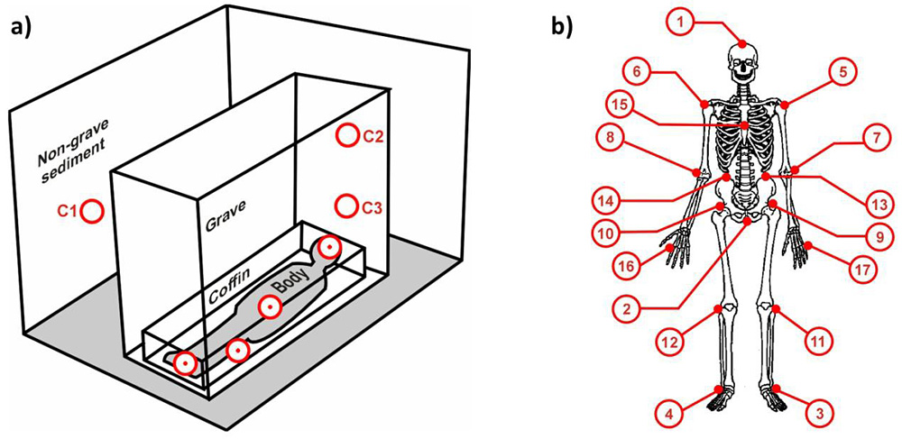
Typically, archaeological excavation of human burials concentrates on recovering the skeleton and associated grave goods, as well as recording grave form. The research reported here investigates whether additional information might be obtained from sampling the burial matrix directly adjacent to human remains. This demands new thinking about the sampling, and the development and testing of a new methodology, combining soil micromorphology and chemical analysis. Such sampling needs to consider both the environment uninfluenced by the grave cut, and positions within the grave, both above and at the level of the remains (Figure 1a). Moreover, such sampling needs to account for variation at different localities and periods, as well as both chemical and micromorphological differences.
a) Soil sampling positions (circles) in relation to the human burial. C1 to C3 represent control samples; b) positions of the maximum number of samples taken for analysis of organic residues.

Research objectives
The primary objective of the project is to contrast the degree of chemical and micromorphological variation that can be associated with grave construction, burial ritual and factors of decomposition with that in the non-burial environment. The nature and range of within-site differences between graves is to be questioned, as well as the possible chronological influences on grave fill consolidation and decomposition processes. Do ritual factors, such as anointing and clothing the body, or using a coffin, influence rates or forms of decay? Last but not least, because of the variation of the body composition along a corpse, can this result in regional anatomical contrasts within the grave?
The sites
The geographical, environmental and temporal spread of our 31 study sites was determined by collaboration with those archaeological groups prepared to permit our sampling methods. We have gained access to sites varying in date from the Neolithic to the early twentieth century, and extending from Iceland to Sudan, and from Portugal to Turkey (List 1). The contrasting deposits into which the graves were cut range from volcanic soils to desert sands and bog.
List 1.
Archaeological sites sampled: Driffield Terrace, York (UK, Roman); Heslington East, York (UK, Iron Age to Roman); Maastricht, (Netherlands, Merovingian); Pill'e Matta (Sardinia, Phoenician, Punic, Roman); Tharros (Sardinia, Roman); St Giovanni, (Sardinia, Roman); Usini, (Sardinia, Bronze Age); El Khilday (Sudan, Neolithic– Mesolithic); Velletri, Rome (Italy, Roman); Hungate, York (UK, Roman–Viking); Thessaloniki (Greece, Iron Age and Roman); Gannì (Sardinia, Chalcolithic); Çatalhöyük (Turkey, Neolithic); Tremini (Sardinia, Phoenician, Punic, Roman); Hofstadir (Iceland, Viking– medieval); Sala (Sweden, medieval); Al Quarassa (Syria, Neolithic); Bamburgh (UK, medieval); Edinburgh (UK, medieval); Cashal, (Ireland, Early Bronze Age bog body) Eindhove (Netherlands, medieval); Fewston, Yorkshire (UK, post-medieval to recent); St. Thomas, Osbaldwick (UK, Medieval to historic); Ridgeway, Dorset (UK, medieval mass graves); Rossio de Carmo (Mertola, Portugal, medieval); Sedgeford (UK, medieval); Basly, (France, Bronze Age); Syningthwaite Priory, Yorkshire (UK, medieval); Mechelen (Belgium, medieval); Fromelles (WWI mass grave).
Methodology
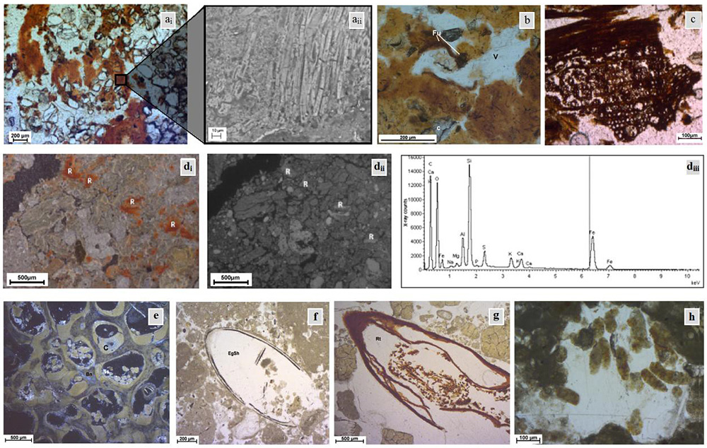
Selection of micromorphological features observed in archaeological grave soils. (ai-aii) Soil from beneath skull in Grave 414 from Mechelen, with (ai) dark brown feature representing fragment of fabric in a white sand-rich grave soil, (aii) SEM image of an inset from (ai) showing phytolith remains aligned in fabric weaving. (b) Fungal activity (Fu) and void (V) in a layer of degraded coffin wood from the sacral region in Grave 114 from Hofstadir (Iceland). (c) Wood fragment from soil adjacent to the skull in a post-medieval grave from Fewston, North Yorkshire. di-diii) soil close to the pelvis in a Chalcolithic burial from Sardinia, with (di) micromorphological image in oblique incident light showing red pedofeatures (R), (dii) SEM-BSE image of red (R) pedofeatures and (diii) X-ray spectrum from SEM- EDS analysis of the red pedofeatures (R) showing enhanced levels of Fe (mean 13.4 per cent Wt). (e– h) Punic grave from Pill'e Mata (Sardinia) with (e) fragment of bone (Bn) from the pelvic area showing sandy-silty clay infilling, (f) eggshell in soil adjacent to the foot, (g) root with excrement in soil near the skull and (h) excrement within soil pore adjacent to the skeleton.
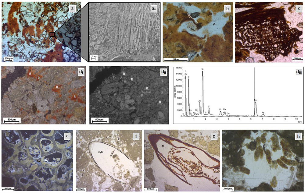
A maximum of 17 samples were collected from specific locations in relation to each skeleton (Figure 1b). Separate samples were taken for chemical and micromorphological analyses, with additional samples collected where possible for plant macro-evaluation and examination for intestinal parasite eggs. Soil macro-evaluations and bulk elemental analysis were also undertaken. Micromorphological analysis necessitated resin impregnation of sediment blocks and the cutting of 30 µm-thin sections both tangentially and perpendicular to the long axis of the skeletal remains. Established micromorphology procedures were employed: for instance, soil voids (Figure 2b, e, h) were assessed using quantitative and qualitative image analysis. Such measurements help determine the movements of fluids and decay products. Microprobe analysis was conducted on selected samples and elemental mapping has permitted identification of areas with distinct chemical signatures (various inorganic elements and organic C, N, S and O). For example, iron-rich features in a Chalcolithic burial from Sardinia (Figure 2di-iii) possibly relate to the presence of ochre. Element leaching and the migration of components through the soil have also been considered (Figure 2d, e). Although basic soil micromorphology (Bullock et al. 1985; Reference FitzpatrickFitzpatrick 1993; Reference UsaiUsai 1996; Reference StoopsStoops 2003) has been used to study burial matrices before, in this study we examine spatial variation within graves to answer specific questions linked to ritual, burial and taphonomy associated with grave construction and use. It was questioned whether spatial changes in soil micromorphology within/between burials result from natural spatial variability, or are induced by burial-related processes. For instance, in the case of microscopic and larger organic remains within or near the body (Figure 2), it was questioned whether bone micro-fragments in the pelvic area (Figure 2e) indicate degraded human bone from the sacrum and pelvis, food debris from the gut, or incidental inclusions from the grave, including plant debris. Similarly, it may be asked whether the plant remains, especially commonly occurring wood particles, represent decayed burial containers (coffins), food debris or ritual offerings (Figure 2c & g). Similarly, shells of soil fauna (Figure 2f) indicate their activity.
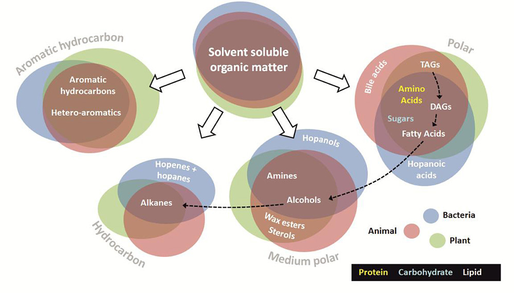
Composition, origins, transformation pathways and chromatographic fractions of organic matter of biological origins commonly present in soils and archaeological sites.

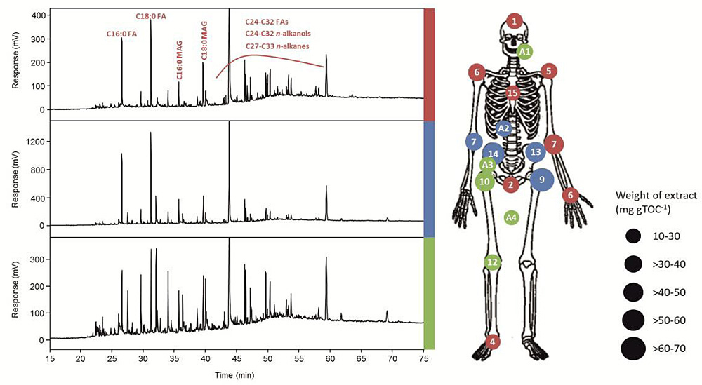
In addition to the micromorphological studies, trace organic analyses have been performed. Organic residues, which originate from animal, plant and microbial sources, were extracted from the soil samples, fractionated according to polarity and each of the four individual fractions (hydrocarbon, aromatic hydrocarbon, medium polar and high polar; Figure 3) analysed by gas chromatography (GC; Figure 4) and/or liquid chromatography (LC). Selected fractions showing significant variation in compound distributions with location in the grave indicate differences in the sources and/or preservation (Figure 4) and received detailed consideration by mass spectrometry (MS, GC-MS and LC-MS). The organic compounds, which are often limited to scarce residues derived from degradation of complex natural product molecules, have the potential to be linked to specific sources including the body, funerary items and the agents of decomposition. In addition, targeted analysis of specific compound classes has been conducted to allow the differentiation of components that may represent, for example, embalming agents, clothing, faecal material and food offerings. Even alkaloids and drug substances have been sought. Signatures from un-extracted (solvent insoluble) organic matter are characterised by pyrolysis GC of sediments after extraction. Matrix assisted laser desorption ionisation mass spectrometric imaging is also being considered for a select number of sections to determine whether the location of specific organic residues can be linked to features apparent from the micromorphological investigation.
Organic signatures around the skeletal remains in a Bronze Age burial from Heslington East, Yorkshire, UK. a) Gas chromatograms showing three distinct total organic extract profiles observed all of which show dominance of fatty acids (FA), presence of mono-acyl glycerols (MAG), n-alkanols and n-alkanes. Colour codes identify chromatograms with the locations on the skeletal diagram where each distribution occurs (numbers refer to sampling positions in Figure 1, prefix A indicates additional sample). The distributions show systematic variations with position around the remains. The sizes of the circles indicate concentration levels.

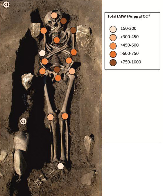
Organic signatures around the skeletal remains in a Bronze Age burial from Heslington East, Yorkshire, UK. b) Levels of low molecular weight fatty acids (LMW FAs) in soils adjacent to each sampling position and in control samples (C1 & C3) represented on the photographic image.

Individual organic compounds (hydrocarbons, lipids, proteins and peptides; Figure 3) have been identified where possible, and there is a need to discriminate between compounds derived from the body, those associated with the burial and the natural background signature from the soil. How refined we can make the identifications is one of the major challenges for the project. After burial, chemical and microbial decomposition and diagenetic changes impact the organic matter to the molecular level, affecting in particular, triacylglycerols, proteins, peptides and polysaccharides (Figure 3). Certain compounds, such as cholesterol and coprostanols of faecal origin, can remain intact in soils over archaeological timescales (Knights et al. 1983; Bull et al. 2003) and specific plant drugs may remain identifiable under ideal burial conditions (arid or frozen; Springfield et al. 1993). Aspects of wood chemistry can at least allow the discrimination of hard and soft woods (Van Bergen et al. 2000) and provide an indication of the state of preservation. This is still a largely unexplored research field and the extent to which recognisable and informative signatures remain is a key question.
Intestinal parasites
A number of gut parasites belonging to the phyla Nematoda and Platyhelminthes have been found in archaeological deposits (Jones et al. 1988) and human remains (Jones et al. 1986). The genera Trichuris and Ascaris are probably the most common, and recent epidemiological evidence makes it likely that earlier human populations were infected to a higher degree than modern populations (Ewers & Jeffrey 1971; Reference TerpstraTerpstra 1972). Usually it is the eggs of these worms which are found in faecal or latrine deposits. There is considerable chance of preservation in graves, and for this reason they are being sought in grave samples and micromorphology slides. A major question is the extent to which they degrade and become unidentifiable in contrasting environments. Bog conditions appear to be the optimal for preservation, but we need to know more about their survival in other conditions.
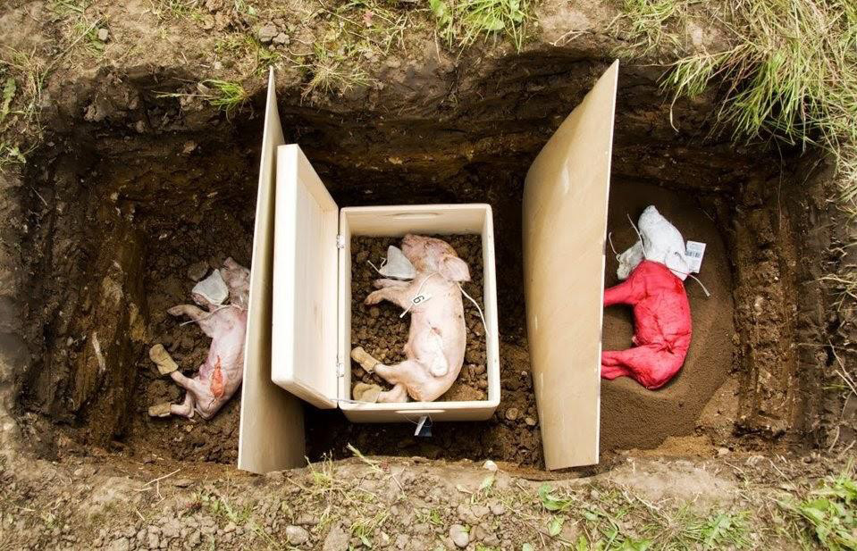
Experimental burials of three piglets at Heslington East, Yorkshire, UK into native soil (left), coffin containing grave fill (centre) and sand (right). Additional materials included in the burials (muslin bags at the neck and leather boots) can be seen. The piglet on the right was painted in an attempt to monitor migration of material away from the body.

Experimental burials
In order to consider our archaeological questions in relation to recent inhumations, it was necessary to conduct a series of experimental burials (Figure 5). Newborn pigs were buried with and without wooden 'coffins' or covering. Additional organic materials were placed in the graves mainly in muslin bags, one placed near the head and one inserted into the abdomen. A range of contrasting organic material, including hair, beeswax, tobacco and oats was weighed out for each burial. The pigs were buried in contrasting environments at five sites: Hovingham (one in controlled clear sand; one in crushed limestone), King's Manor, York (in loamy garden soil), West Heslerton (in a lacustrine sand-rich context), Folkton (peaty soil) and near Heslington (acidic heterogeneous sandy soil). After nearly four years, the burials are currently being excavated, sampled and studied using the procedures outlined above. The experimental burials promise to inform us of early stages of pedogenesis within graves, as well as the decomposition of organic materials and their interaction with the soils. There is also the issue of the early stages of dispersal of organic decomposition products within and even beyond the graves. It is worth noting that experimental studies of this kind have relevance beyond archaeology, and we hope our results will also inform forensic studies.
Conclusions and prospects
The analysed graves reveal considerable variation in chemistry and micromorphology. There is both intra- and inter-site variation, linked to environmental contrasts and taphonomic variation. Post burial changes are even more complex than we originally anticipated. The micromorphology is revealing evidence associated with the burials, their decomposition and the infill compaction, including perishable artefacts, and components formed in situ (Figure 2), and evidence for mobilisation/depletion, transportation and re-deposition of soil/sediment material displaying preferential spatial patterns in relation to the different parts of skeletons and burials (Figure 2).
The organic chemical analyses require careful interpretation, in view of the influence of variable decomposition on the original composition of organic components in burials. There is clearly a need to evaluate these sequences of change in more detail than previously attempted. We also need to interpret the variability within graves in terms of how this might be of relevance and value to forensic investigations. In sum, we believe this research—combining chemical and micromorphological investigations, using multi-site and multi-grave comparisons—points to a new future for burial archaeology.
Research team
York (Archaeology): Don Brothwell, Maria-Raimonda Usai, David Broughton, Carol Lang, Helen Williams, Annika Burns, Sabina Ghislandi, Fabio Serchisu, Allan Hall & Andrew Jones. York (Chemistry): Brendan Keely, Matthew Pickering, Kimberly Green, Adam Pinder & Scott Hicks. Stirling (School of Biological & Environmental Sciences): Clare Wilson.
Acknowledgements
The research received funding from the European Research Council under the European Community's Seventh Framework Programme (FP7/2007-2013) / ERC grant agreement no. 230193. We also wish to express our sincere thanks to the Greek National Department of Antiquities for allowing us to sample in Thessaloniki. We also much appreciate the willingness of the War Graves Commission to allow us to sample soils in relation to their work at a First World War site. Numerous regional archaeological groups have generously allowed us to sample during their cemetery fieldwork. Without the kind cooperation of all these groups, our research could not have been carried out. Full acknowledgements will be given in our final reports. Images were provided by Carol Lang (Figure 2ai– ii), Annika Burns Figure 2b and Sabina Ghislandi Figure 2e– h.