

Introduction
The acute care of a seriously injured patient involves rapid diagnosis and management of life-threatening injuries. Radiographic studies, including x-rays, computed tomography (CT), and other imaging studies, are frequently used in the initial evaluation of the trauma patient in order to delineate and define injuries, as well as to detect injuries that may be occult in nature. While these studies may confirm or reveal serious and life-threatening injuries, they all expose the patient to ionizing radiation, an entity which has been linked to the long-term development of cancer, even at low doses.Reference Brenner, Doll and Goodhead 1 , 2 CT use in the investigation of trauma patients has increased significantly during the past two decades,Reference Nickloff and Alderson 3 which may further increase the burden of radiation-induced malignancy on society. Several recent studiesReference Kim, Gracias and Maidment 4 - Reference Salottolo, Bar-Or and Fleishman 9 have examined the radiation dose to trauma patients during their hospital stay, but only one studyReference Winslow, Hinshaw and Hughes 8 has examined the radiation exposure associated with the patient’s resuscitative phase of care and diagnosis. This study by Winslow et al excluded the most severely-injured patients and did not consider tests done at referring centres, despite evidence to show that patients who are transferred from regional centres have higher total doses of radiation than those imaged at the trauma centre only.Reference Gupta, Greer and Martin 10
The purpose of this study was to use a calculation-based method to determine the radiation dose (termed “radiation exposure”) to which trauma patients were exposed during their initial trauma management at a Canadian tertiary trauma centre (TTC).
Methods
Study design and setting
The study was a retrospective chart review of patients who underwent trauma team activation at the Queen Elizabeth Health Sciences Centre in Halifax, Nova Scotia. The study site is the only adult (age 16≥years) Level I trauma centre in the province, located at an academic facility that sees roughly 250 trauma team activations per year. The annual census of the emergency department for 2009 was 60,088 patients. The study was approved by the hospital’s ethics review board.
Identification of patients
All adult patients who underwent trauma team activation at our centre between March 1, 2008 and March 1, 2009 were included in the study. The decision for trauma team activation was based on physiologic, anatomic, mechanistic and logistic factors (Figure 1).
Criteria for Trauma Team Activation (TTA).

Patients were identified using the Nova Scotia Provincial Trauma Registry, which is maintained by the Nova Scotia Trauma Program. This is a population-based provincial trauma registry that contributes annually to the Canadian National Trauma Registry and has been used in peer-reviewed research.Reference Tallon, Flowerdew and Stewart 11 , Reference Thibault-Halman, Tallon and Ackroyd-Stolarz 12 Subjects initially managed at a regional hospital and subsequently transferred to the trauma team were included. Patients who were under the age of 16, were dead on arrival, or died prior to any investigations were excluded.
Data collection and processing
Using the cohort of patients identified by the trauma registry, data were collected via formal chart review using standard methodology by two abstractors not blinded to the purpose of the study: a primary investigator (PI) and a research assistant trained by the PI.Reference Gilbert, Lowenstein and Koziol-McLain 13 Our province has a centralized Picture Archiving and Communication System (PACS) (Impax v6.5.1.1008; Mortsel, Belgium: Agfa Healthcare) for reviewing and documenting radiographic studies. Radiation dosage data were collected directly from PACS into a standardized spreadsheet. Demographic data were collected by one abstractor using the hospital’s electronic patient records and the trauma registry. These data included patient age, gender, and injury severity score (ISS).
Only the CT investigations done during the patient’s initial trauma work-up (defined as any studies ordered by the trauma team prior to a disposition decision being made) at the TTC were included. It was determined that there could be significant variability in number of subsequent investigations and exposure to radiation from other sources (e.g., fluoroscopy), which could confound results.
Individual radiation dose reports for each examination, including dose length product (DLP), were collected from the CT study data on the PACS system. The DLP was multiplied by conversion factors for each body part imaged to calculate a total-body effective dose. This calculation was done using the ImPACT CTDosimetry software calculator (CTDosimetry v 1.0, 2009; ImPACT, London, UK), under the guidance of a medical physicist. Effective dose was calculated for each CT performed, and were reported in millisieverts (mSv).
Primary data analysis
The primary outcome metric for this study was the mean radiation exposure received per trauma patient within the study cohorts. The data were entered into a spreadsheet, and data analysis was performed using Microsoft Excel (Microsoft Office Excel 2003; Microsoft Corporation, Redmond, WA).
Results
Initial chart review yielded 230 patients who underwent trauma team activation during the study period, 206 (90%) of whom had at least one CT imaging study. Of these 206 patients, 144 had imaging at the TTC only, 40 had imaging at a regional centre only, and 22 had imaging at both a regional centre and the TTC. The patients were 71.4% male. The mean age was 34 years (±17 years). The mean ISS was 17±11.4 (Table 1).
Characteristics of study cohort
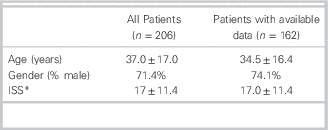
* Abbreviations: ISS – Injury Severity Score
CT dose information was unavailable for all imaging done outside the TTC, which included 40 patients who had imaging performed only at regional centres. For the 22 patients imaged at both a regional centre and the TTC, only data for the diagnostic imaging tests performed at the TTC were available. Data were unavailable for four patients imaged at the TTC (two had invalid identification numbers in the trauma database and two had no dose data recorded on the PACS system). Thus, full imaging details were available for 162 patients (78.6%) (Figure 2).
Derivation of Study Cohort (March 1, 2008 – March 1, 2009).

Of the patients for whom dosage information was available, 101 (62.3%) of the patients reviewed had whole-body CTs (including head, cervical spine, chest, abdomen and pelvis). Nine patients (5.6%) had only a head CT. Sixty-two (38.3%) patients had CTs performed at regional centres before transfer to the TTC. Of these patients, 22 had CTs repeated at the TTC (35.5% of transferred patients). The mean whole-body radiation exposure was 24.4±10.3. When patients were stratified by those who received imaging in a regional centre and those who did not, the mean whole-body radiation exposure for imaging done at the TTC was 17.3±10.1 mSv and 25.5±9.8 mSv, respectively (Table 2).
Radiation dose
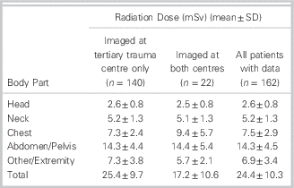
The highest radiation exposure for a single patient in the study was 47.2 mSv.
Discussion
The mean whole-body radiation exposure in this study population was 24.4 mSv, which may correlate to one additional cancer death for every 100 patients scanned, and is comparable to trauma patient radiation exposure presented in the recent literature (11.1–40.2 mSv).Reference Tien, Tremblay and Rizoli 7 - Reference Salottolo, Bar-Or and Fleishman 9 , Reference Sharma, Oswanski and Sidhu 14 , Reference Inaba, Branco and Lim 15 There is mounting evidence that, even at low doses, the ionizing radiation associated with CT imaging may increase lifetime cancer risks. The Biological Effects of Ionizing Radiation (BIER) Report is a comprehensive review of the biological and clinical data relating to health risks from ionizing radiation exposure, compiled by the National Academy of Sciences. The most recent report (BIER VII Phase 2), based largely on observational studies of survivors of the bombing of Hiroshima and Nagasaki, showed that even doses in the 5 to 100 mSv range (mean 29 mSv) can cause fatal solid-organ cancers at a rate of 800 fatal cancers per 100,000 scans, or about 1/100. 2 Added to the lifetime baseline risk of developing cancer in Canada (46/100 for males, 41/100 for females), 16 the relative risk of developing cancer as a result of trauma-team-related imaging is roughly 1.02, compared to the general population. The National Trauma Registry in Canada reported 15,190 major injury cases in 2011 (defined as ISS>12), with 4,973 of those patients under the age of 35. 17 Considering the number of trauma patient investigations that occur nationally and the relatively young age of most trauma victims, this relative risk increase is important on a population health level.
A study conducted by Inaba et alReference Inaba, Branco and Lim 15 compared the number of imaging tests and the radiation exposure in trauma patients over a 5-year period, and found a significant increase in both the number of diagnostic imaging tests (2.1 to 3.2 per patient) and in the radiation exposure (11.5 mSv to 20.7 mSv). The increased number of CTs did not correlate with a change in patient mortality or outcomes. This study reflected patterns presented by other researchers 16 who have also reported a remarkable increase in CT volumes, suggesting that the aforementioned radiation-related cancer risks will continue to increase in the future.
Interestingly, the Inaba study found that among trauma patients with penetrating injuries, for whom centres have more strict protocols and imaging guidelines, the number of CTs remained fairly constant over the five-year period (0.7 vs 1 CT per patient).Reference Inaba, Branco and Lim 15 This raises the possibility that development of and adherence to guidelines for imaging of blunt trauma patients, which has proven difficult so far, may help to decrease the trauma-related radiation burden over time. It has been shown that appropriate application of decision rules, such as the Canadian CT Head Rule and Canadian C-Spine Rule,Reference Smits, Dippel and de Haan 18 , Reference Kerr, Bradshaw and Kelly 19 can significantly reduce the need for CT imaging.
A more recent study published by Rodriguez et al attempted to determine the percentage of chest imaging (x-rays and CTs) as part of a trauma work-up that actually identified significant injury.Reference Rodriguez, Baumann and Raja 20 The study found that only 6.9% (6.2%–7.7%) of chest CTs found a clinically significant injury. After calculating the effective dose of chest imaging, the authors determined that only one significant injury was found per 129 (101.4–217.4) mSv of radiation, suggesting that a substantial number of trauma investigations find no clinically significant injury.
A study published in Korea in 2011 demonstrated successful implementation of a dose-reduction strategy.Reference Kim, Jung and Oh 21 The strategy involved reducing the number of CTs performed by reliance on other modalities (i.e., MRI, ultrasound), eliminating repeat CT scans, and increasing reliance on careful clinical examination. Careful optimization of CT scanning parameters was completed in order to reduce radiation dose. This strategy resulted in a 63% reduction in radiation exposure, from 78.7 mSv to 29.5 mSv, without any increase in number of surgeries, length of stay, mortality, or negative patient outcomes. Although a dose of 78.7 mSv is much higher than those reported in North American studies, the data suggested that radiation exposure in trauma patients can be significantly reduced without negatively affecting patient outcomes.
CT imaging remains an integral part of the initial evaluation of the multiply- injured patient. However, given the increasing volume of imaging studies being ordered and the known risk of cancer related to CT imaging, limiting the radiation dose administered to trauma patients must become a focused area of research. With the awareness of the potential impact of radiation-related cancers on public health, it becomes difficult to justify the routine use of whole-body CT in trauma patients. Clinicians must carefully consider alternate options, such as reliance on decision rules or careful clinical examination, to minimize unnecessary examinations. Further, more research must be completed to develop useful clinical decision rules for trauma patients (especially in blunt chest and abdominal trauma, where no well-supported decision rules currently exist), and to improve strategies to reduce the radiation dose for imaging studies that are performed.
Limitations
This study was limited by its retrospective design, which allows for potential missed data or errors in data collection. The study was also limited by the inability to collect radiation dose data for any CTs performed outside of the single adult TTC, as these data were not routinely documented on the PACS system at institutions other than the TTC. It is quite likely that the whole-body radiation dose for transferred trauma patients is much higher than reported in this study.
Since only CT scans performed during the initial emergency department trauma team activation phase of care were included, the whole-body radiation dose reported underestimates the total radiation exposure to trauma patients during their hospitalization. However, due to the variability of subsequent imaging studies performed during hospital admission, depending on specific injuries and length of stay, the authors decided that evaluation of the initial workup only would encompass the most homogeneous patient population. Also, this study did not include radiation exposure from plain x-ray films and fluoroscopy during the initial workup, which would have further increased our radiation exposure estimate, albeit by a small amount in most cases.
This study was conducted at a single centre, and thus variation in practice patterns and in CT scanner parameters could account for higher or lower whole-body radiation dose at other centres. Additionally, because outcome data were not collected, we were unable to assess the appropriateness of the imaging studies ordered, or to estimate the number of clinically significant radiographic findings that may have changed patient outcome.
This study was conducted using data from 2008-2009. It is possible that current changes in CT ordering practices, scanning parameters and CT machines may result in an over- or underestimate of the radiation dose.
Our estimates of whole-body radiation dose were based on calculations using the CT dose index. Although the ImPACT allows for very precise estimates of individual organ doses, it is possible that a method of direct measurement (e.g., dosimetry) could allow for a more accurate estimation.
Conclusion
In this study cohort, trauma patients were exposed to significant amounts of ionizing radiation during their initial investigation. Particularly in younger patients, ionizing radiation is known to increase the lifetime risk of potentially fatal cancers. Clinicians should carefully consider strategies to limit this radiation exposure, including reduced-dosing strategies, reliance on validated clinical decision rules, and considering alternate imaging modalities or increased reliance on clinical examination, during the initial management of trauma patients.
Competing Interests: None declared.


