





Introduction
According to the American Heart Association Electrocardiography and Arrhythmias Committee, low voltage in electrocardiography is defined as a nadir-to-peak QRS amplitude <0.5 mV in all limb leads and <1.0 mV in all precordial leads. Reference Rautaharju, Surawicz and Gettes1
Low QRS voltages are rare in healthy individuals; however, emerging evidence suggests a higher prevalence in elite athletes compared to both sedentary individuals and recreational athletes. Reference Pelliccia, Drezner, Zorzi and Corrado2
The underlying causes for low QRS voltage can be numerous. A practical approach is to distinguish between and extracardiac causes, both of which share a common pathophysiological basis: either the transmission of generated potentials to the electrocardiography electrodes is impaired or the generation of cardiac electrical impulses is itself is compromised. Reference Valentini, Anselm and Metra3
Pericardial effusion, obesity, pulmonary, or peripheral oedema causing an attenuated transmission of generated potentials to the body surface are typical extracardiac reasons for low QRS voltage. In contrast, the most common cardiac causes for low QRS voltage in childhood and adolescence include cardiomyopathies, myocardial infarction, or myocarditis, leading to in myocardial remodelling and therefore altered generation of electric potentials. Reference Madias4 Left ventricular dilation appears to be associated with low QRS voltage in both limb and precordial leads, whereas isolated low QRS voltage confined to the precordial leads is characteristic of right ventricular dilatation. Reference Kim and Verdino5
The clinical significance of low QRS voltage remains incompletely understood. Existing studies implicate it may serve as a negative prognostic marker in various cardiac conditions, including hypertrophic cardiomyopathy, and it is has been proposed as an independent risk factor for mortality even in healthy individuals. Reference Pelliccia, Tatangelo and Borrazzo6,Reference Usoro, Bradford, Shah and Soliman7 In adult populations, low QRS voltage has been identified as a predictor of higher in-hospital mortality following myocardial infarction. Reference Tan, Goodman and Yan8 Here, we report the unusual case of a young patient presenting with persistently very low QRS voltages and clinical features consistent with cardiomyopathy of unknown aetiology, despite an extensive diagnostic evaluation.
Clinical presentation and medical history
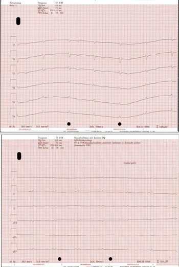
The now 25-year-old male patient first presented to our department of paediatric cardiology in October 2016 at the age of 16. The year prior, the previously healthy boy with no notable medical history had been hospitalised in Turkey following sudden cardiac arrest. At that time, the local healthcare provider established a diagnosis of long QT syndrome and initiated beta-blocker therapy; an implantable cardioverter-defibrillator was subsequently implanted—both decisions made prior to and independent of any involvement of our centre. At the time of his initial presentation to our department, he was largely asymptomatic, despite for episodes of tachycardia attributable to a significant burden of polymorphic ventricular extrasystoles. Throughout follow-up, the patient consistently demonstrated clinically relevant ventricular extrasystoles on electrocardiography along with marked low voltage (see Figure 1).
12-lead electrocardiogram showing marked low voltage across all the limb leads as well as the precordial leads.

Figure 1. Long description
Panel A: An electrocardiogram showing multiple leads (V1 to V6) with annotations indicating various measurements such as heart rate, PQ time, QRS duration, QT/QTc intervals, and T wave axis. The graph displays the electrical activity of the heart over time. Panel B: Another section of the electrocardiogram showing leads I, II, III, aVR, aVL, and aVF with similar annotations and measurements. The graph also depicts the heart’s electrical activity over time.
Initial echocardiography revealed mildly impaired left ventricular function with a reduced ejection fraction of 55%, while right ventricular function remained preserved. Subsequent echocardiographic follow-up demonstrated stable, mildly reduced left ventricular function over the years; however, right ventricular function gradually deteriorated due to progressive tricuspid valve insufficiency following implantable cardioverter-defibrillator device replacement in 2021. The patient subsequently developed worsening symptoms of cardiac insufficiency even with minimal physical exertion, equivalent to a functional class III heart failure according to the New York Heart Association.
Diagnostic approach
As the QT interval remained within normal limits on all electrocardiographies performed at our centre, the initial suspicion of long QT syndrome became progressively less tenable. Comprehensive additional diagnostic testing was therefore pursued. Genetic analysis identified no pathogenic mutations, apart from a heterozygous polymorphism of uncertain clinical significance in the KCNH2 gene, which was also present in the patient’s phenotypically healthy father. No pathogenic variants were detected on extended long QT panel testing, and genetic screening for catecholaminergic polymorphic ventricular tachycardia and arrhythmogenic cardiomyopathy yielded unremarkable results.
Cardiac MRI demonstrated mid-myocardial late gadolinium enhancement within the interventricular septum, raising suspicion of prior myocarditis, with differential diagnoses including dilated and arrhythmogenic cardiomyopathy.
To further characterise the underlying aetiology, an endomyocardial biopsy was performed in September 2017, revealing chronic active lymphocytic myocarditis with interstitial myocardial remodelling. Molecular pathological assessment confirmed persistent or reactivated Parvovirus B19 (PB19) infection with a myocardial viral load of 627 copies/µg. No features of arrhythmogenic cardiomyopathy were identified. Serial endomyocardial biopsies obtained during regular cardiac catheterisation follow-up demonstrated persistent PB19 infection and chronic myocardial damage, without evidence of chronically active myocarditis. Autoimmune-related cardiomyopathy and interstitial lung disease were excluded, and no indicators of a rheumatological systemic condition were identified.
Therapeutic management
Based on the diagnostic findings outlined above, interferon therapy was initiated and administered over a period of six months. An electrophysiological study performed in September 2017 identified a ventricular ectopic focus originating from the right ventricular outflow tract, which was subsequently treated by catheter ablation. Additionally, antiarrhythmic therapy with amiodarone was administered, resulting in a meaningful reduction of ventricular ectopic activity. Guideline-directed heart failure therapy could not be fully implemented due to significant side effects, including dizziness and near-syncopal episodes.
To address the progressively impaired right ventricular function secondary to tricuspid valve insufficiency, tricuspid valvuloplasty was performed, resulting in considerable clinical improvement. Intraoperative macroscopic inspection revealed a marked reduction of myocardial musculature alongside extensive fibrotic areas affecting both the right and left ventricle, consistent with advanced fibrotic myocardial remodelling. Despite these interventions, cardiac function continued to deteriorate, ultimately necessitating implantation of a left ventricular assist device and listing for cardiac transplantation.
Discussion
This 25-year-old patient suffered a sudden cardiac arrest most likely in the context of viral myocarditis. As the initial diagnosis of long QT syndrome—initially established abroad prior to the patient’s first presentation at our centre— became increasingly unlikely, extensive diagnostic efforts were made to systematically exclude relevant differential diagnoses potentially accounting for the clinical presentation.
In athletes, low QRS voltage is frequently associated with ventricular arrhythmias on exertion, and up to 40% of those exhibiting both low QRS voltage and ventricular arrhythmias demonstrate late gadolinium enhancement on cardiac MRI. Reference Pelliccia, Drezner, Zorzi and Corrado2 In dilated cardiomyopathy, the prevalence of low QRS voltages ranges from 6 to 7%, although markedly higher rates of up to 36% have been reported in certain genetic forms of dilated cardiomyopathy. Reference Pelliccia, Drezner, Zorzi and Corrado2 Low QRS voltage has been shown to be independently associated with major cardiac events, such as sudden death, sustained ventricular arrhythmias, and appropriate implantable cardioverter-defibrillator discharge. Reference Chayanopparat, Boonyasirinant and Prapan9
In our patient, no definitive cardiac or extracardiac cause for the persistently low QRS voltage could be identified. Consequently, dilated cardiomyopathy of unknown origin remains the only causative and definitive diagnosis, while the significance of the genetic findings in this context remains uncertain. Despite targeted treatment of all suspected differential diagnoses, the patient continued to exhibit markedly low QRS voltages over the entire follow-up period.
Conclusion
Systematic analysis of the 12-lead resting electrocardiography in patients with established or suspected cardiomyopathy represents a cornerstone of both diagnosis and therapeutic decision-making. Current evidence indicates that low QRS voltage may carry significant diagnostic, differential diagnostic, and prognostic implications in patients with cardiomyopathies, particularly in the context of arrhythmogenic cardiomyopathy and non-ischaemic left ventricular scarring.
Accordingly, low QRS voltage warrants careful clinical attention and should prompt systematic exclusion of arrhythmogenic substrates in healthy individuals, athletes, and patients alike. While further research is needed, it is reasonable to consider low QRS voltage as a trigger for thorough clinical investigation to exclude underlying conditions associated with a risk of life-threatening arrhythmias.
Financial support
This research received no specific grant from any funding agency, commercial, or not-for-profit sectors.
Competing interests
None.
Key learning points
-
1. Low QRS voltages in otherwise healthy individuals or those with pre-existing medical conditions should prompt further clinical investigation.
-
2. Low QRS voltage may represent a diagnostic finding independent of any currently recognised cardiac or extracardiac cause of reduced electrical activity.
-
3. Low QRS voltage is a potential negative prognostic factor following myocardial damage in previously healthy individuals and is associated with sudden cardiac death, sustained ventricular arrhythmias, and appropriate implantable cardioverter-defibrillator discharge.

 Open access

