


1. Introduction
Medical models aim to represent the human body as realistically as possible, often replicating specific pathologies, to support training and research without relying on human test subjects. These models, when used in medical imaging, are referred to as phantoms. Phantoms serve purposes such as quality assessment in radiotherapy imaging and radiation dose measurement. Their design focuses on replicating specific clinical characteristics, like imaging contrast, to meet the needs of medical imaging and therapy (Wegner et al., Reference Wegner, Gargioni and Krause2023). Common imaging techniques in clinical practice include magnetic resonance imaging (MRI), computed tomography (CT), and ultrasound (US), also known as sonography (Wegner et al., Reference Wegner, Gargioni and Krause2023). In phantom design, materials known as surrogates are selected to mimic tissue properties according to the specific imaging characteristics required.
Medical imaging provides a view of the inside of living (in vivo) objects without using invasive techniques. The aim is to detect tissue changes or pathologies at an early stage for diagnosis, treatment and scientific research. CT, MRI and US are commonly used imaging modalities in clinical practice. The choice of imaging modality depends on the diagnostic question (Morgenstern, Reference Morgenstern2014). Quantitative imaging refers to measurements using electromagnetic or mechanical waves, which are then visualised numerically (Filippou & Tsoumpas, Reference Filippou and Tsoumpas2018). It refers to grey-scale images with a metric scale of the measured physical quantities, so that, for example, the attenuation coefficient in CT can be used to determine the status of osteoporosis (Morgenstern, Reference Morgenstern2014).
The quality of images varies in terms of spatial resolution, image noise and image contrast (Smith, Reference Smith2011). These three image characteristics are specific to the medical imaging system (Morgenstern, Reference Morgenstern2014). They are also important for the usefulness of an image for diagnosis (Smith, Reference Smith2011).
The signal-to-noise and contrast-to-noise ratios are particularly important for quantifying the image properties (Kalender, Reference Kalender2006). Individual image properties can be modified by the choice of acquisition settings (Kalender, Reference Kalender2006) or by subsequent image processing (Morgenstern, Reference Morgenstern2014). However, the image properties are mutually dependent, so that, for example, the reduction of image noise causes a deterioration in contrast (Smith, Reference Smith2011). Accordingly, the imaging settings must be weighed against each other, taking into account the diagnostic question (Kalender, Reference Kalender2006).
In medical imaging, phantoms are used to examine imaging modalities or to test diagnostic and therapeutic methods without the need for a test subject (Silvestro et al., Reference Silvestro, Betts, Francavilla, Andronikou and Sze2020). A phantom imitates an object reference, whereby the level of detail varies greatly in terms of geometry, heterogeneity and morphology. Anthropomorphic phantoms are very detailed and take into account the imitation of mechanical and imaging properties (Ruvio et al., Reference Ruvio, Solimene, Cuccaro, Fiaschetti, Fagan, Cournane, Cooke, Ammann, Tobon and Browne2020). A categorization of phantom types can be found in Wegner et al., where phantom literature is analyzed to generate a comprehensive classification framework (Wegner et al., Reference Wegner, Gargioni and Krause2023). Phantoms are categorized based on their application and purpose, which includes key factors such as the phantom type (virtual or physical), purpose (quality assurance, research, or education and training), medical application (imaging modalities and/or therapy), and anatomical focus (Wegner et al., Reference Wegner, Gargioni and Krause2023). Further, this classification considers custom design characteristics that enhance the functionality and specificity of phantoms. These characteristics include: homogeneity, distinguishing between homogeneous and heterogeneous phantoms; realism, which ranges from geometrical to anthropomorphic representations; deformation capability, which differentiates between dynamic (deformable) and static (non-deformable) phantoms; and production method, noting whether phantoms are commercially produced or developed in-house (Wegner et al., Reference Wegner, Gargioni and Krause2023). This structured categorization aids in selecting or designing phantoms that align closely with specific clinical, research, or educational objectives.
Despite the structured categorization of phantom types based on factors like application, anatomy, and production methods, selecting appropriate surrogate materials remains challenging. This difficulty arises since there is a limited basis for decision-making when selecting appropriate materials. Surrogate materials are often insufficiently analyzed for their imaging properties, and current literature offer limited guidance on quantitative material selection. As a result, surrogate materials are frequently chosen on a trial-and-error basis or evaluated qualitatively afterward, since equivalent quantitative values between the reference tissue and surrogate do not always yield reliable imaging outcomes. These gaps underscore the need for a comprehensive, literature-based catalogue of imaging properties that consolidates relevant data to support systematic and informed decision-making in phantom development.
3D printing has impacted the field of medical phantom manufacturing, significantly expanding the range of available material choices and enabling the creation of highly customized and complex phantom geometries, (Wegner et al., Reference Wegner and Krause2024). This technological advancement presents both opportunities and challenges, as the vast array of new 3D printed materials must be systematically evaluated for their suitability in medical imaging applications.
A design catalogue offers a promising solution by consolidating literature-based data on the imaging properties of various surrogates. By systematically organizing relevant material characteristics—such as quantitative imaging properties and specific measurement conditions—a catalogue can streamline decision-making and enhance consistency and accuracy in phantom development. This structured approach would enable phantom designers to select materials, ensuring that phantoms are better suited to their intended diagnostic or therapeutic applications.
The aim of this work is to create a literature-based catalogue that outlines the imaging properties of various surrogates. This catalogue is designed to enhance decision-making when selecting materials for phantom development and includes four key components. First, it provides a brief overview of general material information; second, it details the quantitative imaging properties; third, it documents the relevant recording or measurement conditions; and fourth, it identifies qualitative characteristics that can assist in material selection.
2. Methodology
This chapter outlines the methodology used to develop the phantom material catalogue. Starting with deriving the requirements for the catalogue. Next, a comprehensive review of relevant literature was conducted to collect data on tissue-mimicking materials, focusing on studies that analyze material properties in relation to specific imaging modalities. Finally, categories were identified to structure the catalogue in a way that ensures materials are classified efficiently, making the catalogue a useful and accessible resource for selecting materials based on their imaging properties. This approach ensures that the catalogue will support informed decision-making in phantom development.
2.1. Derive requirements for the phantom material catalogue
A catalogue serves as an organized information repository, providing a structured overview of available data or solutions (Roth, Reference Roth2001). A material catalogue is a task-independent object catalogue that belongs to the broader class of design catalogues (Roth, Reference Roth2001). Its purpose is to support development by assisting in material selection during the design phase, making it a vital tool for efficient and informed decision-making (Roth, Reference Roth2001).
The requirements for an effective catalogue are outlined in the VDI Guideline 2222 (Verein Deutscher Ingenieure, 1997). Key principles include quick access to information, ease of use, adaptability to design methodologies, completeness within defined limits, relevance for a broad user base, expandability, consistency, and recognizability of structural relationships (Verein Deutscher Ingenieure, 1997). These requirements ensure that a catalogue is not only functional but adaptable to evolving needs. Although completeness is often expected in design catalogues, it is recognized that an object catalogue typically represents only a portion of potential solutions (Roth, Reference Roth2001). For the desired phantom material catalogue, “completeness” refers to including a broad yet relevant range of materials with specific properties needed for medical imaging applications, rather than an exhaustive list of all possible materials.
In the context of phantom design, this catalogue must accommodate unique material requirements, such as tissue-mimicking properties and compatibility with various imaging modalities (e.g., MRI, CT, US). These properties guide the selection of surrogate materials, focusing on material parameters that need to be gathered from the phantom literature. Including these parameters as essential fields in the main catalogue ensures that users can easily compare materials and select options that best match the requirements of their specific phantom design.
A catalogue generally comprises three key sections: the structure section, the main section, and the access section (Roth, Reference Roth2001). The structure section defines the organizational framework, allowing users to quickly understand the catalogue’s scope and completeness (Grote & Feldhusen, Reference Grote and Feldhusen2007). This section ideally employs a “one-dimensional” layout for a clear, linear arrangement of objects (Verein Deutscher Ingenieure, 1997). The main section contains the actual catalogue entries—each item enriched with relevant data, possibly including formulas or sketches to facilitate understanding (Grote & Feldhusen, Reference Grote and Feldhusen2007). Given the functional focus of this catalogue, imaging properties are highlighted alongside general material characteristics to assist in accurate surrogate selection. Finally, the access section defines specific characteristics that aid in easy and reliable material selection (Grote & Feldhusen, Reference Grote and Feldhusen2007). Since the catalogue should includes a large selection of materials, it should be subdivided into an overview catalogue and multiple detail catalogues for each imaging modality. Consistency and clarity in design and layout are critical for usability and effectiveness of a design catalogue (Roth, Reference Roth2001).
2.2. Review studies and collect tissue mimicking materials
In general, the search for information that is suitable for the catalogue content is the main task when creating a catalogue (Verein Deutscher Ingenieure, 1997). In order to guarantee ‘completeness within set limits’, as formulated as a requirement for the catalogue, a methodical approach must be taken when searching the catalogue content.
Methodology of the literature search performed

This work therefore requires a concept for collecting material data from the literature. (Figure 1) shows schematically how the literature identification, filtering, screening and selection were carried out.
In this work, literature serves two purposes: first, to gather relevant material data for the catalogue content, and second, to analyze imaging properties specific to CT, MRI, and US, with which the catalogue will be structured. To meet these objectives, three types of publications are of particular interest and form the foundation of the literature selection:
-
Studies analyzing surrogate materials for their imaging properties: These include publications that evaluate potential tissue-mimicking materials under CT, MRI, and US modalities, focusing on analysis like gathering their CT-numbers, relaxation times, and acoustic impedance. These studies provide essential quantitative data needed for material selection.
-
Studies examining material selection in phantom development: Publications that explore material choices within the context of phantom development processes are included. Such studies often provide insights into criteria and challenges in selecting appropriate materials, offering a broader understanding of how imaging properties are matched with intended applications.
-
Studies analyzing which material properties or imaging parameters influence imaging: This category includes research focused on the intrinsic properties of materials—such as elasticity, and density—that directly impact imaging results. These studies are crucial for understanding how specific material characteristics correlate with imaging performance across different modalities, supporting more informed material selection for phantom development.
To narrow the scope of the catalogue and focus on the most common applications, only three imaging modalities, which are CT, MRI, and US, are included. These modalities are chosen since they are the most common application modalities in phantom literature (Wegner et al., Reference Wegner, Gargioni and Krause2023), providing a robust foundation for developing a phantom material catalogue. By focusing on these modalities, the catalogue can be both comprehensive and manageable, addressing the primary needs of current phantom design. However, this structure remains adaptable; if needed, other imaging modalities could be added as detail catalogues in the future to expand the resource for other applications. Also excluded are studies that are not related to phantom development or that only serve to evaluate the imaging process using phantoms, as in these cases the focus is not on the imaging material properties.
In addition to a systematic literature search, a forward and backward citation analysis was conducted to further broaden the pool of relevant publications, see (Figure 1). The systematic literature analysis was conducted in Scopus and Google Scholar, looking for terms like “review phantom material MRI|CT|US”, “material phantom”, “tissue mimicking material” and “imaging phantom material”. These queries were selected to capture a broad range of literature related to phantom materials across different imaging modalities. The search was refined using keyword variations and Boolean operators to ensure a comprehensive collection of relevant studies.
The literature filtering process was structured to minimize potential biases introduced by manual review. To manage the high volume of available literature, the search was limited to studies published between 2016 and 2024. For studies prior to 2021, only those with at least five citations were considered, ensuring the inclusion of influential and relevant works. The quality of the literature was assessed using objective metrics such as the number of citations and references, as these factors strongly determine the reliability and impact of a study. The document type and citation count were extracted from the Scopus database, and the absolute number of citations was normalized by dividing by (2024 - year of publication of the study) to account for the varying timeframes since publication. This approach helped prioritize well-received and impactful studies while ensuring newer, emerging research was not overlooked.
To mitigate selection bias, the screening process followed predefined inclusion and exclusion criteria. During the screening process, abstracts were reviewed, with particular preference given to review studies due to their broad scope and diversity of information, which aligned well with the comprehensive nature of this catalogue. These review studies included among others (Ahmad et al., Reference Ahmad, Suardi, Shukri, Mohammad, Oglat, Alarab and Makhamrah2020; Culjat et al., Reference Culjat, Goldenberg, Tewari and Singh2010; Filippou & Tsoumpas, Reference Filippou and Tsoumpas2018; Garcia et al., Reference Garcia, Yang, Mongrain, Leask and Lachapelle2018; Higgins et al., Reference Higgins, Leung and Radacsi2022; Keenan et al., Reference Keenan, Ainslie, Barker, Boss, Cecil, Charles, Chenevert, Clarke, Evelhoch, Finn, Gembris, Gunter, Hill, Jack, Jackson, Liu, Russek, Sharma, Steckner and Zheng2018; Pogue & Patterson, Reference Pogue and Patterson2006; Tino et al., Reference Tino, Leary, Yeo, Kyriakou, Kron and Brandt2020; Tino et al., Reference Tino, Yeo, Leary, Brandt and Kron2019; Wake et al., Reference Wake, Ianniello, Brown and Collins2022; Wang et al., Reference Wang, Noh, Brown, Roujol, Li, Wang, Housden, Ester, Al-Hamadani, Rajani and Rhode2020; Yusuff et al., Reference Yusuff, Chatelin and Dillenseger2024). Some significant references that did not appear in the systematic search were identified during the review process. To ensure these were not overlooked, a backward citation search was added to expand the screened literature.
The systematic search resulted in 80 studies for initial screening, while the citation analysis added a total of 90 additional studies. Each study was then assessed based on its relevance, quality, and scope, leading to the final inclusion of 68 studies for cataloguing and identifying key material properties. This thorough review process ensured that the catalogue would be both comprehensive and relevant, providing a strong foundation for future phantom development. A potential limitation of the methodology used is the inherent subjectivity in manual screening and the potential influence of existing citation networks, which may lead to the overrepresentation of certain research directions.
2.3. Identify categories used to structure the catalogue
For each imaging modality, CT, MRI and US the relevant qualitative and quantitative material properties and imaging parameters were analysed to determine which aspects need to be considered in phantom design. These considerations are integral to the structure and access section of the catalogue.
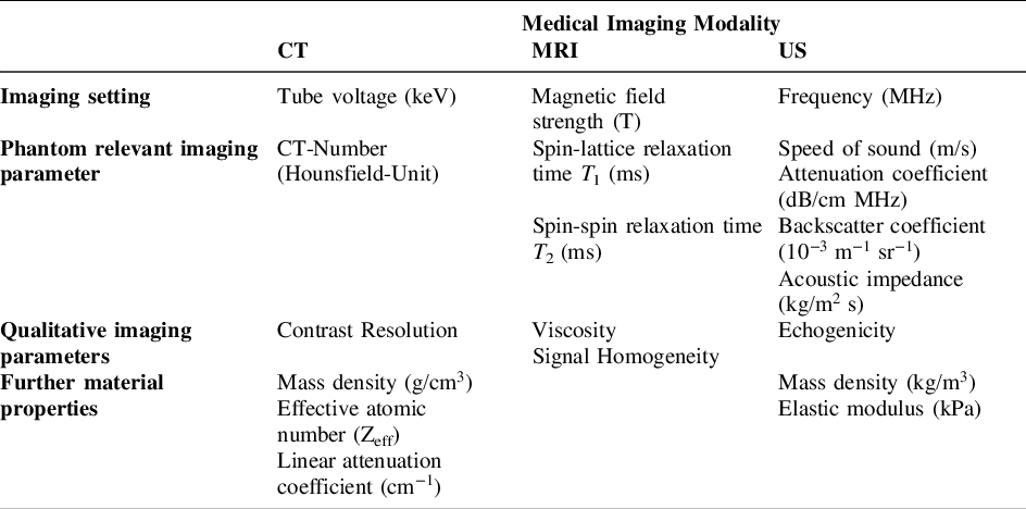
In (Table 1) the selected parameters and settings for the material catalogue are listed. While there are additional factors that could be considered, these represent the most relevant parameters and settings essential for achieving realistic and functional phantom designs and should be integrated into the catalogue.
Relevant image settings and imaging parameters for imaging modalities CT, MRI and US

For CT, the CT number, also known as the Hounsfield Unit (HU), is a primary selection criterion for phantom materials in most cases (Filippou & Tsoumpas, Reference Filippou and Tsoumpas2018). Reference CT values for human tissues can be obtained from clinical imaging or derived from literature sources. In many research studies, reference values for the mass attenuation coefficient from ICRU Report 44 (White et al., Reference White, Booz, Griffith, Spokas and Wilson1989) are frequently used. The CT number depends on various material properties, including the linear attenuation coefficient, the effective atomic number, and the mass density. Visual similarity to in vivo images can also serve as a qualitative measure for evaluating materials, as it helps in assessing how closely the phantom tissue mimics actual tissue appearance.
Other key considerations for CT imaging include contrast resolution—the ability to differentiate slight differences in radiodensity, which is crucial for simulating soft tissue contrasts in organs and lesions. Imaging settings, particularly the tube voltage, are also important factors, as they significantly influence image quality and material response in CT imaging.
For MRI imaging, the primary factor influencing imaging settings is the magnetic field strength of the MRI system. The longitudinal relaxation time (T1) and the transverse relaxation time (T2) are the two most widely used criteria for selecting MRI phantom materials, as these relaxation times are tissue-specific and help create realistic MRI images. Generally, the longitudinal relaxation time (T1) increases and the transverse relaxation time (T2) decreases as the magnetic field strength rises. Reference values for T1 and T2 relaxation times for different tissue types can be obtained from the literature or clinical imaging data. Additionally, material properties like spectral density and viscosity also impact relaxation times, with higher viscosity leading to shorter transverse relaxation times (T2). Temperature could also be considered, as relaxation times can vary with changes in temperature.
Magnetic susceptibility is another important factor; differences in susceptibility between materials can impact local magnetic field uniformity, which affects image quality. Depending on the specific application of the MRI phantom—such as anatomical imaging, functional MRI (fMRI), or diffusion-weighted imaging (DWI)—additional properties, like diffusion characteristics, may be relevant.
Similar to CT imaging, qualitative factors such as tissue contrast and signal homogeneity are essential for MRI phantom materials, as they contribute to realistic tissue differentiation and uniform signal distribution across the image.
In sonography (ultrasound) imaging, key parameters include the speed of sound, attenuation coefficient, backscatter coefficients, and acoustic impedance, as outlined in Table 1. These parameters are critical in selecting materials that accurately mimic the acoustic properties of human tissues. As a qualitative parameter, the echogenicity of a material needs to be considered. Tissues and organs can range from hypoechoic (low echo, darker) to hyperechoic (high echo, lighter) or isoechoic (equal echogenicity), depending on the amount of sound they reflect back to the transducer. Attenuation coefficients are also important, as they determine how much sound energy is absorbed or scattered within different tissue types, impacting the brightness and contrast of the image.
Elasticity, which includes mass density and elastic modulus, is essential for phantoms designed for elastography or to simulate tissue stiffness differences, such as soft tissue versus fibrous or tumorous regions. Homogeneity in tissue-mimicking materials helps reduce artifacts, though in some cases, phantoms may include features that intentionally generate artifacts like shadowing or refraction to enhance realism in training. The primary imaging setting for ultrasound is the frequency, which influences both resolution and penetration depth. Lower frequencies provide greater depth penetration, while higher frequencies enhance resolution for superficial structures. Selecting the appropriate frequency range and ensuring it matches the echogenic and attenuation characteristics of target tissues are important for designing realistic ultrasound phantoms.
3. Design of the phantom material design catalogue
The phantom material design catalogue is implemented as a digital tool in Microsoft Excel (Microsoft Corporation, USA), allowing for easy access, update, and expansion of content as new material data becomes available. A printed version can also be used if needed. To provide a clear overview, facilitate quick information retrieval, and ensure user-friendly navigation, the catalogue is divided into an overview section and three detailed catalogues, each dedicated to one of the primary imaging modalities (CT, MRI, and US), as shown in (Figure 2).
A graphical user interface (GUI) supports navigation through the different sections of the catalogue, providing users with guided access to the data, cf. on the right of (Figure 2). Filtering options are available via a drop-down menu or slicers located at the top of each catalogue sheet, allowing users to efficiently sort and filter information to meet specific needs. This design enables both broad accessibility and a tailored user experience for professionals working in phantom development.
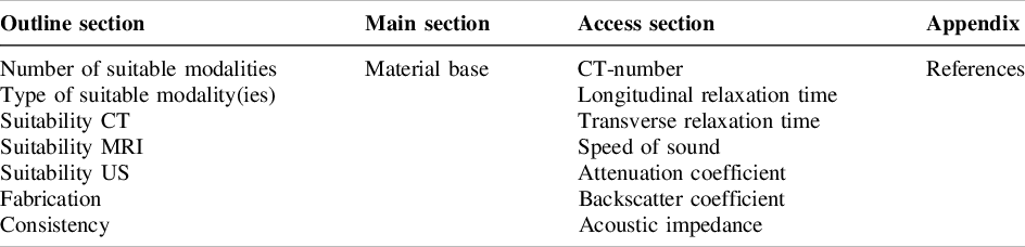
The structure of the overview catalogue is illustrated in (Table 2). When selecting materials for phantoms, the suitability of each material for one or more imaging modalities is of primary importance, so the first classification criteria include the number and type of applicable modalities. Another key criterion is the fabrication method, which is crucial for accurately reproducing geometrically complex organs, with mould casting and additive manufacturing being the most commonly used methods in phantom design. Material consistency is also an important classification factor, as it relates to the imaging, physical, and mechanical properties needed to meet specific phantom requirements. Viscosity was deliberately not chosen to describe consistency as it is rarely reported in the studies, may be less understandable and does not provide a clear added value.
Graphical user interface (left), excerpt of the overview catalogue (right, qualitative image)

The structure is not differentiated to the extent that the individual elements of the main section can be distinguished from each other, as this would no longer serve any useful purpose in terms of material selection. For example, the materials could be further differentiated by the number of different monomers or the molecular weight of a polymer, density, elements contained, classification as a pure substance or a mixture, etc., but these characteristics would be of little use in phantom development.
The main part of the overview catalogue lists various material bases, organized into twenty categories to support practical material selection in phantom development. Examples include gelatine-alginate, elastomeric materials, rigid plastics, silicon polymers, and agarose gel. The categories were chosen according to the current state of research, in particular based on the study by (Ahmad et al., Reference Ahmad, Suardi, Shukri, Mohammad, Oglat, Alarab and Makhamrah2020).
In the access section of the overview catalogue the phantom relevant imaging parameters are listed, cf. (Table 2). Selectable parameters include the CT-number for CT imaging, the longitudinal and transverse relaxation times for MRI, and, for ultrasound, the speed of sound, attenuation, backscatter coefficients, and acoustic impedance. For each parameter, minimum and maximum values are provided where available. For the attenuation and backscatter coefficients in US, only the frequency-independent variant is used, allowing for simpler material comparisons and more precise statements about material behaviour. To improve differentiation between imaging-specific information, background colours are used: yellow for CT, green for MRI, and blue for US.
This structure ensures that the catalogue effectively supports informed decision-making by focusing on material properties aligned with key development and imaging needs.
Structure of the outline, main, and access sections of the overview catalogue

The three detailed catalogues are related to each other by the extended classification section of the general catalogue, ensuring consistency in the catalogue system. The only difference between the detailed catalogues is the focus on a single modality, see for example (Figure 3). However, information on whether a material is suitable for other modalities is intentionally included.
In each detailed catalogue, the access section includes the information from (Table 1). The primary imaging settings are prioritized as access features, followed by imaging parameter values and qualitative selection criteria, cf. (Table 1). Imaging settings were narrowed down to the most significant ones, like tube voltage for CT, magnetic field strength for MRI, and frequency for US. Additionally, the detailed catalogue provides information on other material properties directly related to imaging properties and their adjustability. Possible tissue equivalences are also provided, allowing users to search by specific tissue types, such as bone. In comparison to the main section to the overview catalogue in the main section of the detail catalogues the material base is also specified further and other material components listed.
Following the example of (Roth, Reference Roth2001), each printed catalogue is preceded by a principles sheet that briefly explains the purpose, use, application, definition of the central term, structure, variants and examples. This short introduction to the catalogue allows a quick understanding of the structure and content of the catalogue.
Excerpt from the print version of the US detail catalogue

The catalogue was be validated using the requirements list of VDI 2222 (Verein Deutscher Ingenieure, 1997) including user feedback from medical imaging experts from the university medical center Hamburg-Eppendorf (UKE).
Fast access is ensured through the division into an overview catalogue and detailed catalogues, with imaging properties serving as access features. Standardized structural components, flowcharts, and principle sheets enhance usability. The catalogue is structured to align with the material selection process in phantom development. Imaging modalities define the classification of materials, shaping characteristics guide the manufacturing process, and material consistency reflects key mechanical properties relevant to phantom applications. In the detailed catalogues, materials are further categorized based on their fundamental material bases, reinforcing a structured and systematic approach. This organization establishes a crucial interface between imaging modality, fabrication method, and material selection.
Beyond structural adaptability, the catalogue is also designed to remain aligned with the current state of research. As new surrogate materials are developed, selection parameters refined, and influencing factors on imaging properties investigated, the catalogue remains flexible. The selection of access parameters is based on the latest research insights, ensuring that relevant material characteristics are highlighted. Should research findings evolve, the catalogue’s adaptability will be key in updating selection criteria accordingly.
Completeness is evaluated by comparing the included material bases with those reported in review studies. For example, 11 out of 14 material bases listed by (Wang et al. Reference Wang, Noh, Brown, Roujol, Li, Wang, Housden, Ester, Al-Hamadani, Rajani and Rhode2020) for ultrasound phantoms, 11 out of 12 material bases cited by (Ahmad et al. Reference Ahmad, Suardi, Shukri, Mohammad, Oglat, Alarab and Makhamrah2020) across imaging modalities, and all commonly used materials for additive manufacturing of CT phantoms from (Tino et al. Reference Tino, Leary, Yeo, Kyriakou, Kron and Brandt2020) are represented in the catalogue. While the catalogue is not exhaustive, it presents a broad and representative spectrum of phantom materials.
The catalogue also serves as a tool for inspiring further research and refinement. By identifying gaps in material studies, it encourages targeted investigations. One scenario is that if a general material base is suitable for a specific imaging modality, it suggests that its subcategories may also be viable, prompting further testing. Another possibility is the identification of modifying material components that influence imaging properties, allowing for novel material combinations. Additionally, systematic material investigations can address knowledge gaps, improving reproducibility by maintaining consistent influencing factors in phantom development.
The medical imaging experts from the UKE were consulted through a questionnaire, they rated the catalogue with the highest points possible, reflecting their strong approval and acknowledging its effectiveness and utility in their field.
By providing a structured, adaptable, and research-driven approach to material selection, the catalogue serves as a valuable resource for researchers and practitioners, supporting the ongoing development and optimization of phantoms across various imaging modalities.
4. Conclusion and outlook
Phantom material reviews provide valuable overviews of available materials for phantom design. However, these reviews are often limited in scope, typically focusing on just one imaging modality or a very specific tissue type. This narrow focus can restrict the comprehensive applicability of the materials reviewed, as phantoms are frequently used across multiple modalities, each with distinct imaging characteristics and requirements. Furthermore, many of these reviews do not account for the influence of image acquisition settings, which play a crucial role in determining the quality and accuracy of the recorded data. Parameters such as tube voltage in CT, magnetic field strength in MRI, and frequency in US can significantly impact the imaging properties of the materials and, consequently, the phantom’s imaging parameters. This limitation highlights the need for a more integrated approach that combines material properties with the specific imaging conditions to ensure the selection of the most appropriate surrogates for each use case.
A design catalogue, like it is presented in this work, enhances this process by providing a structured overview of phantom materials, incorporating not only material properties but also imaging settings and their influence on performance. By offering detailed, organized information, the catalogue supports the material selection process and improve accessibility to phantom data. This enhances decision-making, allowing for more informed and precise choices when designing phantoms for various imaging modalities and applications.
While the designed catalogue supports material selection, it is essential that the selected materials still undergo testing for suitability in the specific application and under the specific imaging settings. The testing results should be integrated into the catalogue to continually expand and improve its accuracy and utility. Nevertheless, the catalogue is just one element of the support tool for phantom design. Other supportive approaches include material testing and the use of additional tools to refine the design and validation of phantoms.
Limitations in generating the catalogue were encountered during entering material data, primarily due to the lack of available information. Many studies did not report all of the factors necessary for a comprehensive catalogue, which resulted in numerous empty fields within the database. One of the primary exclusion criteria for the included data was the absence of crucial imaging parameters, such as tube voltage for CT or magnetic field strength for MRI. These variables have a significant impact on imaging properties and are essential for accurate material selection. Missing parameters for CT often included details such as slice thickness, pitch, object thickness, material density, and effective atomic number, which can influence the recorded imaging characteristics. Similarly, for MRI, the temperature during the measurement, as well as the choice of pulse sequences and times, were often not specified, despite their importance in determining relaxation times and signal intensity. In ultrasound imaging, temperature was also rarely reported, and the acoustic properties measured varied greatly between studies, with different combinations of parameters such as attenuation, backscatter coefficients, and speed of sound being used across different publications. These inconsistencies in the reporting of imaging parameters highlight the challenges in creating a fully comprehensive and standardized catalogue. Despite these limitations, the designed catalogue includes over 100 material combinations and serves as a valuable tool by consolidating available data and providing a foundation for future work, where more complete datasets can be integrated as further studies provide more detailed and consistent reporting. This is also of interest for the emerging 3D printing materials in this field, offering a framework to systematically document their suitability for medical imaging applications.
Acknowledgments
Marie Wegner is supported by the Add-on Fellowship of the Joachim Herz Foundation.




 Open access

