


Introduction
Culture-negative endocarditis requires urgent treatment despite incomplete microbiologic data. Tropheryma whipplei is an increasingly recognized cause, often presenting as isolated cardiac disease rather than classic gastrointestinal Whipple disease. Published series emphasize middle-aged men, aortic-valve involvement, and heart failure, and molecular studies suggest that T. whipplei is an important contributor to culture-negative endocarditis cohorts. Reference Ioannou, Kourtidis, Mytilinis, Psyllaki, Baliou and Kofteridis1–Reference Geissdorfer, Moos and Moter4 Presentation may be dominated by cardiac manifestations, lowering clinical suspicion. Yet routine blood cultures are usually negative and blood polymerase chain reaction has limited sensitivity, so confirmation often depends on valve tissue obtained late in the clinical course. Reference McGee, Brienesse, Chong, Levendel and Lai2,Reference Knol, Nijhuis, Geeraedts and Linssen3 This delay can prolong exposure to broad-spectrum therapy and defer organism-directed management. In patients who receive empiric antibiotics before diagnostic sampling is complete, noninvasive molecular assays may offer earlier organism-directed guidance. Reference Eichenberger, Degner and Scott5
Clinical case
A 56-year-old man with heart failure presented with progressive lower-extremity edema, worsening fatigue, and 3 to 6 months of orthopnea. He was admitted to the medical intensive care unit with acute decompensation manifested by transaminitis, coagulopathy, elevated lactate, leukocytosis, thrombocytopenia, pleural effusions, acute kidney injury, and ascites. His history included hypertension, hyperlipidemia, and osteoarthritis. He denied injection drug use but reported remote tobacco use and occupational exposure to animals and animal feces.
Point-of-care ultrasonography showed an echogenic aortic-valve lesion. Transthoracic and transesophageal echocardiography demonstrated a dilated left ventricle with mildly reduced ejection fraction, bicuspid aortic-valve morphology, a large mobile vegetation, severe eccentric aortic regurgitation, and a flail leaflet (Figure 1).
Transthoracic and transesophageal echocardiography of aortic-valve infective endocarditis. (A) Transthoracic parasternal long-axis view showing a large mobile aortic-valve vegetation (yellow arrow). (B) Color Doppler image showing severe eccentric aortic regurgitation (white arrow). (C) Additional transthoracic view demonstrating bicuspid aortic-valve morphology. (D) Transesophageal view showing a flail aortic leaflet with attached vegetation (yellow arrow). (E) Color Doppler transesophageal view confirming severe eccentric aortic regurgitation (white arrow). (F) Three-dimensional transesophageal echocardiography showing the abnormal bicuspid aortic valve.

Because infective endocarditis was strongly suspected, empiric ceftriaxone, meropenem, and vancomycin were started. Blood cultures were obtained only after the first ceftriaxone dose, potentially lowering culture yield. Plasma microbial cell-free DNA sequencing (Karius assay; Karius, Redwood City, CA) was collected on hospital day 2. Serologic and microbiologic evaluation for Brucella, Coxiella, Bartonella, and Lyme or rickettsial infection was unrevealing. Four days later, the assay detected a high burden of T. whipplei.
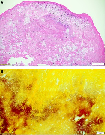
Therapy was narrowed to ceftriaxone after that result. Ten days after echocardiographic diagnosis, the patient underwent surgical aortic-valve replacement with a bovine pericardial valve and debridement and repair of infected tissue involving the aorto-mitral curtain. Surgical pathology showed destructive vegetation with mixed inflammation, neovascularization, organizing fibrin thrombi, and calcification; Gram stain demonstrated numerous gram-positive bacilli within the vegetation (Figure 2). Grocott-Gomori methenamine silver, Kinyoun acid-fast bacilli, and Fite stains were negative. He completed 4 weeks of ceftriaxone after surgery and then began a planned 18-month course of trimethoprim-sulfamethoxazole to reduce relapse risk. He was discharged home in stable condition nearly 3 weeks after admission and remained adherent to therapy at follow-up.
Surgical pathology of the excised valvular tissue. (A) Hematoxylin and eosin stain showing destruction of valvular tissue by vegetation, mixed inflammation, neovascularization, organizing fibrin thrombi, and calcification. (B) Gram stain at 60x magnification showing numerous gram-positive bacilli within the vegetation.

A diagnostic stewardship framework for culture-negative endocarditis
This case suggests 3 stewardship questions when considering advanced diagnostics in culture-negative endocarditis. First, is the organism plausible in the clinical syndrome? T. whipplei deserves consideration in middle-aged men with culture-negative aortic-valve endocarditis, heart failure, or otherwise unexplained destructive valvular disease, even without diarrhea or other classic extracardiac features. Reference Ioannou, Kourtidis, Mytilinis, Psyllaki, Baliou and Kofteridis1–Reference Knol, Nijhuis, Geeraedts and Linssen3 The bicuspid aortic valve in this patient may also have increased susceptibility to endocarditis.
Second, will the result change management before invasive confirmation is available? Here, the patient was critically ill, and the blood cultures were likely compromised by antibiotic exposure. The plasma sequencing result supported the earlier discontinuation of meropenem and vancomycin and enabled narrower therapy before operative pathology was available. The assay was especially useful because invasive tissue confirmation was not immediately available. That earlier narrowing was clinically relevant because he presented with severe valvular dysfunction, heart failure, acute kidney injury, and multiorgan derangements.
Third, how should the test complement rather than replace standard evaluation? Plasma microbial cell-free DNA did not replace echocardiography, surgery, histopathology, or clinicopathologic correlation; instead, it provided microbiologic clarity when conventional blood culture was least informative. Reference Eichenberger, Degner and Scott5 Published experience with plasma microbial cell-free DNA sequencing in T. whipplei endocarditis remains limited. In a focused review of the English-language literature, we identified 3 earlier reports, including 1 conference abstract and 2 full case reports. Reference de Vries, Macintyre and Buggy6–Reference Rabinowitz, Walia, Pottinger and Lieberman8 Across those reports and the present case, patients were men older than 50 years, aortic or mitral valve involvement predominated, and plasma sequencing established or strongly supported the diagnosis before or without valve-tissue confirmation. Our case presents with fulminant valvular dysfunction, prior antibiotic exposure, and subsequent pathology demonstrating abundant gram-positive bacilli.
Conclusion
For ASHE readers, the central lesson is not that plasma microbial cell-free DNA should replace foundational endocarditis diagnostics. Rather, in carefully selected patients with suspected culture-negative endocarditis, especially after antibiotic exposure or when invasive sampling will be delayed, it can support earlier organism-directed therapy and reduce unnecessary broad-spectrum antimicrobial exposure while definitive tissue-based studies are pending.
Data availability statement
Deidentified clinical details supporting this case report are not publicly available because of patient privacy considerations.
Acknowledgments
The authors thank the clinical teams involved in the care of this patient.
Author contributions
Conceptualization: A.J., M.A.A. Data curation: A.J. Formal analysis: A.J. Investigation: A.J., S.K., M.R.S., L.C.L., E.G.S., K.K.L., A.H. Methodology: T.L. Project administration: M.A.A. Supervision: M.A.A. Writing-original draft: A.J. Writing-review and editing: T.L., L.C.L., E.G.S., K.K.L., S.K., M.R.S., A.H., M.A.A. All authors reviewed and approved the final manuscript.
Financial support
None reported.
Competing interests
S.K. and M.R.S. report honoraria and consulting fees from Karius Inc. A.J., L.C.L., E.G.S., K.K.L., A.H., T.L., and M.A.A. report no conflicts of interest relevant to this article.
Artificial intelligence assistance
Artificial intelligence tools were used solely for minor editorial assistance related to grammar and language clarity. All content, analysis, and conclusions were developed by the authors.
Patient consent
This case report was reviewed by the Baylor College of Medicine Institutional Review Board and determined to meet criteria for exemption; separate patient consent for publication was not required. No identifiable patient information is included.


 Open access

